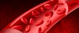
Thrombosis in pregnant women is a pathological condition characterized by the formation of blood clots in the lumen of blood vessels, which are called thrombi. They do not allow blood to circulate freely through the circulatory system, seriously impeding blood flow and causing a number of serious complications. Vein thrombosis in pregnant women is not uncommon. It appears against the backdrop of a number of changes occurring in a woman’s body during this period, and requires special attention both from the woman herself and from doctors.
Prevention of thrombosis in pregnant women and its treatment is one of the areas of work of the CELT Phlebology Department. Our clinic has been operating since 1993 and is multidisciplinary, so phlebologists work closely with gynecologists, achieving the best results. They have at their disposal a powerful diagnostic and treatment base, as well as modern gentle techniques that allow them to maintain the patient’s health and give birth to a healthy baby.
At CELT you can get advice from a phlebologist.
- Initial consultation – 3,000
- Repeated consultation – 2,000
Make an appointment
How does blood clotting occur?
The actual clot is formed by creating a fibrin network that strengthens and stabilizes it. This is due to the activation of the coagulation cascade - inactive blood coagulation factors circulating in the blood plasma begin to activate each other.
Under the influence of mechanical damage, platelets secrete thrombokinase, which triggers a number of processes leading to the formation of the corresponding factor that initiates blood clotting - calcium ions and plasma protein factors are important in this process.
Blood coagulation diagram
As a result of the blood clotting cascade, factor X together with factor Va forms a complex called prothrombinase, which converts prothrombin into thrombin. Thrombin, in turn, converts fibrinogen (a plasma protein that circulates in the blood) into fibrin (a water-insoluble protein), which forms a network of fibers that form the backbone of the clot.
Causes of blood clots
Thrombosis is promoted, in particular:
- surgical intervention;
- extensive injuries;
- respiratory failure;
- heart failure;
- oral contraceptives;
- use of certain hormonal drugs;
- phlebeurysm;
- obesity;
- stroke;
- age > 40 years.
The risk of thrombosis increases significantly during pregnancy and the postpartum period.
The risk of developing thrombosis during pregnancy is many times higher in people with congenital or acquired thrombophilia. It is estimated that 20-30% of cases of thromboembolic disease are associated with a genetic predisposition to its development.
The main genetic causes of the disease are the carrier of the Leiden variant of the factor V gene of the blood coagulation system (F5 gene, mutation p.Arg534Gln) or a mutation of the prothrombin gene (F2 gene, mutation c. * 97G>A). Other known predispositions are hereditary deficiencies of blood clotting inhibitors (antithrombin, protein C or protein S) and hereditary hyperhomocysteinemia (associated, for example, with mutations in the MTHFR gene). In families with thrombosis, factor V Leiden mutation and prothrombin gene mutation are detected in approximately 60% of cases.
Diagram of blood clot formation
Why is a blood clot dangerous?
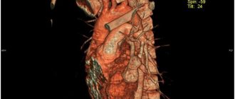
Thrombosis is life-threatening when a blood clot breaks away from the wall of a vein. A clot that travels through the bloodstream may travel into the atrium or ventricle or into the pulmonary artery, causing shock, cardiac arrest, and respiratory arrest due to a heart attack or pulmonary embolism. These conditions pose a direct threat to the life of a pregnant woman and can cause her sudden death.
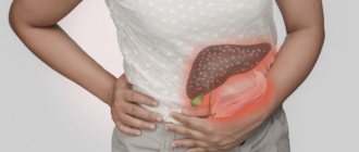
Other consequences of thrombosis include: post-thrombotic syndrome, which manifests itself as changes in skin color, pulmonary hypertension and leg ulcers. In pregnant women, thrombosis can cause miscarriage, and pregnancy and the postpartum period significantly increase the risk of its occurrence.
The risk of thrombosis during pregnancy is associated with pressure on the iliac veins, which increases as the baby grows. In this case, embolism most often develops in the iliofemoral region of the deep veins, and in 90% of women, symptoms appear only on the left leg.
Treatment
The choice of therapy depends on the location and severity of the disease. If a life-threatening condition occurs in a patient, he undergoes emergency surgery; in other cases, conservative therapy is chosen.
Conservative therapy includes groups of drugs:
- anticoagulant;
- NSAIDs (non-steroidal anti-inflammatory drugs);
- glucocorticoid;
- phlebotonic;
- disaggregant;
- preparations for local therapy - ointments, gels with heparin or in combination with non-steroidal anti-inflammatory drugs;
- infusion vascular therapy to improve microcirculation and blood rheology.
The patient is required to wear compression garments or elastic bandaging of the sore limb. And to accelerate thrombus lysis and improve hemodynamics in the lower extremities, you can use hardware pneumocompression (lymphopressotherapy).
Surgery
It is performed in case of lack of effect from conservative methods of therapy, increasing symptoms of vessel occlusion, development of ascending phlebothrombosis, and if there is a suspected risk of blood clot rupture. The following surgical interventions can be performed:
- Installation of a vena cava filter in the IVC to prevent thromboembolism.
- Catheter-directed thrombolysis.
- Crossectomy.
- Thrombextraction
- Bypass or prosthetic replacement of the affected vein.
Causes of thrombosis during pregnancy
During pregnancy and the perinatal period, the cause of thrombosis can be:
- hormonal changes necessary to maintain pregnancy (hormones cause vasodilation and stagnation of blood in the veins, increase blood viscosity, which contributes to the formation of blood clots);
- difficult outflow of blood from the legs due to pressure on the veins by the enlarging uterus;
- C-section;
- prolonged immobilization after childbirth, contributing to venous stagnation.
Stagnation of blood in the veins
The above risk factors, coexisting with an individual genetically determined predisposition to thrombosis, significantly increase the risk of thrombosis during pregnancy and its dangerous consequences.
Main risk factors for the development of thrombophlebitis
Among the risk factors for the development of thrombophlebitis are the following:
- varicose veins;
- prolonged immobility of the body during a long trip by plane or car;
- prolonged bed rest (for example, after surgery);
- stroke;
- venous catheterization;
- pregnancy, childbirth, abortion and other gynecological operations;
- hormone therapy;
- blood disorders (for example, increased blood clotting);
- obesity;
- malignant diseases;
- dehydration (including while taking diuretics);
- decreased immunity;
- infectious diseases, etc.
The presence of one or even more than one factor increases the risk of developing thrombophlebitis. Contact a phlebologist!
Symptoms of thrombosis in pregnant women
Thrombosis in pregnant women is often asymptomatic. The symptoms that may appear with thrombosis during pregnancy can be ambiguous and can occur with other diseases, and when they do occur, they are often underestimated and considered typical pregnancy symptoms.
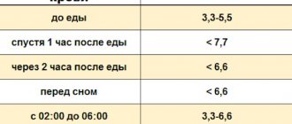
The most common symptoms that appear several days after a clot forms include:
- swelling, usually in one leg, around the ankle, then swelling in the calf or the whole leg (usually one leg is affected);
- pain in the legs, aggravated by walking, with increased pain and tenderness when touched, disappearing when the limb is immobilized;
- redness and increased warmth of the skin of the painful limb.
Dangerous symptoms of thrombosis appear when a blood clot breaks off and migrates along with the bloodstream. During complications of thrombosis (pulmonary embolism), difficulty breathing occurs with shortness of breath and chest pain. Difficulty in breathing progresses rapidly and can kill the patient within seconds.
Diagnosis of thrombosis
Diagnostic tests for thrombosis include blood chemistry tests (especially D-dimer levels) and duplex Doppler ultrasound of the venous vessels, which will determine the correct venous blood flow in the extremities and the presence of blood clots. These tests can confirm the occurrence of thrombosis only when symptoms first appear.
Ultrasound examination of venous vessels
A test to determine the risk of thrombosis is to perform DNA tests that look for abnormal gene mutations, and most often mutations that predispose to congenital thrombophilia, especially during pregnancy and the puerperium.
Determining the risk of the disease in women during the precontraceptive period will allow the doctor to begin appropriate prevention and treatment of thrombosis during pregnancy. To determine whether you are at risk of developing thrombosis, it is necessary to carry out thrombophilia tests, that is, tests that detect mutations in the genes encoding: factor V Leiden (Leiden-FVL mutation), prothrombin, methylenetetrahydrofolate reductase (MTHFR), factor PAI-I. / SERPINE1 Factor V R2.
In pregnant women, thrombosis most often occurs in the form of FVL mutation (2-10%), mutation of the methylenetetrahydrofolate reductase gene MTHFR (8-16%), mutation of the prothrombin gene (2-6%), deficiency of proteins C and S (0.2-1% ) and the presence of anticardiolipin antibodies (1-7%).
Both congenital (hereditary) thrombosis and acquired thrombosis increase the risk of pregnancy loss. An estimated 15% of pregnancies end in miscarriage, and this affects 0.4–2% of couples. The results of a clinical study conducted by the Nimes Obstetrics and Hematology Department (NOHA) showed a strong correlation between unexplained pregnancy loss and the presence of prothrombin and factor V mutations in the heterozygous system.
The Leiden mutation is a genetic defect that affects approximately 5% of white people, is associated with a 3- to 7-fold increased risk of venous thrombosis, and is predominantly inherited.
This means that a person who has a mutation in one copy of the gene (known as a heterozygote) has an increased risk of thrombophilia. The risk is even higher for people who are homozygous, i.e. have a mutation in both copies of the F5 gene. Heterozygotes for the Leiden mutation have a 2-3 times increased risk of pregnancy loss and other complications during pregnancy (for example, eclampsia, fetal malnutrition, premature placental abruption).
Thrombophilia associated with factor V Leiden mutation and thrombophilia associated with resistance to active protein C are inherited in an autosomal dominant manner. The resistance of factor V to activated protein C accelerates the blood clotting process and does not inhibit the growth of a blood clot.
Activated coagulation factor V is a component of the factor X enzyme complex that converts prothrombin to thrombin during blood clotting. Factor V deficiency is inherited in an autosomal recessive manner and promotes a slower blood clotting process due to decreased amounts of factor V.
Factor II of the prothrombin F2 gene (G20210A) causes thrombophilia associated with thrombin deficiency. The mutation increases the concentration of prothrombin in the blood and is inherited in an autosomal dominant manner. Prothrombin is activated to thrombin during blood clotting, which allows fibrinogen to be converted into fibrin; the presence of mutations disrupts this process.
The MTHFR gene encodes the enzyme methylenetetrahydrofolate reductase, which catalyzes the formation of 5-methyltetrahydrofolate, which is necessary for the conversion of the potentially toxic amino acid homocysteine to methionine by methionine synthase. This mutation is manifested by an increase in the concentration of homocysteine in the blood serum. Excess homocysteine in the body can damage the endothelium of blood vessels and, as a result, lead to atherosclerosis and venous and arterial thrombosis.
Thrombophilia associated with prothrombin is predominantly inherited and is characterized by symptoms of venous thromboembolism, in adults mainly deep vein thrombosis or pulmonary embolism. Thrombophilia associated with the presence of the c.20210G>A variant of the F2 gene has been shown to increase the risk of pregnancy loss.
Deep vein thrombosis
The risk of thrombosis during pregnancy and pulmonary embolism is reduced by any measures to improve blood flow and prevent its stagnation. In pregnant women with a genetically confirmed predisposition to the disease, prophylactic anticoagulants (low molecular weight heparins) are used under strict medical supervision. These women should wear special tights, maintain a healthy body weight, eat a healthy low-fat diet, hydrate regularly, be physically active and avoid prolonged sitting.
By performing genetic tests for thrombophilia and gene mutations that cause thrombosis, a pregnant woman can prevent serious complications (mainly pulmonary embolism and miscarriage) by administering prophylactic or anticoagulant treatment in close collaboration with her doctor. Only this procedure guarantees the correct course of pregnancy, childbirth and the postpartum period for both mother and child.
Once performed, genetic testing will provide lifelong information and enable you to make better decisions, including preventative measures and reducing symptoms of thrombosis during pregnancy and throughout the fertile period.
MAKE AN APPOINTMENT
[contact-form-7 id=”296" title=”Untitled”]
Abortion and contraception clinic in St. Petersburg - department of the medical gynecological association "Diana"
Make an appointment, tests or ultrasound via the contact form or by calling +8 (812) 62-962-77. We work seven days a week from 09:00 to 21:00.
We are located in the Krasnogvardeisky district, next to the Novocherkasskaya, Ploshchad Alexander Nevsky and Ladozhskaya metro stations.
The cost of a medical abortion in our clinic is 3,300 rubles. The price includes all pills, an examination by a gynecologist and an ultrasound to determine the timing of pregnancy.
Our doctors
Malakhov Yuri Stanislavovich
Doctor - cardiovascular surgeon, phlebologist, Honored Doctor of the Russian Federation, Doctor of Medical Sciences, doctor of the highest category
Experience 36 years
Make an appointment
Drozdov Sergey Alexandrovich
Cardiovascular surgeon, phlebologist, Doctor of Medical Sciences
47 years of experience
Make an appointment