The article was checked by a cardiologist of the highest category Kurbatova I.V. , is for general informational purposes only and does not replace specialist advice. For recommendations on diagnosis and treatment, consultation with a doctor is necessary.
At the Clinical Hospital on Yauza, a range of diagnostics is carried out for paroxysmal tachycardia - ECG, 24-hour ECG monitoring, echocardiography, if indicated - bicycle ergometry, stress ECHO-CG, EPI - electrophysiological study of the heart, CT coronary angiography, cardiac MRI, etc. Experienced cardiologists will select an effective one drug therapy regimen. If necessary, surgical treatment is possible - radiofrequency ablation, implantation of a cardioverter-defibrillator, pacemaker, which is performed by leading cardiac surgeon Prof. A.V. Ardashev.
Paroxysmal tachycardia is a paroxysmal increase in heart rate from 140 to 220 beats per minute or more. Occurs when the normal sinus rhythm (that is, set by the sinus node) is suppressed by a pathological source of impulses. If the source is located in the atria or in the transition from the atrium to the ventricles, they speak of supraventricular or supraventricular tachycardia. If the source is located in the ventricles of the heart, we are talking about ventricular tachycardia.
Make an appointment with a cardiologist
Diagnostics
- Electrocadiography, including 24-hour Holter monitoring, ECG with stress - to identify the fact and type of arrhythmia.
- Echocardiography, less often - MRI and CT studies of the heart - to search for intracardiac causes of rhythm disturbances.
- Additional studies according to an individually developed examination plan to identify non-cardiac causes of arrhythmia (laboratory tests, ultrasound, etc.).
- According to indications, intracardiac electrophysiological examination (EPI) is performed followed by surgical treatment of this type of arrhythmia.
Treatment
- Emergency care for paroxysmal tachycardia. There are a number of ways to reflexively slow down the rhythm, the so-called vagal tests, which the cardiologist teaches the patient. An urgent call for a cardiac emergency medical team is urgent.
- Conservative therapy . Performed by experienced cardiologists. Individual selection of medications to relieve paroxysmal tachycardia and prevent attacks, as well as to reduce the risk of thrombosis, improve myocardial condition, etc.
- Surgery . Depending on the patient’s condition and the nature of the attacks, various interventions are used: radiofrequency ablation of arrhythmogenic zones or implantation of a cardioverter-defibrillator.
At the Yauza Clinical Hospital, the decision on surgical intervention, its choice and the operation itself are carried out by cardiac surgeon-arrhythmologist Professor A.V. Ardashev. The cardiology department of our hospital carries out dynamic monitoring of patients with paroxysmal tachycardia and timely correction of their treatment, reducing the risk of dangerous conditions and complications.
Cost of services
Make an appointment
You can see prices for services
Clinical variants and incidence of supraventricular tachycardias in children
Supraventricular (supraventricular) tachycardias (SVT) account for 95% of all tachycardias in children and are often paroxysmal in nature. In most cases, SVTs are not life-threatening rhythm disturbances, but may be accompanied by complaints of a sharp deterioration in health and have a pronounced clinical picture.
The term “supraventricular tachycardia” refers to three or more consecutive heart contractions with a frequency exceeding the upper limit of the age norm in children and more than 100 beats per minute in adults, if the participation of the atria or atrioventricular (AV) junction is required for the occurrence and maintenance of tachycardia.
SVT includes tachycardias that occur above the bifurcation of the His bundle, namely in the sinus node, atrial myocardium, AV junction, trunk of the His bundle, emanating from the mouths of the vena cava, pulmonary veins, and also associated with additional conduction pathways.
Supraventricular tachycardia is a common form of cardiac arrhythmia in children and adults. The prevalence of paroxysmal SVT in the general population is 2.25 cases per 1000 people, with 35 new cases occurring per year per 100,000 population [1]. The incidence of SVT in children, according to various authors, varies significantly and ranges from 1 case in 25,000 children to 1 case in 250 children [2, 3].
In practice, it is convenient to use the clinical and electrophysiological classification of SVT, which systematizes individual nosological forms of tachycardias, indicating their localization, electrophysiological mechanism, various subtypes and variants of the clinical course [4].
Clinical and electrophysiological classification of supraventricular tachycardias in children:
I. Clinical options for SVT:
1. Paroxysmal tachycardia:
- stable (duration of attack 30 s or more);
- unstable (duration of attack less than 30 s).
2. Chronic tachycardia:
- constant;
- permanently returnable.
II. Clinical and electrophysiological types of SVT:
1. Sinus tachycardia:
- sinus tachycardia (functional);
- chronic sinus tachycardia;
- sinoatrial reciprocal tachycardia.
2. Atrial tachycardia:
- focal (focal) atrial tachycardia;
- multifocal or chaotic atrial tachycardia;
- incisional atrial tachycardia;
- atrial flutter;
- atrial fibrillation.
3. Tachycardia from the AV junction:
- atrioventricular nodal reciprocal tachycardia: - typical; - atypical;
- focal (focal) tachycardia from the AV junction: - postoperative; - congenital; - “adult” form.
4. Tachycardia with the participation of accessory pathways (Wolf–Parkinson–White syndrome (WPW), atriofascicular tract and other accessory pathways (ADP)):
- paroxysmal orthodromic AV reciprocal tachycardia with the participation of AP;
- chronic orthodromic AV reciprocal tachycardia with the participation of “slow” AP;
- paroxysmal antidromic AV reciprocal tachycardia with the participation of AP;
- paroxysmal AV reciprocal tachycardia with pre-excitation (involving several APs).
According to the nature of the course, tachycardias are divided into paroxysmal and chronic. Paroxysmal tachycardia has a sudden onset and end of an attack. Attacks of tachycardia are considered stable if they last more than 30 seconds, and unstable if their duration is less than 30 seconds. The clinical picture of paroxysmal tachycardia is quite diverse. In children of the first year of life, during an attack of tachycardia, anxiety, lethargy, refusal to feed, sweating during feeding, and pallor may be observed. In young children, attacks of tachycardia may be accompanied by pallor, weakness, sweating, drowsiness, and chest pain. In addition, children quite often emotionally and figuratively describe attacks, for example, as “heart in the tummy”, “jumping heart”, etc. School-age children can usually talk about all the clinical manifestations of an attack of tachycardia. Often attacks of tachycardia are provoked by physical and emotional stress, but they can also occur at rest. When asked about the heart rate during an attack of tachycardia, children and their parents usually answer that the pulse “cannot be counted”, “cannot be counted”. Sometimes attacks of tachycardia occur with a pronounced clinical picture, accompanied by weakness, dizziness, darkening of the eyes, syncope, and neurological symptoms. Loss of consciousness occurs in 10–15% of children with SVT, usually immediately after the onset of a paroxysm of tachycardia or during a long pause in the rhythm after its cessation.
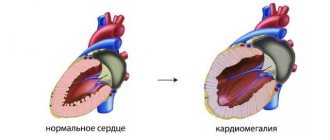
Chronic tachycardia does not have an acute beginning and end of the attack; it drags on for a long time and can last for years. Chronic tachycardias are divided into constant (continuous) and constant-recurrent (continuously recurrent). Tachycardia is said to be permanent if it accounts for most of the time of day and represents a continuous tachycardic chain. With the constantly recurrent type of tachycardia, its circuits are interrupted by periods of sinus rhythm, but tachycardia can also occupy a significant portion of the day. This division of chronic tachycardia into two forms is somewhat arbitrary, but has a certain clinical significance, since the more time of day the tachycardia occupies and the higher the heart rate, the higher the risk of the child developing secondary arrhythmogenic cardiomyopathy and progressive heart failure. Quite often, chronic forms of SVT occur without distinct symptoms and are diagnosed after the first signs of heart failure appear.
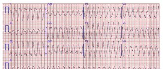
In most cases with SVT, the QRS complexes are narrow, but with aberrant impulse conduction they can widen. The heart rate (HR) during tachycardia depends on the age of the children. In newborns and children of the first years of life, heart rate during paroxysmal tachycardia is usually 220–300 beats/min, and in older children it is 180–250 beats/min. In chronic forms of tachycardia, heart rate is usually somewhat lower and amounts to 200–250 beats/min in children of the first years of life and 150–200 beats/min in older ages.
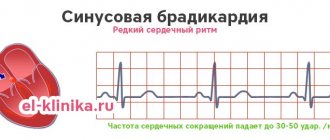
Most often, SVT must be differentiated from functional sinus tachycardia, which is usually a normal physiological response to physical and emotional stress due to an increase in sympathetic influences on the heart. At the same time, sinus tachycardia can signal serious illnesses. It is a symptom and/or compensatory mechanism of the following pathological conditions: fever, arterial hypotension, anemia, hypovolemia, which can be the result of infection, malignant processes, myocardial ischemia, congestive heart failure, pulmonary embolism, shock, thyrotoxicosis and other conditions. It is known that the heart rate has a direct relationship with body temperature; for example, when the body temperature of a child older than two months increases by 1 °C, the heart rate increases by 9.6 beats/min [5]. Sinus tachycardia is provoked by various stimulants (caffeine, alcohol, nicotine), the use of sympathomimetic, anticholinergic, some antihypertensive, hormonal and psychotropic drugs, as well as a number of toxic and narcotic substances (amphetamines, cocaine, ecstasy, etc.). Functional sinus tachycardia usually does not require special treatment. The disappearance or elimination of the cause of sinus tachycardia in most cases leads to the restoration of the normal frequency of sinus rhythm. Sometimes children experience chronic sinus tachycardia, in which the sinus rate does not correspond to the level of physical, emotional, pharmacological or pathological influence. Sinoatrial reciprocal tachycardia is extremely rarely recorded, usually having an unstable paroxysmal course.
Atrial tachycardia in children is often chronic and difficult to treat with medication, can lead to congestive heart failure, and is the most common cause of secondary arrhythmogenic cardiomyopathy. Therefore, despite the relatively low incidence, atrial tachycardia is a serious problem in pediatric arrhythmology. The most common type of atrial tachycardia is focal (ectopic) atrial tachycardia, which accounts for 15% of all SVT in children under one year of age and 10% in children aged one to five years [6].
Focal atrial tachycardia is a relatively regular atrial rhythm with a rate above the upper normal for age, usually between 120 and 300 bpm. In this case, frequent P waves of non-sinus origin, located in front of the QRS complexes, are recorded on the ECG. The morphology of P waves depends on the location of the tachycardia focus. With the simultaneous functioning of several atrial rhythm sources, multifocal (multifocal) atrial tachycardia occurs. This rather rare form of tachycardia is well known as “chaotic atrial tachycardia”. Chaotic atrial tachycardia is an irregular atrial rhythm with a continuously varying frequency from 100 to 400 beats per minute with variable AV conduction of atrial impulses with a frequency of an also irregular ventricular rhythm of 100–250 beats/min. Atrial flutter is a regular, regular atrial rhythm, usually between 250 and 450 beats per minute. With typical atrial flutter on the ECG, instead of P waves, “sawtooth” F waves are recorded with the absence of an isoline between them and with a maximum amplitude in leads II, III and aVF. Incisional (postoperative) atrial tachycardia occurs in 10–30% of children after correction of congenital heart disease (CHD), during which surgical manipulations were performed in the atria. Incisional tachycardias can appear both in the early postoperative period and several years after surgery. They are a serious problem and largely determine mortality after cardiac surgery. Quite rarely, children experience atrial fibrillation, which is chaotic electrical activity of the atria with a frequency of 300–700 per minute, while f waves of different amplitudes and configurations are recorded on the ECG without an isoline between them. Atrial fibrillation leads to a decrease in cardiac output due to loss of atrial systole and arrhythmia itself. Another serious danger of atrial fibrillation is the risk of thromboembolic complications.
The AV junction is associated with the occurrence of two tachycardias that are different in electrophysiological mechanism and clinical course: AV nodal reciprocal tachycardia and focal tachycardia from the AV junction. Paroxysmal AV nodal reentrant tachycardia accounts for 13–23% of all SVTs [2, 6]. Moreover, the incidence of this form of tachycardia increases with age - from isolated cases in children under two years of age to 31% of all SVT in adolescents [6, 7]. The occurrence of this tachycardia is based on the division of the AV connection into zones of fast and slow impulse conduction, which are called the “fast” and “slow” paths of the AV connection. These pathways form a re-entry circle, and depending on the direction of movement of the impulse, typical and atypical forms of AV nodal reciprocal tachycardia are distinguished. Focal tachycardia from the AV junction is associated with the occurrence of foci of pathological automatism or trigger activity in the area of the AV junction and is quite rare.
The most common type of SVT in children in all age groups is paroxysmal AV reentrant tachycardia involving an accessory AV junction (AVJ), which is a clinical manifestation of WPW syndrome. In half of the cases, this type of tachycardia occurs in childhood; it accounts for up to 80% of all SVT in children under one year of age and 65–70% in older age [6, 8].
In orthodromic AV reciprocal tachycardia, the impulse is conducted anterogradely (from the atria to the ventricles) through the AV node, and retrogradely (from the ventricles to the atria) returns through the AP. The ECG shows tachycardia with narrow QRS complexes. In children of the first year of life, the heart rate during tachycardia is usually 260–300 beats/min, in adolescents it is lower - 180–220 beats/min. Attacks of tachycardia can begin either at rest or be associated with physical and emotional stress. The beginning, like the end of the attack, is always sudden. The clinical picture is determined by the age of the child, heart rate, and duration of attacks. In a more rare variant, antidromic AV reciprocal tachycardia, the impulse is conducted anterogradely along the AP and returns through the AV node. In this case, the ECG shows tachycardia with wide, deformed QRS complexes.
We analyzed the incidence of various types of tachycardia in 525 children with SVT examined during the period 1993–2010. in the department of surgical treatment of complex cardiac arrhythmias and cardiac pacing in St. Petersburg State Healthcare Institution “City Clinical Hospital No. 31” (table).
Pathological forms of sinus tachycardia were diagnosed in 25 (4.7%) children, atrial tachycardia - in 75 (14.3%) children, tachycardia from the AV junction - in 163 (31.1%) children, tachycardia involving the APP - in 262 (49.9%) children. According to the nature of the course, 445 (84.8%) children had paroxysmal tachycardia, 80 (16.2%) had chronic tachycardia. It should be noted that functional sinus tachycardia is included in the classification of SVT, but is not taken into account when analyzing the structure of SVT due to the absolute prevalence over other forms of tachycardia, because it is observed in every child with normal sinus node function, for example, during physical activity or emotional stress .
257 (48.9%) children had WPW syndrome and its clinical manifestation was paroxysmal AV reciprocal tachycardia with the participation of an additional AV connection.
157 (29.9%) children were diagnosed with AV nodal reentrant tachycardia: 149 had the typical form, 8 had variants of the atypical form. 6 (1.1%) children had chronic focal tachycardia from the AV junction.
In 75 (14.3%) children, various types of atrial tachycardia were observed: chronic focal atrial tachycardia in 36 children, paroxysmal focal atrial tachycardia - in 15 children, incisional atrial tachycardia - in 2 children, atrial fibrillation - in 11 children, atrial flutter - in 9 children, chaotic atrial tachycardia - in 2 children. 2 (0.4%) children had sinoatrial reentrant tachycardia, 23 (4.4%) had chronic sinus tachycardia.
Considering that in St. Petersburg the overwhelming number of children with tachycardia were examined in our center, we can imagine an approximate epidemiological situation in the city according to SVT. 800 thousand children live in St. Petersburg. Approximately 32 children presented with new cases of SVT per year, which amounted to 1 case per 25,000 children.
From 2000 to 2010, during each year, from 14 to 28 children with WPW syndrome (average 19.4 ± 4.2 children) and from 8 to 16 children with paroxysmal AV nodal reentrant tachycardia (average 10.7 ± 2.8 children). Based on the child population of St. Petersburg, an average of 2 new cases of WPW syndrome and 1 case of paroxysmal AV nodal reentrant tachycardia per 80,000 children were detected per year.
Thus, supraventricular tachycardias in children have many clinical and electrophysiological variants. The first place in terms of frequency of occurrence is occupied by WPW syndrome, the second place is paroxysmal AV nodal reciprocal tachycardia, the third place is atrial tachycardia. According to our study, supraventricular tachycardias in children had the following structure: according to localization of occurrence: 4.7% - sinus tachycardias, 14.3% - atrial tachycardias, 31.1% - tachycardias from the AV junction, 49.9% - tachycardias with participation of the DPP; according to the clinical course: 84.8% - paroxysmal tachycardia, 15.2% - chronic tachycardia. Paroxysmal AV reciprocal tachycardia with the participation of an additional AV connection (WPW syndrome) accounted for 48.9% of all SVTs and 57.8% of paroxysmal forms of SVT. Systematization of various forms of tachycardia in children is of great clinical importance, as it allows one to navigate their diversity and helps to make a consistent differential diagnosis. Accurate verification of the type of tachycardia plays a decisive role in predicting the course of the disease, choosing antiarrhythmic therapy and assessing the effectiveness and safety of catheter ablation.
Literature
- Orejarena LA, Vidaillet HJ, DeStefano F. et al. Paroxysmal supraventricular tachycardia in general population // J. Am. Coll. Cardiol. 1998. Vol. 31, No. 1. P. 150–157.
- Ludomirsky A. Garson A. Supraventricular tachycardia // Pediatric Arrythmias: Electrophysiology and Pacing. Ed. by PC Gillette, A. Garson. Philadelphia, WB Saunders, 1990, pp. 380–426.
- Bauersfeld U., Pfammatter J.-P., Jaeggi E. Treatment of supraventricular tachycardias in the new millennium - drugs or radiofrequency catheter ablation? //Eur. J. Pediatr. 2001. V. 160. P. 1–9.
- Kruchina T.K., Vasichkina E.S., Egorov D.F. Supraventricular tachycardia in children: educational manual. Ed. prof. G. A. Novik. St. Petersburg: SPbGPMA, 2011. 60 p.
- Hanna C., Greenes D. How much tachycardia in infants can be attributed to fever? //Ann. Emerg. Med. 2004. V. 43. P. 699–705.
- Ko JK, Deal BJ, Strasburger JF, Benson DW Supraventricular tachycardia mechanisms and their age distribution in pediatric patients // Am. J. Cardiol. 1992. Vol. 69, No. 12. P. 1028–32.
- Kruchina T.K., Egorov D.F., Gordeev O.L. et al. Features of the clinical course of paroxysmal atrioventricular nodal reciprocal tachycardia in children // Bulletin of Arrhythmology. 2004. No. 35, appendix. B, p. 236–239.
- Rodriguez L.-M., de Chillou C., Schlapfer J. et al. Age at onset and gender of patients with different types of supraventricular tachycardias // Am. J. Cardiol. 1992. Vol. 70. P. 1213–1215.
T. K. Kruchina *, **, Candidate of Medical Sciences G. A. Novik ***, Doctor of Medical Sciences, Professor D. F. Egorov *, ****, Doctor of Medical Sciences, Professor
*Research Laboratory of Surgery, **St. Petersburg State Healthcare Institution “City Clinical Hospital No. 31”, ***St. Petersburg State Pediatric Medical Academy, ****National Research Center of St. Petersburg State Medical University named after. Academician I. P. Pavlov, St. Petersburg
Contact information for authors for correspondence
Reviews
Andrey
The state of health during attacks of paroxysmal tachycardia leaves much to be desired. and complications of the disease cause panic and overwhelming fear. It is important to diagnose the pathology in a timely manner in order to choose effective treatment. Thanks to the doctors of the clinic on Yauza for the quick and informative examination. Thanks to you, I avoided dangerous complications and managed to maintain my health.
Alexander
I was sincerely admired by the technical capabilities of the hospital. I never thought that one hospital could have so many different devices for detecting disease. The professionalism of the doctors also left the impression of no panic, no unnecessary movements. They quickly stopped the attack, ordered examinations, identified the cause and even selected treatment. I feel great only thanks to you.
3. Symptoms and diagnosis
Symptoms of tachycardia
Some people with tachycardia do not experience any unpleasant symptoms. But others have signs of tachycardia
may include:
- Well felt increased heartbeat. Feeling that your heart is pounding and pounding;
- Dizziness;
- Fainting (loss of consciousness) and semi-fainting;
- Chest pain;
- Sweating;
- Feeling of pressure in the throat.
Diagnosis of tachycardia
The diagnosis of tachycardia is made by a doctor after analyzing the symptoms of the disease, a general examination and, if necessary, special tests. The doctor will usually ask if there are any factors that cause heart palpitations, if they start and stop suddenly, and if the heart palpitations are regularly felt.
To diagnose tachycardia, an ECG may be performed, a test that measures the electrical activity of the heart and records the episode of tachycardia. There are special portable ECG machines
that you need to carry with you. With this device, an ECG can be done anywhere and at any time - you just need to activate it during an attack of tachycardia.
About our clinic Chistye Prudy metro station Medintercom page!