To ensure safe passage through the mother's birth canal, there are six fontanelles on the baby's head. Thanks to them, the shape of the skull changes during childbirth. Interestingly, after birth, small fontanelles close within a few days. Only the parietal fontanelle remains open, which is a kind of marker of the child’s condition. In certain situations, the crown may sink or, conversely, protrude. Such changes often indicate the presence of serious pathologies. Therefore, parents should know what to do if the fontanel has changed shape.
Why does the fontanel swell
Swelling in the crown area means that intracranial pressure has increased. Regardless of the child’s behavior, this condition of the fontanel should alert parents, since it can develop against the background of the following pathologies:
- encephalitis;
- meningitis;
- hydrocephalus;
- cerebral malaria;
- tick-borne borreliosis;
- traumatic brain injury;
- poisoning;
- polio;
- leukemia;
- heart failure;
- a brain tumor.
The fontanel protrudes in small children with anemia and metabolic disorders. This symptom may indicate vitamin A deficiency and hemorrhage. A short-term pulsation of the large fontanel is observed in children under one year of age with prolonged crying and coughing.
But, regardless of the reason that caused changes in this area, parents should definitely show their baby to a pediatric neurologist. The Kaliningrad Edkarik clinic employs the best specialists who can quickly identify the cause of a pulsating or sunken fontanel.
Intracranial pressure in a child
The brain is washed inside and outside by cerebrospinal fluid, or cerebrospinal fluid. It serves as protection for the brain, washes it, nourishes and protects it from mechanical stress, helps maintain constant intracranial pressure, and also supports metabolic processes between the blood and the brain. This fluid constantly circulates inside the skull, in the ventricles of the brain and in other spaces of the brain and spinal cord.
Cerebrospinal fluid is formed in certain areas of the brain, flows out from there, is distributed to other areas, and is then absorbed back.
There is a certain balance between the release and absorption of this fluid. If more fluid is released than can be absorbed back, or less is absorbed than is released, or somewhere along the way there are obstacles to the outflow of fluid, then it accumulates in a certain place, thereby causing intracranial pressure. Since the head is a closed cavity, the brain is inside the skull and has nowhere to go, the accumulated fluid begins to put pressure on the brain, squeezing it, and it, for its part, begins to resist and, thereby, causes an increase in intracranial pressure. In this case, the child may constantly or occasionally complain of headaches.
Increased intracranial pressure in infants is not a rare case. Such a child grows up with increased intracranial pressure from an early age, but does not complain of a headache, because he no longer feels it, since he grew up with it. The brain itself does not have pain receptors, but they are present in the meninges. Prolonged intracranial pressure leads to stretching of the meninges and headaches occur. If a child has suffered intrauterine hypoxia or infection, neuroinfection, trauma at birth, or there are any congenital defects, prematurity, then increased intracranial pressure is possible from the moment of birth.
Such children, as a rule, do not complain of headaches, because this is their normal condition. But at the same time, the child may be overly excitable or cannot maintain concentration for a long time, or the child is slowly developing some individual mental functions: speech, walking, writing, memory, logic, counting, phonemic hearing. Therefore, for almost any dysfunction of the child’s nervous system, it is recommended to check the nervous system for the presence of increased intracranial pressure. It often happens that a child allegedly has psychological problems, but in fact, the child is simply very uncomfortable due to problems with high intracranial pressure.
If cerebrospinal fluid presses on the brain for a long time, the volume of the brain under pressure decreases, and the volume of the fluid cavities of the brain increases, and this is called hydrocephalus. Partial replacement of brain matter with fluid occurs. In severe cases, disruption of the formation of nervous system functions in newborns, even cerebral palsy, can occur. Severe cases of hydrocephalus are operated on, a shunt is performed and excess fluid is drained through the shunt. But, in most cases, with the help of neurological and osteopathic treatment, it is possible to cope with such diseases without surgery.
When should you contact a neurologist?
Babies under one year of age are under constant supervision of a pediatrician. At each scheduled examination, a specialist must evaluate the condition of the fontanel. If the pediatrician sees a deviation from the norm, he will refer the child for a consultation with a neurologist. But there are situations when a child needs emergency medical care. You should immediately consult a doctor if the retraction or protrusion of the fontanel is accompanied by the following signs:
- rapid rise in temperature to 40C;
- uncontrollable vomiting;
- convulsions;
- loss of consciousness;
- drowsiness and lethargy;
- refusal to eat;
- dry skin;
- appearance of the “marble skin” effect.
Attentive parents will always notice the changed state of the fontanel. But only a certified doctor can determine the cause of the problem. In Kaliningrad, you can consult a pediatric neurologist at the Edkarik clinic. A competent specialist will competently assess the baby’s condition and prescribe adequate therapy.
Norm and deviations
Well-known pediatrician Evgeny Komarovsky from Ukraine believes that a change in the appearance of the fontanel does not indicate pathology if this is the only sign. The painful condition in children under one year of age is always accompanied by a number of other symptoms:
- If the body is dehydrated, the child will be lethargic, and his skin and mucous membranes will be dry. Urine becomes dark in color and has a pungent odor. Severe dehydration is life-threatening for the child.
- With infections that affect the brain, in addition to the spring protruding beyond the normal boundaries, fever, severe headaches, and hydrocephalus occur.
- With genetic disorders, a concomitant manifestation is a significant lag in physical and mental development.
Rickets also affects the condition of the fontanel and the timing of its closure. The baby experiences increased sweating, is developmentally delayed, and becomes drowsy. A characteristic sign of rickets is baldness of the back of the head.
As for the norm, a sunken crown occurs in post-term infants. The state of this zone can also change during crying and when standing in an upright position for a long time.
Komarovsky about ICP and hydrocephalic syndrome
For yourself, so as not to lose. Maybe it will be useful to someone else
Komarovsky about ICP and hydrocephalic syndrome
It is simply impossible to imagine a woman interested in children's health issues who has not heard of intracranial pressure - ICP.
Phrases like “we have intracranial” or “we treat intracranial pressure” have become so firmly entrenched in the vocabulary of the average visitor to a children's clinic that many have simply stopped thinking about the meaning of these words.
Nevertheless, the frequency of conversations, the frequency of diagnosis and the frequency of treatment do not at all indicate that the very concept of “intracranial pressure” or the diagnosis of “increased intracranial pressure”, in turn, is understandable to the broad masses of workers.
Although at first glance everything seems obvious. And the essence of the problem (from the point of view of the average person) looks something like this. There is a head. There is a brain inside, blood vessels, there is pressure in the blood vessels - everyone knows this - both grandmother and grandfather have pressure. But grandparents have damaged hearts, but for a child everything is different. The heart is healthy, but the pregnancy was unsuccessful, there was not enough oxygen during childbirth, or the umbilical cord got wrapped around, or some other illness happened, or he hit his head, or they gave him the wrong medicine - so the vessels were damaged, now the pressure in the head is high, hence the heap problems: headache, crying, doesn’t listen to mom, doesn’t sleep well, trembles chin, jerks leg, walks on tiptoes, speaks poorly (wrongly), fights in the sandbox, sucks thumb, refuses to eat and dozens, if not hundreds of other consequences of these injuries -promotions. And since the above complaints and symptoms are possible to one degree or another in almost every child, the presence of, in fact, an epidemic of intracranial pressure becomes easily explained, and this epidemic is gaining momentum. Of course, doctors are actively fighting this, and most children recover safely - thanks to medicine, or as the classic used to say: “Glory, glory to Aibolit! Glory to the good doctors!
A doctor’s attempt to approach the problem of intracranial pressure competently, in a modern way, and to treat it like in the best clinics in the world cannot be implemented. Because the epidemic of ICP treatment that has swept the CIS countries is limited to these countries. That is, our overseas friends are somehow disconnected from this topic - either they misunderstand and do not care about the neurological health of children, or they do not diagnose it, or their children are different?
There must be something wrong here: how can there be a disease that pediatric neurologists in our clinics detect in at least 50% of children (this is the most optimistic figure), and at the same time a disease that is completely absent outside the CIS .
No, the phrase ICP exists, its increase is discussed in scientific articles, moreover, tactics to combat this very dangerous phenomenon are being studied, but the list of conditions accompanied by an increase in ICP is very small, and these are increasingly such terrible horror stories that can be easily made conclusion: having increased ICP, you can sooner end up in the intensive care unit than wait in line for an appointment with a pediatric neurologist at the district clinic.
That is, globally, here and there, the approaches to ICP are fundamentally different: there it is a very rare, very dangerous (life and health threatening) condition, usually requiring hospitalization and emergency care, but in our country it is an extremely common disease, easily diagnosed, almost always easily treatable and almost always on an outpatient basis.
No, something is definitely wrong here. And it seems that we need to figure it out: either we don’t understand something, or we are being misled together, or our children are special - not like those in the rest of the world. Since the last statement seems extremely unlikely, and you don’t really want to be lost and misunderstood, let’s consider the topic slowly and in order.
So, what is ICP and where does it come from? What is the pressure on what and how does it all come out?
In the cranial cavity there is a brain, there is blood, there is a special fluid called cerebrospinal fluid (CSF). Liquor is formed from the blood in special choroid plexuses, circulates, washing the brain and spinal cord, after which it is again absorbed into the blood through special venous sinuses. Liquor performs a number of important functions; without these functions, normal brain function is simply impossible.
Liquor does not stand still, but just like blood, it moves all the time. There are vessels for blood movement. For the movement of cerebrospinal fluid, there are special anatomical cavities - the ventricles of the brain and the spinal canal.
This is, so to speak, elementary, or, to be more precise, superficially primitive anatomical and physiological information.
But now we can understand where intracranial pressure comes from. So, a certain liquid is constantly being formed and is constantly being absorbed. You probably already remembered school mathematics with problems about a swimming pool and two pipes - the same is true with liquor. It flows out of one pipe (choroid plexuses), and flows into another pipe (venous sinuses). While it flows, it puts pressure on the walls of the pool (the inner surface of the ventricles of the brain and the spinal canal). That, in fact, is all.
Now for some obvious conclusions. Everyone has intracranial pressure, just like everyone has noses, hands and butts. The phrase “my child has an intracranial condition” is at least ridiculous and certainly does not indicate that this child has something that others do not have.
Another question is that a specific figure indicating the value of ICP in a specific period of time is not a stable concept, which, in fact, follows from the fact that ICP changes all the time. Both the formation of CSF, and the speed of its movement, and the activity of absorption depend on many factors: whether the child is sleeping or awake, lying, sitting or standing, silent or screaming, normal body temperature or elevated, and in general what the temperature around is - comfortable, or hot, or Cold. The connection between the ICP level and all of the above parameters does not seem obvious at first glance, but an elementary illustration: if the room is hot and the child is actively sweating, blood thickening occurs, as a result, the rate at which the choroid plexuses produce cerebrospinal fluid decreases. It is clear that many manifestations of a wide variety of diseases will, in turn, affect the level of ICP - vomiting, coughing, prolonged crying, painful sitting on the potty due to constipation, and much, much more.
And in this aspect, the analogy between blood pressure and intracranial pressure may be quite appropriate.
In an absolutely healthy child who does not suffer from hypertension at all, the level of blood pressure can fluctuate within a fairly wide range. I went for a run, cried, laughed, got scared - it got better; fell asleep, calmed down, caught my breath - it went down. But the concrete and obvious physiological fact of fluctuations in blood pressure does not make anyone want to run after a child with a tonometer and constantly correct this pressure.
The situation with ICP is exactly the same, but logic and common sense do not answer the elementary question: why is so much attention paid to the level of ICP and its fluctuations? Why is talk about ICP so popular, and its supposed treatment so widespread?
We will give the answer a little later, but now let's talk about really increased intracranial pressure (synonym - intracranial hypertension).
From the point of view of modern, civilized, evidence-based medicine, increased intracranial pressure is one of the manifestations of a number of diseases. Rare and very serious diseases. I emphasize once again: intracranial hypertension is not a disease, not an independent disease, but only a symptom of other very specific and definite diseases. In order for ICP to increase significantly, certain preconditions must be met, for example, a sharp increase in the production of cerebrospinal fluid, which occurs with meningitis and encephalitis. Any damage to the brain substance: stroke, tumor, abscess, injury - also affects all three factors that determine the level of ICP - the production of cerebrospinal fluid, its absorption, and its circulation. Excessive production of cerebrospinal fluid can also be observed in some very serious metabolic disorders, for example in very severe forms of diabetes.
Nevertheless, there is a very specific disease when the increase in ICP is quite noticeable - hydrocephalus. Hydrocephalus, as a rule, is associated with congenital abnormalities of the brain, when either there is a very active production of CSF, or the reabsorption of cerebrospinal fluid is impaired, or due to certain anatomical defects its circulation is impaired, or when a combination of these factors occurs. Sometimes hydrocephalus is not congenital, but occurs as a complication after very serious diseases (meningoencephalitis, for example) and neurosurgical interventions.
With hydrocephalus, excess or non-exitable CSF puts pressure on the ventricles of the brain, they seriously expand, the consequence of all this is a rapid increase in the size of the head, a corresponding increase in the size of the fontanelles, and separation of the sutures between the bones of the skull. Hydrocephalus comes in varying degrees of severity. Compensated forms, when mental development does not suffer and symptoms appear moderately, are treated conservatively, with special medications that reduce the production of cerebrospinal fluid and activate its outflow, but in severe cases of the disease, rather complex neurosurgical operations are performed.
It is clear that hydrocephalus does not happen suddenly - that is, a normal child in all respects was walking, and suddenly hydrocephalus happened to you - out of the blue. Hydrocephalus is a congenital disease, and its symptoms appear in the first months of life.
Since the main symptom of hydrocephalus is a rapid increase in head size, measuring head circumference is included in the standards of any preventive examination, starting, of course, from the moment of birth. It is very important to emphasize here that it is not the specific size expressed in centimeters that matters, but rather the dynamics of this indicator. That is, stating the fact that the boy Petya at 3 months has a head circumference of as much as 45 cm is not at all a reason to become depressed and urgently save this boy. But the fact that the head circumference has grown by 7 cm over the past month is already alarming and dangerous, and requires both serious attitude and active control. Let me emphasize once again - not immediate treatment, but control. And if the trend continues, then measures should be taken.
Nevertheless, hydrocephalus, to which we devoted four whole paragraphs, is a rather rare disease and occurs with a frequency of 1 case in 2-4 thousand children. And problems with intracranial pressure are detected in almost every second child - a paradoxical situation...
Here another problem looms. When a child’s head quickly increases in size, the increase in ICP is visible to everyone - it’s so pressing... And when everything seems to be normal, and the doctor looks and says - high blood pressure, it needs to be treated, then how did he know about it? Based on what parameters, indicators, symptoms?
When it comes to an increase in blood pressure in a grandmother, everything seems to be clear here - they took out a device (tonometer) and measured it - yes, hypertension - 190 to 120. We treated it, measured it again - we see that it has definitely become better - 160 to 90 - that means they were treated for good reason and with the right medications... Plus, the improvement was not limited to just changing the numbers. Grandma was really unwell - she had a headache, she couldn’t even get up, but now where is she, exactly? I ran to the store to get some potatoes - well, that certainly means it helped...
And what to do with ICP - where can I get a magic device to show - here, mommy, look how high the ICP is. Here are the medicines - save yourself. Come back in a week, we'll try it on again, we'll see.
And here we have to sadly admit: there is no such device! Neither magical, nor real, nor expensive, nor cheap - there is none!
With all the amazing progress of medical science, with all the variety of special equipment, ICP can be reliably measured in only one way: insert a needle either into the spinal canal (lumbar puncture) or into the ventricles of the brain. After the cerebrospinal fluid begins to flow out of the needle, a simple pressure gauge is connected - a graduated glass tube. The measurement is carried out according to the same principle as in a regular home alcohol or mercury thermometer: the level of liquid (CSF) corresponds to a specific line and a specific number on a glass tube. Cerebrospinal fluid pressure is usually measured in millimeters of water. By the way, it should be noted that until now there is no clear opinion among scientists regarding what ICP should be considered normal. Some say that the norm is from 80 to 140 mm of water. Art., others insist that the normal limits are much wider and the pressure may well fluctuate from 60 to 200 mm of water. Art. The given standards are for a horizontal body position. If the patient is sitting, the norms are completely different.
But the main thing for us is not a specific figure, but a statement of the fact that there are no simple, accessible, convenient and at the same time reliable methods for measuring ICP. It is clear that any talk about punctures in a clinic is simply not serious.
However, there are examination methods that allow one to draw a conclusion about the value of ICP based on a number of indirect signs.
One such method is ultrasound examination of the brain. This method is not used in adults because ultrasound cannot penetrate the skull bones. In children, the situation is completely different, since there is a fontanel - a wonderful window for ultrasound. Neurosonography, which is what ultrasound of the brain is called, is an affordable and absolutely safe method. It allows you to estimate the size of the ventricles of the brain, and an increase in these sizes can well be regarded as an indirect sign of increased ICP. At the same time, as with head circumference, it is not so much the width of the ventricles of the brain that matters, but the dynamics of this indicator.
After the fontanel is closed, the size of the ventricles of the brain can only be seen and assessed using tomography - computed tomography (CT) or magnetic resonance imaging (MRI). At the same time, tomography is a serious, unsafe, expensive method, it is used infrequently - only in cases of real suspicion of serious intracranial pathology.
Another method - outdated, but still widely used - is echoencephalography (EchoEG). Using a special device (echoencephalograph), using the same ultrasound, a number of parameters are assessed, including the pulsation of the blood vessels of the brain. In this case, the amplitude of oscillations of the ultrasonic signal is considered as an indicator capable of assessing ICP.
We emphasize once again: all of the listed methods are not reliable, they do not state, do not confirm, but only admit the possibility, suggest, allow one to suspect an increase in ICP.
This is the result: the examination methods that exist today only give the doctor additional information to think about, but they cannot dot the i’s. This means that you have to rely mainly on specific symptoms. This has its own problems: this is not your grandmother, who, when her blood pressure is high, lies down, and when her blood pressure is normal, runs around shopping. This is a young child, or rather a little month old, who is unreasonable and does not complain about anything in particular.
But the problems are not only due to age and the inability to point with a finger to the place where it hurts. The main problem is that almost all the symptoms that make it possible to suspect an increase in ICP in a child can occur in completely healthy children.
For example, a child’s restlessness, trembling of limbs, and screaming may be manifestations of increased ICP, but may well have nothing to do with ICP at all. And any mother can confirm this, because it is simply impossible to find a child who is always calm and who never shakes anything. Another symptom of increased ICP is strabismus, but it is well known that in children of the first year of life, the extraocular muscles have not yet formed and infant strabismus is often a physiological phenomenon, that is, absolutely normal.
It should, however, be recognized: words such as “anxiety,” “trembling,” “screams,” and “squint” are not capable of seriously frightening the average domestic mother, since everyone hears them and often uses them in everyday life.
It’s a completely different matter when such terrible expressions as “Graefe’s symptom” or “spontaneous Moro reflex” are heard from the doctor’s lips or found in the outpatient card - there is no time for jokes and no time for calm: it is clear that the situation is serious.
Let us try to explain the essence of these wise words. The essence of Graefe's symptom is the lag of the upper eyelid when the eyeball moves downward. In an additional translation into Russian, this means that when a child looks down and gets scared, several millimeters of the white of the eye are visible above the iris. Looks like a bulging eye. If the child is looking straight, then everything is fine.
The German ophthalmologist Graefe, who lived in the 19th century, described this symptom as typical for patients with goiter (damage to the thyroid gland). In people who do not have goiter, Graefe's symptom can also occur and be a constitutional feature; it can be found in premature infants.
The Moro reflex, or hug reflex, refers to the physiological reflexes of the newborn period. Occurs when hitting the table on which the child is lying, with a sudden loud sound, or when patting the baby on the buttocks or thighs. The reflex consists of two phases. In the first, the child leans back, turns his shoulders, and spreads his arms to the sides. In the second phase, he brings his arms together on his chest. It is clear that the spontaneous Moro reflex is when there were no special external stimuli, and the child throws back his hands... But the absence of “special external stimuli” is a conditional concept. For such a not at all “special”, but quite significant irritant, could well be a doctor’s office - a new environment, an unfamiliar table, a strange aunt-doctor...
It seems that we are completely confused: we promised to explain why the diagnosis of increased ICP and its treatment are so common, but we came to conclusions that were completely opposite. It turns out that additional research methods and examination data in the vast majority of cases do not allow one to confidently diagnose increased ICP. And in situations where such confidence is present, we are almost always talking about extremely dangerous diseases (hydrocephalus, meningitis, tumors, traumatic brain injury) and extremely disturbing symptoms (sharp bulging of the fontanel, disorders of consciousness, vomiting, paralysis).
Let's summarize the main results. 1. Increased ICP is not a disease, but a symptom of certain diseases. 2. Increased ICP is a rare and very dangerous symptom of rare and very dangerous diseases. 3. Treatment of increased ICP has nothing to do with outpatient medicine and almost always requires hospitalization and emergency care.
Negative consequences
Illiterate actions of parents with a changed crown in a child can lead to very serious consequences, including death. Therefore, this deviation must be considered in conjunction with other clinical manifestations. It is equally important that the child receives qualified medical care in a timely manner. It is especially dangerous for a child if a bulging fontanelle signals brain damage due to infection. Long-term consequences of such pathologies can lead to the following pathologies:
- epilepsy;
- mental disorders;
- developmental delay (intellectual and physical).
Therefore, parents should regularly monitor the condition of the fontanel. This is best done during daytime sleep, when the baby is in a horizontal position.
Fever in a child: how to bring down the temperature
Why does the temperature rise during illness?
The body has its own “climate control” system, which is responsible for the constancy of body temperature. During inflammation, substances are produced that change the settings of the thermoregulation center, and the temperature begins to be maintained at a higher level. This makes a certain sense: in conditions of elevated temperature, bacteria and viruses feel less comfortable, and the immune system, on the contrary, begins to work more actively. This is a fever - with its help the body fights infection. Fever should be distinguished from heatstroke - there is no protective meaning in it, and the temperature rises due to the fact that the body simply does not have time to cool down (usually due to heat, physical stress and lack of water).
What temperature is considered normal?
Body temperature is very variable and depends on age (the younger the child, the higher his temperature), time of day (usually higher in the evening), physical activity, and also on the method of measurement - thus, the 36.6 ° C familiar to everyone is very arbitrary and There is simply no single standard.
How to measure temperature?
We are accustomed to measuring temperature under the armpit; in many countries, measurements are taken in the mouth or rectum (these indicators are higher). Electronic thermometers are preferable to mercury thermometers - but it must be borne in mind that in order to get an accurate result after the sound signal, you need to leave the thermometer for a few more minutes. Not everyone does this, and therefore the indicators of an electronic thermometer are sometimes unfairly considered to be underestimated. There are also ear and non-contact infrared thermometers, but their readings are not highly accurate.
When should the temperature be lowered?
In itself, high temperatures up to 41 ° C are not life-threatening. The temperature needs to be brought down if the child feels unwell, and the numbers on the thermometer are of secondary importance. If a child has chronic diseases, then it is worth discussing this issue with the attending physician in advance, and if a high temperature appears in a baby under three months old, this is a reason to call an ambulance. In all other cases, you should focus on the child’s well-being: if he is lethargic, complains of a headache and general malaise already at 37.9 ° C, you have the right to give an antipyretic drug. And if a child can barely sit still with a thermometer, you should not run after him with medicine, even if the thermometer reads almost 39 °C.
Many people think that the higher the child’s temperature, the more dangerous and severe the infection he faces, but this is not entirely true. The immune system has not yet fully formed and attacks any infections with all its might. This is why a child’s temperature can rise significantly with a banal ARVI, while this usually does not happen in adults.
How to lower the temperature correctly?
First of all, you need to make sure that the child drinks a lot and does not overheat additionally - to do this, you need to remove excess clothes and blankets from him and open the window slightly so that the room is not stuffy. You can wipe with warm (not cold) water - the cooling effect is achieved due to the evaporation of water from the surface of the skin. And finally, if you feel unwell, you can give an antipyretic drug - it will return the thermoregulation center to maintaining the original, normal temperature. The most effective method is to give an antipyretic, and after half an hour, wipe the child with water.
The only two medications allowed for children with fever are ibuprofen and paracetamol (acetaminophen). They are available in the form of suppositories and syrups under different trade names.
The dose is calculated by weight, not by age of the child.
Ibuprofen: single dose - 10 mg per 1 kg of child's weight. Can be given every 6 hours no more than 4 times a day.
Paracetamol: single dose - 10-15 mg per 1 kg of child's weight. Can be given every 4-6 hours, but no more than 4 times a day.
The drugs begin to act 30-60 minutes after administration and reduce the temperature by about 1-2 °C (and not up to 36.6 °C), while improving well-being. If more than 3 hours have passed after taking one of them, and there is no effect, you can change the drug and start using another, but do not alternate them each time.
When to call an ambulance?
Fever itself is not a reason to panic. It is better to seek emergency medical help if there are so-called red flags:
- disturbance of consciousness, convulsions;
- dehydration (lack of urination, tears when crying, dry mucous membranes);
- breathing problems (sharp increase in frequency, difficulty breathing, appearance of uncharacteristic sounds);
- the appearance of a rash that does not fade when pressed (if you press a glass glass tightly to the site of the rash, the rash remains bright);
- severe headache even after taking antipyretics;
- incessant vomiting;
- persistence of poor health and fever of more than 40 ° C after taking antipyretics;
- acute abdominal pain;
- age up to 3 months.
All other situations most likely do not require emergency assistance. So, if ear pain appears, you can give your child a painkiller (the same ibuprofen) and calmly wait for the pediatrician. But if you think that something is wrong with your child and he is behaving unusually, you should take your child to the doctor as soon as possible.
What not to do?
It is strictly forbidden to use aspirin as an antipyretic in children due to the risk of a fatal complication - Reye's syndrome. In addition, you cannot:
- give analgin and soluble combination drugs “against colds” (like Theraflu) as an antipyretic;
- rub with alcohol and vinegar due to the risk of poisoning (the permeability of children's skin is higher than that of adults);
- wrap the child up and wait for him to sweat;
- use mustard plasters due to the risk of burns;
- take antibiotics on the third day of fever. There are many viral infections in which the temperature lasts longer than 5 days. If the fever persists, you need to see a pediatrician rather than take antibiotics.
Don’t just take your child’s temperature, just in case he’s healthy. If something is wrong with your child, you will notice it without a thermometer. Believe me, it is very easy to see that a healthy child’s temperature is above 37 °C. Coping with your anxiety and making sure that this temperature is normal for him is much more difficult.
And finally, there is no need to panic. Fever is an everyday thing. A sick child needs a calm mother who will give him something to drink if necessary, give him medicine, wipe him off with a damp towel and just be there.
Diagnostics
When contacting our children's clinic, parents can be sure that their baby will receive qualified medical care in full. Modern diagnostic methods performed by specialists at the Edkarik polyclinic include:
- checking reactions;
- Ultrasound of the brain (only done up to 6 months);
- CT or MRI;
- encephalogram;
- general and biochemical tests.
If the presence of neuroinfections is suspected, the cerebrospinal fluid is examined. The doctor will choose effective treatment methods based on the examination results.
Facts and misconceptions of perinatal neurology
neurologist S.V. Zaitsev
Key words: perinatal encephalopathy (PEP) or perinatal damage to the central nervous system (PP CNS), hypertensive-hydrocephalic syndrome (HHS); dilatation of the cerebral ventricles, interhemispheric fissure and subarachnoid spaces, pseudocysts on neurosonography (NSG), muscular dystonia syndrome (MSD), hyperexcitability syndrome, perinatal convulsions.
It turns out... more than 70-80%! Children of the first year of life come for consultation to neurological centers about a non-existent diagnosis - perinatal encephalopathy (PEP):
Child neurology is a relatively new field, but is already going through difficult times. At the moment, many doctors practicing in the field of infant neurology, as well as parents of infants with any changes in the nervous system and mental sphere, find themselves “between two fires.” On the one hand, the school of “Soviet child neurology” is excessive diagnosis and incorrect assessment of functional and physiological changes in the nervous system of a child in the first year of life, combined with long-outdated recommendations for intensive treatment with a variety of medications. On the other hand, there is often an obvious underestimation of existing psychoneurological symptoms, ignorance of general pediatrics and the fundamentals of medical psychology, some therapeutic nihilism and fear of using the potential of modern drug therapy; and as a result - lost time and missed opportunities. At the same time, unfortunately, a certain (and sometimes significant) “formality” and “automaticity” of modern medical technologies lead, at a minimum, to the development of psychological problems in the child and his family members. The concept of “norm” in neurology at the end of the 20th century was sharply narrowed; now it is intensively and not always justifiably expanding. Probably the truth is somewhere in the middle...
According to data from the Perinatal Neurology Medical Clinic and other leading medical centers in Moscow (and probably in other places), so far, more than 80%!!! Children in their first year of life are referred by a pediatrician or neurologist from a district clinic for a consultation regarding a non-existent diagnosis - perinatal encephalopathy (PEP):
The diagnosis of “perinatal encephalopathy” (PEP) in Soviet child neurology very vaguely characterized almost any dysfunction (and even structure) of the brain in the perinatal period of a child’s life (from about 7 months of intrauterine development of the child and up to 1 month of life after childbirth), arising as a result pathologies of cerebral blood flow and oxygen deficiency.
Such a diagnosis was usually based on one or more sets of any signs (syndromes) of a possible disorder of the nervous system, for example, hypertensive-hydrocephalic syndrome (HHS), muscular dystonia syndrome (MDS), hyperexcitability syndrome.
After conducting an appropriate comprehensive examination: clinical examination in combination with analysis of data from additional research methods (ultrasound of the brain - neurosonography) and cerebral circulation (Dopplerography of cerebral vessels), fundus examination and other methods, the percentage of reliable diagnoses of perinatal brain damage (hypoxic, traumatic, toxic-metabolic, infectious) is reduced to 3-4% - this is more than 20 times!
The most bleak thing about these figures is not only a certain reluctance of individual doctors to use the knowledge of modern neurology and conscientious delusion, but also a clearly visible psychological (and not only) comfort in the pursuit of such “overdiagnosis.”
Hypertension-hydrocephalic syndrome (HHS): increased intracranial pressure (ICP) and hydrocephalus
Until now, the diagnosis of “intracranial hypertension” (increased intracranial pressure (ICP)) is one of the most commonly used and “favorite” medical terms among pediatric neurologists and pediatricians, which can explain almost everything! and at any age, complaints from parents.
For example, a child often cries and shudders, sleeps poorly, spits up a lot, eats poorly and gains little weight, eyes widen, walks on tiptoes, his arms and chin tremble, there are convulsions and there is a lag in psycho-speech and motor development: “it’s only his fault - increased intracranial pressure." Isn't that a convenient diagnosis?
Quite often, the main argument for parents is “heavy artillery” - data from instrumental diagnostic methods with mysterious scientific graphs and figures. Methods can be used either completely outdated and uninformative / echoencephalography (ECHO-EG) and rheoencephalography (REG) /, or examinations “from the wrong opera” (EEG), or incorrect, in isolation from clinical manifestations, subjective interpretation of normal variants during neurosonodopplerography or tomography.
Unhappy mothers of such children unwittingly, at the suggestion of doctors (or voluntarily, feeding on their own anxiety and fears), pick up the flag of “intracranial hypertension” and for a long time end up in the system of monitoring and treatment of perinatal encephalopathy.
In fact, intracranial hypertension is a very serious and quite rare neurological and neurosurgical pathology. It accompanies severe neuroinfections and brain injuries, hydrocephalus, cerebrovascular accidents, brain tumors, etc.
Hospitalization is mandatory and urgent!!!
Intracranial hypertension (if it really exists) is not difficult for attentive parents to notice: it is characterized by constant or paroxysmal headaches (usually in the morning), nausea and vomiting not associated with food. The child is often lethargic and sad, is constantly capricious, refuses to eat, he always wants to lie down and cuddle with his mother.
A very serious symptom can be strabismus or difference in pupils, and, of course, disturbances of consciousness. In infants, bulging and tension of the fontanelle, divergence of the sutures between the bones of the skull, as well as excessive growth of the head circumference are very suspicious.
Without a doubt, in such cases the child must be shown to specialists as soon as possible. Quite often, one clinical examination is enough to exclude or preliminarily diagnose this pathology. Sometimes additional research methods are required (fundus examination, neurosonodopplerography, computed tomography or magnetic resonance imaging of the brain)
Of course, expansion of the interhemispheric fissure, cerebral ventricles, subarachnoid and other spaces of the cerebrospinal fluid system on neurosonography (NSG) images or brain tomograms (CT or MRI) cannot serve as evidence of intracranial hypertension. The same applies to cerebral blood flow disorders isolated from the clinic, identified by vascular Dopplerography, and “finger impressions” on a skull x-ray
In addition, there is no connection between intracranial hypertension and translucent vessels on the face and scalp, walking on tiptoes, trembling hands and chin, hyperexcitability, developmental disorders, poor academic performance, nosebleeds, tics, stuttering, bad behavior, etc. and so on.
That’s why, if your baby has been diagnosed with “PEP, intracranial hypertension”, based on “goggle” eyes (Graefe’s symptom, “setting sun”) and walking on tiptoes, then you shouldn’t go crazy in advance. In fact, these reactions may be characteristic of easily excitable young children. They react very emotionally to everything that surrounds them and what happens. Attentive parents will easily notice these connections.
Thus, when diagnosing PEP and increased intracranial pressure, it is naturally best to contact a specialized neurological clinic. This is the only way to be sure of the correct diagnosis and treatment.
It is absolutely unreasonable to begin treatment of this serious pathology on the recommendations of one doctor based on the above “arguments”; in addition, such unreasonable treatment is not at all safe.
Just look at the diuretic drugs that are prescribed to children for a long time, which has an extremely adverse effect on the growing body, causing metabolic disorders.
There is another, no less important aspect of the problem that must be taken into account in this situation. Sometimes medications are necessary and the wrongful refusal of them, based only on the mother’s (and more often than not the father’s) own conviction that medications are harmful, can lead to serious troubles. In addition, if there really is a serious progressive increase in intracranial pressure and the development of hydrocephalus, then often incorrect drug therapy for intracranial hypertension entails the loss of a favorable moment for surgical intervention (shunt surgery) and the development of severe irreversible consequences for the child: hydrocephalus, developmental disorders, blindness , deafness, etc.
Now a few words about the equally “adored” hydrocephalus and hydrocephalic syndrome. In fact, we are talking about a progressive increase in intracranial and intracerebral spaces filled with cerebrospinal fluid (CSF) due to the existing! at that moment of intracranial hypertension. In this case, neurosonograms (NSG) or tomograms reveal dilations of the ventricles of the brain, interhemispheric fissure and other parts of the cerebrospinal fluid system that change over time. Everything depends on the severity and dynamics of the symptoms, and most importantly, on the correct assessment of the relationships between the increase in intracerebral spaces and other neural changes. This can be easily determined by a qualified neurologist. True hydrocephalus, which does require treatment, like intracranial hypertension, is relatively rare. Such children must be observed by neurologists and neurosurgeons at specialized medical centers.
Unfortunately, in ordinary life such an erroneous “diagnosis” occurs in almost every fourth or fifth baby. It turns out that some doctors often incorrectly call a stable (usually slight) increase in the ventricles and other cerebrospinal fluid spaces of the brain hydrocephalus (hydrocephalic syndrome). This does not manifest itself in any way through external signs or complaints and does not require treatment. Moreover, if the child is suspected of having hydrocephalus based on a “large” head, translucent vessels on the face and scalp, etc. - this should not cause panic among parents. The large size of the head in this case plays practically no role. However, the dynamics of head circumference growth is very important. In addition, you need to know that among modern children it is not uncommon to have so-called “tadpoles” whose heads are relatively large for their age (macrocephaly). In most of these cases, infants with large heads show signs of rickets, less often macrocephaly due to the family constitution. For example, dad or mom, or maybe grandpa has a big head, in a word, it’s a family matter and doesn’t require treatment.
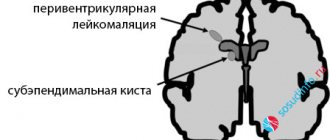
Sometimes, when performing neurosonography, an ultrasound doctor finds pseudocysts in the brain - but this is not a reason to panic at all! Pseudocysts are single round tiny formations (cavities) containing cerebrospinal fluid and located in typical areas of the brain. The reasons for their appearance, as a rule, are not reliably known; they usually disappear by 8-12 months. life. It is important to know that the existence of such cysts in most children is not a risk factor for further neuropsychic development and does not require treatment. However, although quite rare, pseudocysts form at the site of subependymal hemorrhages, or are associated with perinatal cerebral ischemia or intrauterine infection. The number, size, structure and location of cysts provide specialists with very important information, taking into account which, based on a clinical examination, final conclusions are formed.
Description of NSG is not a diagnosis! and not necessarily a reason for treatment.
Most often, NSG data provide indirect and uncertain results, and are taken into account only in conjunction with the results of a clinical examination.
Once again, I remind you of the other extreme: in difficult cases, sometimes there is a clear underestimation on the part of parents (less often, doctors) of the child’s problems, which leads to a complete refusal of the necessary dynamic observation and examination, as a result of which the correct diagnosis is made late, and treatment does not lead to the desired result.
Undoubtedly, therefore, if increased intracranial pressure and hydrocephalus are suspected, diagnosis should be carried out at the highest professional level.
What is muscle tone and why is it so “loved”?
Look at your child’s medical record: is there no such diagnosis as “muscular dystonia”, “hypertension” and “hypotension”? — you probably just didn’t go with your baby to the neurologist’s clinic until he was a year old. This is, of course, a joke. However, the diagnosis of “muscular dystonia” is no less common (and perhaps more common) than hydrocephalic syndrome and increased intracranial pressure.
Changes in muscle tone can be, depending on the severity, either a variant of the norm (most often) or a serious neurological problem (this is much less common).
Briefly about external signs of changes in muscle tone.
Muscular hypotonia is characterized by a decrease in resistance to passive movements and an increase in their volume. Spontaneous and voluntary motor activity may be limited; palpation of the muscles is somewhat reminiscent of “jelly or very soft dough.” Severe muscle hypotonia can significantly affect the rate of motor development (for more details, see the chapter on movement disorders in children of the first year of life).
Muscular dystonia is characterized by a condition where muscle hypotonia alternates with hypertension, as well as a variant of disharmony and asymmetry of muscle tension in individual muscle groups (for example, more in the arms than in the legs, more on the right than on the left, etc.)
At rest, these children may experience some muscle hypotonia during passive movements. When trying to actively perform any movement, during emotional reactions, when the body changes in space, muscle tone increases sharply, pathological tonic reflexes become pronounced. Often, such disorders subsequently lead to improper development of motor skills and orthopedic problems (for example, torticollis, scoliosis).
Muscular hypertension is characterized by increased resistance to passive movements and limitation of spontaneous and voluntary motor activity. Severe muscle hypertension can also significantly affect the rate of motor development.
Violation of muscle tone (muscle tension at rest) can be limited to one limb or one muscle group (obstetric paresis of the arm, traumatic paresis of the leg) - and this is the most noticeable and very alarming sign, forcing parents to immediately consult a neurologist.
It is sometimes quite difficult for even a competent doctor to notice the difference between physiological changes and pathological symptoms in one consultation. The fact is that changes in muscle tone are not only associated with neurological disorders, but also strongly depend on the specific age period and other characteristics of the child’s condition (excited, crying, hungry, drowsy, cold, etc.). Thus, the presence of individual deviations in the characteristics of muscle tone does not always cause concern and require any treatment.
But even if functional disorders of muscle tone are confirmed, there is nothing to worry about. A good neurologist will most likely prescribe massage and physical therapy (exercises on large balls are very effective). Medicines are prescribed extremely rarely.
Hyperexcitability syndrome
(syndrome of increased neuro-reflex excitability)
Frequent crying and whims with or without cause, emotional instability and increased sensitivity to external stimuli, sleep and appetite disturbances, excessive frequent regurgitation, motor restlessness and shuddering, trembling of the chin and arms (etc.), often combined with poor growth weight and bowel dysfunction - do you recognize such a child?
All motor, sensitive and emotional reactions to external stimuli in a hyperexcitable child arise intensely and abruptly, and can fade away just as quickly. Having mastered certain motor skills, children constantly move, change positions, constantly reach for and grab objects. Children usually show a keen interest in their surroundings, but increased emotional lability often makes it difficult for them to communicate with others. They are very impressionable, emotional and vulnerable! They fall asleep extremely poorly, only with their mother, they constantly wake up and cry in their sleep. Many of them have a long-term reaction of fear when communicating with unfamiliar adults with active reactions of protest. Typically, hyperexcitability syndrome is combined with increased mental exhaustion.
The presence of such manifestations in a child is just a reason to contact a neurologist, but in no case is it a reason for parental panic, much less drug treatment.
Constant hyperexcitability is not causally specific and can most often be observed in children with temperamental characteristics (for example, the so-called choleric type of reaction).
Much less frequently, hyperexcitability can be associated and explained by perinatal pathology of the central nervous system. In addition, if a child’s behavior is suddenly disrupted unexpectedly and for a long time for virtually no apparent reason, and he or she develops hyperexcitability, the possibility of developing an adaptation disorder reaction (adaptation to external environmental conditions) due to stress cannot be ruled out. And the sooner the child is examined by specialists, the easier and faster it is possible to cope with the problem.
And, finally, most often, transient hyperexcitability is associated with pediatric problems (rickets, digestive disorders and intestinal colic, hernia, teething, etc.).
There are two extremes in the tactics of monitoring such children. Or an “explanation” of hyperexcitability using “intracranial hypertension” and intense drug treatment often using drugs with serious side effects (diacarb, phenobarbital, etc.). Or complete neglect of the problem, which can subsequently lead to the formation of persistent neurotic disorders (fears, tics, stuttering, anxiety disorders, obsessions, sleep disorders) in the child and his family members, and will require long-term psychological correction.
Of course, it is logical to assume that an adequate approach is somewhere in between...
Separately, I would like to draw the attention of parents to seizures - one of the few disorders of the nervous system that really deserves close attention and serious treatment. Epileptic seizures do not occur often in infancy, but they are sometimes severe, insidious and disguised, and immediate drug therapy is almost always necessary.
Such attacks can be hidden behind any stereotypical and repetitive episodes in the child’s behavior. Incomprehensible shudders, head nods, involuntary eye movements, “freezing,” “squeezing,” “limping,” especially with a fixed gaze and lack of response to external stimuli, should alert parents and force them to turn to specialists. Otherwise, a late diagnosis and untimely prescribed drug therapy significantly reduce the chances of treatment success.
All circumstances of the seizure episode must be accurately and completely remembered and, if possible, recorded on video for further detailed description at the consultation. If convulsions last a long time or are repeated, call “03” and urgently consult a doctor.
At an early age, the child’s condition is extremely changeable, so developmental deviations and other disorders of the nervous system can sometimes be detected only during long-term dynamic monitoring of the baby, with repeated consultations. For this purpose, specific dates for planned consultations with a pediatric neurologist in the first year of life have been determined: usually at 1, 3, 6 and 12 months. It is during these periods that most serious diseases of the nervous system of children in the first year of life can be detected (hydrocephalus, epilepsy, cerebral palsy, metabolic disorders, etc.). Thus, identifying a specific neurological pathology in the early stages of development makes it possible to begin complex therapy on time and achieve the maximum possible result.
And in conclusion, I would like to remind parents: be sensitive and attentive to your kids! First of all, it is your meaningful participation in the lives of children that is the basis for their future well-being. Do not treat them for “supposed illnesses,” but if something worries and concerns you, find the opportunity to get independent advice from a qualified specialist.
Treatment methods
- If a protruding or sunken fontanelle indicates the course of a pathological process, treatment of the infant should be carried out in a hospital setting.
- If the baby is dehydrated, infusion therapy is prescribed.
- If the change in the fontanel is caused by an infection, then antibiotics, antiviral and anti-inflammatory drugs are used, and symptomatic treatment is prescribed.
- To relieve intracranial pressure, the child is given diuretics.
- In case of poisoning, enterosorbents are prescribed - drugs that cleanse the body of toxins.
In each individual case, the pediatric neurologist at our clinic decides on the issue of treating the baby, taking into account his individual characteristics.
Preventive actions
To maintain a stable condition of the large fontanelle, it is necessary:
- regularly walk with your child in the fresh air:
- establish a drinking regime;
- dress your baby according to the season;
- ensure that the room has a comfortable microclimate.
Popular traditional methods of treatment for an infant are not allowed. If alarming symptoms appear, parents living in Kaliningrad can immediately call the Edkarik private clinic to make an appointment with a pediatric neurologist. You can also fill out an online application on the official website, choosing a time convenient for visiting a doctor.