Historical reference
In the past, when scientists did not yet have informative instruments at hand that could study physiological processes in a living organism, the greatest scientists were forced to search for anatomical features in corpses. Naturally, the heart of a deceased person does not contract, so some nuances had to be figured out on their own, and sometimes simply fantasized. Thus, back in the second century AD, Claudius Galen, studying from the works of Hippocrates himself, assumed that the arteries contained air instead of blood in their lumen. Over the next centuries, many attempts were made to combine and link together the existing anatomical data from the standpoint of physiology. All scientists knew and understood how the circulatory system works, but how does it work?
Miguel Servetus and William Harvey made a tremendous contribution to the systematization of data on the work of the heart in the 16th century. Harvey, the scientist who first described the systemic and pulmonary circulation , in 1616 determined the presence of two circles, but he could not explain in his works how the arterial and venous beds were connected. And only later, in the 17th century, Marcello Malpighi, one of the first to use a microscope in his practice, discovered and described the presence of tiny capillaries, invisible to the naked eye, which serve as a connecting link in the blood circulation.
William Harvey vs. Claudius Galen: How does the human circulatory system work?
How is blood formed? Where does it come from and where does it flow? Who discovered the human circulatory system and how?
Today even a schoolchild can answer these and many other questions about the “river of life”. But, as with many other “evidences,” the story of blood and its “circulation” in the body has been at the center of fierce scientific debate and battles.
Today's date in the calendar - April 16 - is notable because on this day a contribution was made to medicine that radically changed ideas about the circulatory system. The man who made it was William Harvey, an English physician, the founder of physiology and embryology.
To stand for humanity. William Harvey is a doctor who changed the world. Read more
What was so outrageous about this discovery that it caused a storm of criticism from the European scientific elite?
First, let's take a short excursion into the question of what the human circulatory system is needed for and how it works.
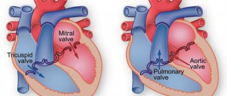
To maintain life, the body needs many substances. The circulatory system is used to deliver them to all organs and tissues. Conceptually, it is represented by the heart and blood vessels. The heart consists of chambers - two atria and two ventricles (right and left) - and, like a pump, “drives” blood through the vessels. The vessels themselves consist of two large sections - the arterial and venous beds. Arteries carry oxygen-rich blood from the heart - from its left ventricle - to organs and tissues. Having given them oxygen and taken away carbon dioxide, the blood collects in the veins and returns through them to the heart, or more precisely, to the right atrium. This circle of blood circulation is called large.
Next, blood from the right ventricle passes through the pulmonary arteries through the lungs, and through the pulmonary veins into the left atrium. This is the pulmonary circulation.
As we can see, a closed system of vessels is formed, leaving the heart and returning to it.
At the very beginning of his experiments, Harvey, like his contemporaries, was certainly familiar with the works of his predecessors.
The unquestioned authority at that time was the ancient Roman physician Galen (or rather, his works), and this authority was well-deserved. Galen's theories dominated European medicine for over a thousand years.
The data on anatomy that he obtained by dissecting monkeys and pigs was used until the publication of Andreas Vesalius’s work “On the Structure of the Human Body” around the middle of the 16th century. Future doctors studied the works of Galen until the 19th century inclusive. His theory that the brain controls movement through the nervous system remains relevant today.
Why did Harvey's model of blood circulation come into confrontation with Galen's earlier ideas?
If the provisions of Galen's theory were based on purely speculative conclusions, that would be half the battle. However, Galen objectified his conclusions by dissecting the corpses of animals and people (gladiators). During the autopsy, he noticed that there was no blood in the left side of the heart. He also believed that blood passes from the right half of the heart to the left through a special hole (holes) in the septum (according to some data - between the ventricles, another - between the atria). Even though anatomists could not detect the foramina mentioned, Galen's authority was such that his assertion was not questioned. Galen's followers did not find blood in the arteries of the opened bodies, while the veins were full.
Galen considered arterial and venous blood to be different fluids, and attributed different functions to them. In addition, he believed that once blood reaches the periphery, it is “destroyed.”
Individual representatives of subsequent generations of scientists in one way or another clarified the provisions of Galen’s theory of blood circulation, but Harvey’s works brought complete clarity to it.
It is possible that this discovery was due to the skepticism attributed to the doctor. Is it appropriate to even approximate comparison of Harvey with Turgenev’s Bazarov and his “denial of authorities” is a rhetorical question. However, it is known that Harvey himself was interested in understanding issues of physiology not only from books, but also by conducting experiments on his own. This is how he came to his conclusions - not least thanks to surgical experiments on living animals (the so-called vivisection). Accusations of this can still be heard today.
Harvey made the first mention of the fact that “blood circles in the body” in 1616 in one of his lectures. However, only after twelve years of work he published the results of his work, called “Anatomical studies on the movement of the heart and blood of animals.”
Remarkable fact:
Harvey's treatise was published extremely carelessly. To avoid significant costs, the author sent the manuscript of the work to a little-known German publishing house. The work was printed on the cheapest paper; only a few copies have reached us. There are many typos in the book: apparently neither the printing house workers nor the author himself proofread the text.
***
“It will not be easy for them, those who rely on the truth of authority, instead of relying on the authority of truth.” These words, attributed in one form or another to the English poet and amateur Egyptologist Gerald Massey, may be somewhat consonant with the story of Harvey's discovery.
After the scientist published the results of his work, he was attacked by the scientific community.
“Paradoxical, useless, false, impossible, incomprehensible, absurd...” It was these epithets that Guy Patin, the physician of Louis XIV, one of the famous representatives of medical science of that time, awarded Harvey’s work. “We are living through an era of incredible inventions, and I don’t even know if our descendants will believe in the possibility of such madness.”
For some period of time, the Paris Faculty of Medicine itself was a hotbed of conservative views; the authority of Galen and Avicenna was secured by parliamentary decree.
Be that as it may, there were also those who supported Harvey. The first of them was Descartes, which greatly contributed to the triumph of Harvey's ideas.
Ultimately, the main goal of science is to establish objective truth. Any previous knowledge, even if it is (in some way) erroneous, is a source of an idea when a researcher, noticing some inconsistencies in it, takes upon himself the courage to follow an already “trodden path.” And, having finally reached the truth, not only refute or clarify/improve the existing erroneous theory, but also be grateful for the very idea that appeared thanks to this theory. As Galen himself said: “A physician should be lenient towards his predecessors.”
Time puts everything in its place. The same thing happened with Harvey’s doctrine of blood circulation.
Other related articles:
Nobel laureate without a high school diploma: X-ray as a proper name
Expelled. Scolded. Acquitted by pathologists. Crusade of the physician Ozias-Turenne
Phylogeny, or the evolution of blood circulation
Due to the fact that, as animals of the vertebrate class evolved, they became more and more progressive in anatomical and physiological terms, they required a complex structure of the cardiovascular system. Thus, for faster movement of the liquid internal environment in the body of a vertebrate animal, the need for a closed blood circulation system arose. Compared to other classes of the animal kingdom (for example, arthropods or worms), the rudiments of a closed vascular system appear in chordates. And if the lancelet, for example, does not have a heart, but there is an abdominal and dorsal aorta, then in fish, amphibians (amphibians), reptiles (reptiles) a two- and three-chambered heart appears, respectively, and in birds and mammals a four-chambered heart appears, the peculiarity of which is is the focus in it of two circles of blood circulation that do not mix with each other.
Thus, the presence of two separated circulatory circles in birds, mammals and humans, in particular, is nothing more than the evolution of the circulatory system, necessary for better adaptation to environmental conditions.
How to keep blood vessels and heart healthy?
The narrowing of the lumen of the arteries leads over time to atherosclerosis, a disease in which the vessels become denser and less elastic.
Atherosclerosis can cause even more serious diseases - such as coronary heart disease, hypertension, angina pectoris, myocardial infarction, and so on. These diseases are practically untreatable, so prevention is extremely important for every person. It is advisable to start improving the circulatory system by changing your lifestyle. This is especially true for overweight people. They should make every effort to normalize it. Moderate and regular physical activity and a proper diet will help you quickly cope with extra pounds, normalize your metabolism and make your circulatory system an effective mechanism for actively supplying the body with all the necessary substances. As for eating habits, a person striving for healthy heart function should exclude animal fats from the diet, which saturate the body with cholesterol and triglycerides. It is also important to limit the consumption of products such as margarine and palm oil (as a result, most confectionery products). Preference should be given to olive oil and fatty sea fish - products rich in polyunsaturated omega-3 fatty acids. A healthy circulatory system is a guarantee of your excellent health, vigor and full functioning of all internal organs. Do you want to be healthy? So, take care of the circulatory system! Photo: flickr.com Tags:
- Heart and blood vessels
- Circulation
To leave a comment you must be an authorized user
Anatomical features of the blood circulation
The circulatory system is a set of blood vessels, which is a closed system for the supply of oxygen and nutrients to the internal organs through gas exchange and nutrient exchange, as well as for the removal of carbon dioxide and other metabolic products from cells. The human body is characterized by two circles - the systemic, or large circle, and the pulmonary, also called the small circle.
Video: blood circulation circles, mini-lecture and animation
Human circulatory system: structure and functions
Normal life activity is impossible without effective blood circulation: it maintains the constancy of the internal environment, transports oxygen, hormones, nutrients and other vital substances, takes part in the cleansing of toxins, waste, decay products, the accumulation of which would sooner or later lead to the death of an individual organ or the whole organism. This process is regulated by the circulatory system - a group of organs, thanks to the joint work of which blood is consistently moved throughout the human body.
Let's look at how the circulatory system
, and what functions it performs in the human body.
The structure of the human circulatory system
At first glance, the circulatory system is structured simply and clearly: it includes the heart and numerous vessels through which blood flows, successively reaching all organs and systems. The heart is a kind of pump that stimulates the blood, ensuring its smooth flow, and the vessels play the role of guiding tubes that determine the specific path of blood movement throughout the body. That is why the circulatory system is also called the cardiovascular or cardiovascular system.
Let's talk in more detail about each organ that belongs to the human circulatory system.
Organs of the human circulatory system
Like any organismal complex, the circulatory system includes a number of different organs, which are classified depending on their structure, location and functions:
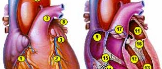
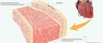
- The heart is considered the central organ of the cardiovascular complex. It is a hollow organ formed primarily by muscle tissue. The cardiac cavity is divided by partitions and valves into 4 sections - 2 ventricles and atria (left and right). Thanks to rhythmic successive contractions, the heart pushes blood through the vessels, ensuring its uniform and continuous circulation.
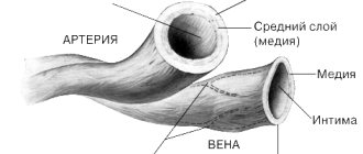
- Arteries carry blood from the heart to other internal organs. The further from the heart they are located, the thinner their diameter: if in the area of the heart sac the average width of the lumen is the thickness of a thumb, then in the area of the upper and lower extremities its diameter is approximately equal to a simple pencil.
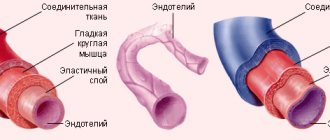
Despite the visual difference, both large and small arteries have a similar structure. They include three layers - adventitia, media and intima. The adventitium, the outer layer, is formed by loose fibrous and elastic connective tissue and includes many pores through which microscopic capillaries pass, feeding the vascular wall, and nerve fibers that regulate the width of the lumen of the artery depending on the impulses sent by the body.
The media, which occupies the median position, includes elastic fibers and smooth muscles, thanks to which the firmness and elasticity of the vascular wall is maintained. It is this layer that largely regulates the speed of blood flow and blood pressure, which can vary within an acceptable range depending on external and internal factors affecting the body. The larger the diameter of the artery, the higher the percentage of elastic fibers in the middle layer. According to this principle, vessels are classified into elastic and muscular.
The intima, or inner lining of the arteries, is represented by a thin layer of endothelium. The smooth structure of this tissue facilitates blood circulation and serves as a passageway for media nutrition.
As the arteries thin, these three layers become less pronounced. If in large vessels the adventitia, media and intima are clearly distinguishable, then in thin arterioles only muscle spirals, elastic fibers and a thin endothelial lining are noticeable.
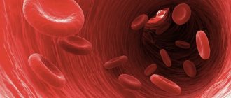
- Capillaries are the thinnest vessels of the cardiovascular system, which are an intermediate link between arteries and veins. They are localized in the areas most distant from the heart and contain no more than 5% of the total blood volume in the body. Despite their small size, capillaries are extremely important: they envelop the body in a dense network, supplying blood to every cell of the body. This is where the exchange of substances between the blood and adjacent tissues occurs. The thinnest capillary walls easily allow molecules of oxygen and nutritional components contained in the blood to pass through, which, under the influence of osmotic pressure, pass into the tissues of other organs. In return, the blood receives the breakdown products and toxins contained in the cells, which are sent through the venous bed back to the heart and then to the lungs.
- Veins are a type of vessels that carry blood from internal organs to the heart. The walls of veins, like arteries, are formed by three layers. The only difference is that each of these layers is less pronounced. This feature is regulated by the physiology of the veins: blood circulation here does not require strong pressure from the vascular walls - the direction of blood flow is maintained due to the presence of internal valves. A greater number of them are found in the veins of the lower and upper extremities - here, with low venous pressure, without alternating contraction of muscle fibers, blood flow would be impossible. Large veins, on the other hand, have very few or no valves.
During the circulation process, part of the fluid from the blood seeps through the walls of capillaries and blood vessels to the internal organs. This liquid, visually somewhat reminiscent of plasma, is lymph that enters the lymphatic system. Merging together, the lymphatic pathways form fairly large ducts, which in the area of the heart flow back into the venous bed of the cardiovascular system.
Systemic circulation
The main function of the large circle is to ensure gas exchange in all internal organs except the lungs. It begins in the cavity of the left ventricle; represented by the aorta and its branches, the arterial bed of the liver, kidneys, brain, skeletal muscles and other organs. Further, this circle continues with the capillary network and venous bed of the listed organs; and through the entry of the vena cava into the cavity of the right atrium it ends in the latter.
So, as already said, the beginning of the great circle is the cavity of the left ventricle. Arterial blood flow, which contains more oxygen than carbon dioxide, is sent here. This flow enters the left ventricle directly from the circulatory system of the lungs, that is, from the small circle. The arterial flow from the left ventricle is pushed through the aortic valve into the largest great vessel - the aorta. The aorta can be figuratively compared to a kind of tree that has many branches, because arteries extend from it to the internal organs (to the liver, kidneys, gastrointestinal tract, to the brain - through the system of carotid arteries, to skeletal muscles, to the subcutaneous fat fiber, etc.) Organ arteries, which also have numerous branches and bear names corresponding to their anatomy, carry oxygen to each organ.
In the tissues of internal organs, arterial vessels are divided into vessels of smaller and smaller diameter, and as a result, a capillary network is formed. Capillaries are the smallest vessels, practically without a middle muscular layer, and are represented by an inner membrane - intima, lined with endothelial cells. The gaps between these cells at the microscopic level are so large compared to other vessels that they allow proteins, gases and even formed elements to easily penetrate into the intercellular fluid of the surrounding tissues. Thus, intense gas exchange and exchange of other substances occurs between the capillary with arterial blood and the liquid intercellular medium in a particular organ. Oxygen penetrates from the capillary, and carbon dioxide, as a product of cell metabolism, enters the capillary. The cellular stage of respiration occurs.
After more oxygen has passed into the tissues and all carbon dioxide has been removed from the tissues, the blood becomes venous. All gas exchange occurs with each new influx of blood, and during the period of time while it moves along the capillary towards the venule - a vessel that collects venous blood. That is, with each cardiac cycle, in one or another part of the body, oxygen enters the tissues and carbon dioxide is removed from them.
These venules unite into larger veins, and a venous bed is formed. Veins, similar to arteries, are named according to the organ in which they are located (renal, cerebral, etc.). From large venous trunks, tributaries of the superior and inferior vena cava are formed, and the latter then flow into the right atrium.
What harms blood vessels?
The scourge of modern man is the deposits of fat on the walls of blood arteries (mainly “bad” cholesterol), as a result of which atherosclerotic changes in blood vessels occur. Fat accumulations form atheromas and cholesterol plaques on the walls of blood vessels, which narrow the patency of blood vessels and impair blood flow. The heart has to work harder, which leads to its premature aging, while little oxygenated blood reaches the tissues. As a result, the body faces the threat of oxygen starvation.
Features of blood flow in the organs of the systemic circle
Some of the internal organs have their own characteristics. So, for example, in the liver there is not only a hepatic vein, which “carries” the venous flow away from it, but also a portal vein, which, on the contrary, brings blood to the liver tissue, where blood purification is performed, and only then the blood collects in the tributaries of the hepatic vein to enter to a big circle. The portal vein brings blood from the stomach and intestines, so everything that a person eats or drinks must undergo a kind of “purification” in the liver.
In addition to the liver, certain nuances exist in other organs, for example, in the tissues of the pituitary gland and kidneys. Thus, in the pituitary gland the presence of a so-called “wonderful” capillary network is noted, because the arteries that bring blood to the pituitary gland from the hypothalamus are divided into capillaries, which then collect into venules. The venules, after the blood with the molecules of releasing hormones are collected, are again divided into capillaries, and then veins are formed that carry the blood from the pituitary gland. In the kidneys, the arterial network is divided twice into capillaries, which is associated with the processes of excretion and reabsorption in the kidney cells - in the nephrons.
Pulmonary circulation
Its function is to carry out gas exchange processes in the lung tissue in order to saturate the “waste” venous blood with oxygen molecules. It begins in the cavity of the right ventricle, where venous blood flow with an extremely small amount of oxygen and a large content of carbon dioxide enters from the right atrial chamber (from the “end point” of the great circle). This blood moves through the pulmonary valve into one of the large vessels called the pulmonary trunk. Next, the venous flow moves along the arterial bed in the lung tissue, which also breaks up into a network of capillaries. By analogy with capillaries in other tissues, gas exchange occurs in them, only oxygen molecules enter the lumen of the capillary, and carbon dioxide penetrates into the alveolocytes (cells of the alveoli). With each act of breathing, air enters the alveoli from the environment, from which oxygen penetrates through the cell membranes into the blood plasma. When exhaling, the carbon dioxide that enters the alveoli is expelled with the exhaled air.
After being saturated with O2 molecules, the blood acquires the properties of arterial blood, flows through the venules and ultimately reaches the pulmonary veins. The latter, consisting of four or five pieces, open into the cavity of the left atrium. As a result, venous blood flows through the right half of the heart, and arterial blood flows through the left half; and normally these flows should not mix.
Lung tissue has a double network of capillaries. With the help of the first, gas exchange processes are carried out in order to enrich the venous flow with oxygen molecules (relationship directly with the small circle), and in the second, the lung tissue itself is supplied with oxygen and nutrients (relationship with the large circle).
Main functions of the system
The work of the circulatory system begins with the enrichment of blood with oxygen. “Depleted” blood enters the heart through the veins: first into the first chamber of the right atrium, then into the right ventricle of the heart. From there, the more powerful heart muscles push oxygen-deprived blood into the pulmonary trunk, divided into two pulmonary arteries. Next, the blood enters the lungs through numerous pulmonary vessels, where it is enriched with oxygen and returns through the pulmonary vessels to the heart - but this time to the left atrium and ventricle. The left ventricle of the heart is responsible for supplying blood to the entire body, therefore, the muscles of the left ventricle are more developed. Human blood circulation consists of two circles: small (pulmonary) and large. The small circle is responsible for enriching the blood with oxygen, and the large circle is responsible for transporting blood throughout the body. Despite the fact that two atria and two ventricles contract simultaneously, the thick-walled left ventricle experiences a load six times greater, because it has to circulate blood in a large circle, supplying all limbs with useful substances.