Both arteries and veins are types of blood vessels in the cardiovascular system
An artery carries blood away from the heart, and a vein brings blood back to the heart.
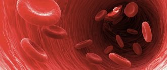
Blood vessels are necessary to transport blood throughout the body. Blood carries oxygen and nutrients to various tissues in the body, allowing them to function.
The heart and blood vessels make up the cardiovascular system. This system contains a complex network of vessels with different structures and functions.
In this article, we discuss the differences between arteries and veins. We also highlight the different types of blood vessels and how they work as part of the cardiovascular system.
Arteries and veins: definitions
Arteries and veins are types of blood vessels that transport blood throughout the body.
Blood vessels form two systems, leading to and from the heart. These two systems form the circulatory system.
The systemic circulation supplies oxygen and other vital substances to organs, tissues and cells.
The systemic arteries transport oxygen-rich blood from the left ventricle to the rest of the body. The low-oxygen blood then collects in the systemic veins and is sent to the right atrium.
The pulmonary circulation is where fresh oxygen enters the blood.
The pulmonary arteries transport low-oxygen blood from the right ventricle to the lungs. The pulmonary veins then transport oxygen-rich blood back to the heart through the left atrium.
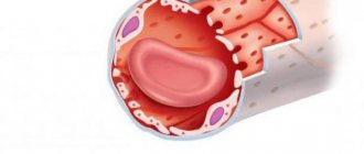
Capillaries are the third type of blood vessel in the body. They supply blood directly to the organs.
Circulation
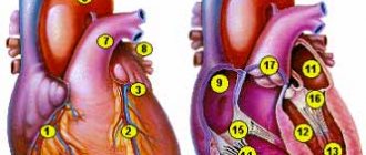
Venous blood (with a low oxygen content) enters the right atrium through the superior and inferior vena cava. The blood then enters the right ventricle, which contracts and pumps it into the pulmonary trunk. The trunk soon divides into the right and left pulmonary arteries, which carry blood to both lungs. The arteries, in turn, break up into lobar and segmental branches, which are further divided into arterioles and capillaries. In the lungs, venous blood is cleared of carbon dioxide and, enriched with oxygen, becomes arterial. It enters the left atrium through the pulmonary veins and then into the left ventricle. From there, under high pressure, the blood is pushed into the aorta, then goes through the arteries to all organs. The arteries branch into smaller and smaller ones and eventually become capillaries. The speed of blood flow and its pressure by this time are significantly reduced. Oxygen and nutrients enter the tissues through the walls of the capillaries from the blood, and carbon dioxide, water and other metabolic products enter the blood. After passing through the network of capillaries, the blood becomes venous. The capillaries merge into venules, then into increasingly larger veins, and eventually the two largest veins - the superior and inferior vena cava - flow into the right atrium. As long as we are alive, this cycle repeats itself over and over again.
Types of arteries
There are three types of arteries:
Elastic arteries
Elastic arteries are large vessels leaving the heart. For example, they include the pulmonary artery and aorta. The aorta is the main artery that carries blood away from the heart.
The heart forcibly pumps out blood to move it throughout the body. Elastic arteries must be flexible to cope with the rush of blood. They expand when the heart pumps out blood.
Elastin is a protein found in many tissues that provides flexibility to organs, including elastic arteries.
Muscular arteries
Elastic arteries carry blood to muscular arteries such as the femoral or coronary arteries.
Smooth muscle fibers make up the walls of muscle arteries. Muscles allow these arteries to expand and contract. These changes in size control how much blood moves through the arteries.
Arterioles
Arterioles are the smallest type of arteries. They distribute blood from larger arteries through networks of capillaries.
The outer layer of arterioles also contains smooth muscle, which regulates expansion and contraction.
What connections between extra- and intracranial veins do you know? Their meaning
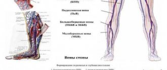
Extracranial: superficial, deep
Intracranial - GM veins, open in the sinuses of the dura mater, do not collapse, there are no valves. From the sinuses through the internal jugular vein is the main route for the outflow of venous blood from the cranial cavity.
Pathways of communication between intra- and extracranial veins:
- Through the venous outlet: 3 pairs, hole in the bones of the skull, blood from inside to outside
- The ophthalmic vein connects the cavernous sinus with the facial vein
- Through the diploe veins (the spongy substance of the skull bones between the plates)
- Through the foramen magnum into the vertebral venous plexuses
Types of Veins
Arteries and veins are designed roughly the same, but veins are thinner and have less muscle, which allows them to hold more blood. Veins typically contain about 70% of the body's blood at one time.
Venules are the smallest type of vein. They have very thin walls in order to hold a lot of blood. They pump low-oxygen blood through capillaries from the arteries directly into the veins. The blood then returns to the heart through a series of veins of increasing size and muscle.
There are two main types of veins: pulmonary and systemic.
Systemic veins are further classified into:
- Deep veins : These veins usually have a corresponding artery nearby and are found in muscle tissue. These veins may have a one-way valve to prevent blood from flowing backwards.
- Superficial veins : These veins do not have an artery of the same name nearby and are found close to the surface of the skin. They may also have a one-way valve.
- Connecting veins : These small veins allow blood to flow from the superficial veins to the deep veins.
Differences
How are arteries different from veins? These blood vessels differ significantly in many ways.
Arteries and veins, first of all, differ in the structure of the wall
According to the structure of the wall
Arteries have thick walls, they have a lot of elastic fibers, smooth muscles are well developed, they do not fall off unless they are filled with blood. Due to the contractility of the tissues that make up their walls, oxygenated blood is quickly delivered to all organs. The cells that make up the layers of the walls ensure the smooth passage of blood through the arteries. Their inner surface is corrugated. The arteries must withstand the high pressure that is created by powerful surges of blood.
The pressure in the veins is low, so the walls are thinner. They fall off when there is no blood in them. Their muscle layer is not able to contract like arteries. The surface inside the vessel is smooth. Blood moves through them slowly.
In veins, the thickest membrane is considered to be the outer one, in arteries it is the middle one. Veins do not have elastic membranes, arteries have an internal and an external one.
By shape
The arteries have a fairly regular cylindrical shape, they are round in cross section.
Due to the pressure of other organs, the veins are flattened, their shape is tortuous, they either narrow or expand, which is due to the location of the valves.
In count
In the human body there are more veins and fewer arteries. Most middle arteries are accompanied by a pair of veins.
According to the presence of valves
Most veins have valves that prevent blood from flowing backwards. They are located in pairs opposite each other throughout the entire length of the vessel. They are not found in the portal cava, brachiocephalic, iliac veins, as well as in the veins of the heart, brain and red bone marrow.
In arteries, valves are located as vessels exit the heart.
By blood volume
Veins circulate approximately twice as much blood as arteries.
By location
The arteries lie deep in the tissues and approach the skin only in a few places, where the pulse is heard: on the temples, neck, wrist, and instep of the feet. Their location is approximately the same for all people.
Veins are mostly located close to the surface of the skin
The location of the veins may vary from person to person.
To ensure blood movement
In the arteries, blood flows under the pressure of the force of the heart, which pushes it out. At first the speed is about 40 m/s, then gradually decreases.
Blood flow in the veins occurs due to several factors:
- pressure forces depending on the push of blood from the heart muscle and arteries;
- the suction force of the heart during relaxation between contractions, that is, the creation of negative pressure in the veins due to the expansion of the atria;
- suction effect on the chest veins of respiratory movements;
- contractions of the muscles of the legs and arms.
In addition, approximately a third of the blood is in the venous depots (in the portal vein, spleen, skin, walls of the stomach and intestines). It is pushed out from there if it is necessary to increase the volume of circulating blood, for example, during massive bleeding or during high physical exertion.
Anatomy
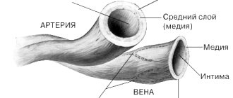
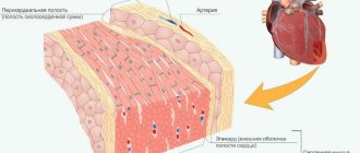
Veins and arteries consist of three layers:
- Tunica adventitia : The outer layer of a blood vessel, composed of collagen and elastin. This layer allows the vessel to expand or contract, depending on the type of vein or artery. This function is important for controlling blood pressure.
- Tunica media : This is the middle of the blood vessel. Elastin and muscle fibers form the shell of the carriers. The amount of elastin or muscle varies depending on the type of blood vessel. For example, elastic arteries contain few muscle fibers in their membranes.
- Tunica Intima: This name refers to the inner layer of a blood vessel. It mainly contains elastic membranes and tissues and may include valves that help blood move in the right direction.
Functions of the venous system
Functions of veins:
- Transport of blood to the heart.
- Blood reservoir - deposition is facilitated by thin-walled, numerous veins (many in the abdominal cavity, they hold 80% of the blood in case of shock => GM, heart, liver, kidneys). In shock, blood pressure drops down to 0. The veins of the lungs can accumulate up to 28% of the blood, and swelling is possible due to inflammation.
- Regulates hemodynamics: blood flow speed, blood pressure provide blood supply, carried out reflexively. There are many receptors in the walls of the veins: chemo, mechano, baro. When they are irritated, reflexes are triggered:
- venovenous;
- venoarthrial (increased pressure, decreased arterial blood flow);
- venolymphatic.
- Participation in metabolic processes between blood and tissue. The walls of the capillaries are well permeable, metabolic processes are ensured by thin-walled venules, post-capillaries; with inflammation, permeability (swelling) increases, since the walls of the venules react to histamine.
The cardiovascular system
The cardiovascular system connects the heart and blood vessels together. The system forms a closed circuit of vessels that transport blood throughout the body.
The cardiovascular system is essential for maintaining life. This is the first major network of organs to develop in embryos.
All body tissues need oxygen and nutrients to survive. They also require the removal of waste, which is a byproduct of metabolism.
Blood is needed both to provide oxygen and nutrients and to remove waste from tissues.
The heart pumps blood throughout the body. It must work constantly and with sufficient force so that all tissues of the body receive enough blood to function. Cardiovascular disorders can have serious consequences.
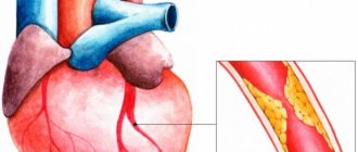
Cardiovascular diseases are a group of disorders that affect the heart and blood vessels, such as coronary heart disease.
These diseases are the leading cause of death worldwide, accounting for approximately 17.9 million deaths in 2021.
Vienna
They are non-muscular and muscular.
The walls of muscleless veins consist of endothelium and connective tissue of a loose structure. Such vessels are found in bone tissue, placenta, brain, retina, and spleen.
Muscular veins, in turn, are divided into three types depending on how the myocytes are developed:
- poorly developed (neck, face, upper body);
- medium (brachial and small veins);
- strongly (lower body and legs).
Veins, in addition to the umbilical and pulmonary veins, carry blood, which gives up oxygen and nutrients and takes away carbon dioxide and breakdown products as a result of metabolic processes. It moves from the organs to the heart. Most often, she has to overcome the force of gravity and her speed is lower, which is due to the peculiarities of hemodynamics (lower pressure in the vessels, the absence of a sharp drop, a small amount of oxygen in the blood).
Structure and its features:
- Larger in diameter compared to arteries.
- The subendothelial layer and elastic component are poorly developed.
- The walls are thin and fall off easily.
- The smooth muscle elements of the middle layer are rather poorly developed.
- Pronounced outer layer.
- The presence of a valve apparatus, which is formed by the inner layer of the vein wall. The base of the valves consists of smooth myocytes, inside the valves there is fibrous connective tissue, and on the outside they are covered by a layer of endothelium.
- All wall membranes are endowed with vascular vessels.
The balance between venous and arterial blood is ensured by several factors:
- a large number of veins;
- their larger caliber;
- density of the vein network;
- formation of venous plexuses.
Arteries and veins: conclusions
Arteries are a type of blood vessel that transports blood away from the heart. Veins carry blood back to the heart. Along with capillaries, these blood vessels are responsible for moving blood to the tissues around the body.
The heart pumps blood through a complex system of blood vessels. There are several types of arteries and veins with different functions. For example, some contain more muscle to change the amount of blood they carry.
The cardiovascular system is essential to life. Changes in the heart or blood vessels can be serious and sometimes fatal.
Different functions - different structure.
The highest blood pressure will be at the blood outlet from the heart (in the left ventricle), slightly lower pressure will be in the arteries, even lower in the capillaries, and the lowest in the veins and at the entrance of the heart (in the right atrium).
The arteries carrying the oxygenated blood pushed out by the heart must resist the high pressure in the circulatory system. That's why they have an elastic membrane. In addition, they must also change their lumen in order to vary the level of blood flow in different organs in response to the actions of the autonomic nervous system - for this they have a well-developed layer of smooth muscle tissue. Therefore, the walls of the arteries are much thicker than the venous ones, they are much more elastic and contain a large number of muscle elements.
The walls of the veins, in turn, are thin and pliable, contain virtually no muscle elements, and ensure the return of blood to the heart. The veins in the lower body have valves that prevent blood from flowing back. Thus, the vascular bed adapts to changing load levels mainly due to changes in the lumen of the arteries.
Around which organs are the periorgan venous plexuses more developed? Their meaning
In many places there are well-developed venous plexuses:
- Small pelvis,
- spinal canal,
- Around the bladder
The significance of these plexuses can be seen in the example of the intravertebral plexus. When filled with blood, it occupies those free spaces that are formed when cerebrospinal fluid is displaced when changing body position or during movements. Thus, the structure and location of the veins depends on the physiological conditions of blood flow in them.