How and why to count your pulse?
Pulse detection is the simplest and most accessible in any conditions, a method for studying the state of the cardiovascular system, its reserve capabilities and pathological changes. That's why every person should learn to count it.What is pulse? If you put your finger on those parts of the body where large arteries are close (on the neck, in the temples, on the back of the foot, etc.), then under the finger you will feel jerky rhythmic vibrations of the walls of these arteries, which are usually called arterial pulse . It occurs with each contraction of the heart, when, at the moment of expulsion of blood, the pressure in the aorta rises and the pulse wave spreads along the walls of the arteries down to the smallest capillaries.
The most convenient point for determining the pulse in extreme situations is on the main cervical artery - the carotid artery. It is easy to find if you run your fingers from the back corner of the lower jaw down the neck until your fingers rest in the depression next to the windpipe, where the pulsation of the carotid artery can be clearly felt.
But most often the pulse is determined on the radial artery at the base of the thumb. To do this, the index, middle and ring fingers are placed slightly above the wrist joint, the artery is felt and pressed against the bone. In this case, the hand of the examined hand should be located at the level of the heart.
If the pulse beats follow at regular intervals, then the pulse is considered rhythmic (correct); otherwise - arrhythmic (irregular).
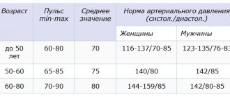
Another important characteristic of the pulse is its frequency . To determine it, count the number of beats in 10 seconds , and the result is multiplied by 6. If the rhythm is incorrect, the number of beats (frequency) of the pulse must be counted within a minute . Moreover, each person has his own pulse rate. In healthy people, it always corresponds to the heart rate 60-80 beats per minute at rest in men , and 5-10 beats more often in women. But even normally, under the influence of various factors, the pulse rate fluctuates within a fairly wide range. Thus, in a healthy adult, the lowest pulse rate is observed in a lying position; in a sitting position, the pulse increases by 4-6 beats per minute, and in a standing position – by 10-14 beats per minute. In winter, the pulse is usually slightly lower than in summer. Its frequency also changes during the day: from 8 to 12 o'clock it is maximum, then until 14 o'clock the pulse gradually slows down, from 15 o'clock it increases somewhat again, reaching its highest value by 18-20 o'clock, and in the middle of the night, when a person is sleeping, the pulse the slowest. The pulse increases noticeably after drinking hot liquids and food, but cold drinks, on the contrary, slow it down.
Fluctuations in heart rate are also associated with physical activity : the more intense the muscle work, the faster the pulse. It is very important to know the maximum permissible heart rate value. To do this, subtract your age from 220. And to find out your personal optimal heart rate , you need to multiply the difference by 0.7 (or by 0.6 if you are over sixty or in poor physical shape). To determine the lower limit of the heart rate, you need to multiply this difference by 0.5. For example, if you are a healthy 50-year-old man, subtract 50 from 220 and get a maximum allowable heart rate of 170 beats per minute. Multiplying 170 by 0.7 gives the result 119 - the optimal indicator that you should adhere to during any physical activity. Multiplying 170 by 0.5 we get the minimum heart rate for your age - 85 beats per minute.
A pulse less than 60 beats per minute is called bradycardia . The cause of its occurrence may be a decrease in the function of the thyroid gland, some heart diseases, etc. However, a decrease in heart rate, achieved through regular physical training, is a beneficial factor for health, since the heart in this case works more economically: the stroke volume of the blood increases, and cardiac pauses increase.
The pumping capacity of the heart at the moment of contraction determines the filling of the pulse , that is, the filling of the artery with blood ejected by the heart during one systole (contraction) of the ventricle, which is determined by the degree of increase and decrease in the volume of the artery at the moment of passage of the pulse wave. In accordance with this, a distinction is made between a full pulse , which gives a feeling of abundant filling, as well as an empty and thread-like pulse , which are often observed in acute cardiovascular failure.
A.A.
Pulse
Pulse is the vibration of arterial walls that is associated with cardiac cycles. Such oscillations are jerky. In clinical practice, a distinction is made between capillary, venous and arterial pulses . The normal pulse of a healthy person is from 60 to 80 beats per minute.
Doctors were aware of the importance of measuring pulse back in ancient times. Scientists created treatises on the pulse and expressed various theories and assumptions. For example, a doctor from Constantinople described the connection between the pulse and malaria, dehydration, and anemia . Doctors from Tibet and Ancient China paid special attention to the pulse. Pulse diagnostics was used in surgical practice and was part of the clinical examination. There was even a rule according to which only a man who had been studying for at least thirty years could learn pulse diagnostics. Many centuries ago, a method of palpation of the pulse was invented, which is still used today.
Today, there are several methods that allow you to measure your pulse. All methods are in one way or another related to pulse wave analysis and heartbeat. hardware techniques are being developed , when various devices are used for analysis: an electrocardiograph, pulse oximeter, heart rate monitor, and traditional approaches , which are similar to those used in traditional medicine. Thus, all research methods can be divided into two groups:
- Hardware research methods
- Manual research methods
Today, practical medicine distinguishes a number of areas that are associated with the analysis of the rhythm of heart functions:
- Diagnosis of conduction disorders
- Screening for severe cardiac pathologies and various cardiomyopathies
- Monitoring cardiac function in the operating room and in critically ill patients
- Functional control in sports and general medical practice
- Monitoring the cardiotoxicity of drugs and other substances
The study of heart rhythm is also widespread for assessing stress levels. The cognitive aspects of the pulse are studied, which connect the structure of the heart rhythm and the mental sphere.
Arterial pulse
The arterial pulse is an oscillation of the arterial walls, which is associated with the fact that the arteries change their blood supply. Arterial pulse can be studied using the following methods:
- Inspection
- Palpation
In some cases, the pulsation of the arteries is pronounced and can be seen even during examination. An example is the so-called carotid dance - pronounced pulsation in the neck in the area of the carotid artery.
Palpation, with all the variety of hardware methods for studying the pulse, is the simplest and most reliable method, since it does not require special preparation before measurement. Palpation can be carried out in several places of the human body, where superficial arteries can be felt.
In the upper extremities, the pulse can be measured at the axillary artery - this is the axillary pulse, the brachial pulse is measured at the brachial artery, near the elbow, this method is usually used as an alternative to the carotid pulse measured in infants. The ulnar pulse is measured on the medial part of the wrist - on the ulnar artery. The radial artery allows you to measure the radial pulse, which is palpated on the lateral aspect of the wrist.
During palpation, the doctor is opposite the patient, feeling the pulsations on the left and right hands. After this, he simultaneously grasps the pulsation area of the patient’s right hand with three fingers of his left hand, and with his right hand, respectively, on the left. Based on his own sense of touch, the specialist determines the absence or presence of the arterial pulse in size and filling, thus determining the symmetry of the pulse. Then the doctor gives the remaining characteristics: shape, height, tension, rhythm. There are different ways to count the pulse, but it is recommended to carry out a full count within a minute, as the frequency can change sharply with arrhythmias . The next stage of pulse palpation is to determine the absence or presence of pulse deficiency. This type of research is carried out simultaneously by two people. One counts your heart rate and the other counts your pulse rate . Next, the results obtained are compared. Normally, they should be equal, but for various diseases, such as arrhythmias, for example, they differ.
In addition to the upper extremities, the pulse can be measured on the head and neck (temporal pulse - on the superficial temporal artery, facial pulse - on the facial artery on the lower edge of the jaw, carotid pulse - on the carotid artery, which is located in the neck, however, excessive compression of such an artery can lead to to cerebral ischemia or fainting), torso (the apical pulse is measured, which is palpated outside the midclavicular line).
Pulse frequency is a value that reflects the number of oscillations of arterial walls per unit time. There are frequent pulses - over 90 beats per minute, rare - less than 60, and moderate - 60-80 beats per minute.
In addition, there is an arthymic pulse , the intervals between successive waves are different, and a rhythmic pulse with the same intervals.
Based on filling, that is, the volume of blood in the artery, the following types of pulse are distinguished: thread-like, that is, a barely perceptible pulse; an empty pulse that is difficult to palpate; a full pulse, in which the artery is filled beyond normal, and a moderate filling pulse.
Venous pulse
The venous pulse is the pulsation of the veins in the neck, as well as other large veins that are located directly near the heart. Such a pulse cannot be traced in the peripheral veins.
In clinical practice, a distinction is made between negative and positive venous pulses. The filling of the arteries is normally accompanied by the collapse and emptying of the veins; in this case, a negative venous pulse occurs. When the tricuspid valve has any pathology, the filling of the veins can be combined with the filling of the arteries - this is a positive venous pulse.
Capillary pulse
Capillary pulse is a change in the intensity of the color of the nail bed, hyperemic skin, which occurs synchronously with the arterial pulse. Since the blood flow in the capillaries in a healthy person is continuous, the presence of such a pulse is not the norm. Its appearance is associated with a large difference between diastolic and systolic pressure, so the precapillary sphincters cannot cope with their work. Many pathological conditions are accompanied by this deviation, but first of all, capillary pulse is observed with aortic valve insufficiency.
There are several methods that allow you to detect the presence of this type of pulse:
- With slight pressure on the end of the nail bed in a healthy person, half of the pressed part turns pale, and in addition, a clear boundary appears that does not change its position until the pressure is relieved. With aortic valve insufficiency, rhythmic redness and blanching of the pressed nail bed occurs.
- Also, the presence of a capillary pulse can be detected by pressing a cover slip to the mucous membrane of the lip. If there is a rhythmic contraction, then there is a capillary pulse.
- The capillary pulse is also detected by rubbing the skin on the forehead. If paleness or redness is observed in the hyperemic area, this is a capillary pulse.
Arterial pulse examination
Pulse
are called
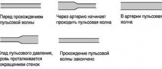
rhythmic oscillations of the arterial wall, caused by the release of blood from the heart into the arterial system and the change in pressure in it during systole and diastole.
The propagation of the pulse wave is associated with the ability of arterial walls to elastically stretch and collapse. The speed of propagation of the pulse wave ranges from 4 to 13 m/s, i.e. significantly exceeds the linear speed of blood flow, which even in large arteries does not exceed 0.5 m/s.