Take the first step
make an appointment with a doctor!
Many women are faced with such a dangerous phenomenon as fetal bradycardia during pregnancy. This is an abnormal decrease in heart rate, which provokes insufficient saturation of the brain and other organs of the unborn child with oxygen. This could potentially lead to the death of the embryo or irreversible changes in its brain. How dangerous is fetal bradycardia, what factors contribute to its occurrence, and is it possible to reduce the risk? We will try to answer these questions in this article, also describing ways to solve this problem.
Types of disease
Starting from 8 weeks of pregnancy, regular visits to the doctor make it possible to determine at an early stage a decrease in the heart rate of the fetus in the womb.
This is important because it allows you to recognize any deviation and say with confidence what type it is and how serious it is. There are 2 types of bradycardia in the fetus:
- Basal. Manifests itself as a decrease in heart rate to 110 beats/minute. This type of disease is practically harmless and does not entail any irreversible changes if it is treated promptly and correctly. Sometimes caused by hypotension in a woman, compression of the baby's head.
- Decelerant. In this case, the heart rate decreases to 90 beats. In addition to a number of reasons, the most common cause of occurrence is hypoxia, that is, lack of air in the womb. Already indicates the suffering of the embryo and requires immediate treatment.
Causes of the disease
Fetal bradycardia can develop due to the following reasons:
- Leading a harmful lifestyle when the expectant mother does not exclude the harmful effects of alcohol and nicotine. This also includes failure to follow doctor’s recommendations regarding a healthy lifestyle. Low levels of vitamins in food, fast food, a small number of walks - all this directly affects the child in the womb.
- The presence of chronic diseases in the mother’s body, especially the heart and lungs, as well as their exacerbation.
- Anemia, including that manifested in position.
- Infectious diseases.
- Taking certain drugs that have a toxic effect on the embryo. Therefore, before use, consult a specialist and review the instructions.
- Severe stress condition.
- Anomalies in the development of the embryo. This includes any abnormalities that arise in the baby: defects in the development of the brain, heart, other organs, and even Rhesus conflict.
- Long-term toxicosis in severe form.
- Multiple pregnancy.
- Premature placental abruption.
- Umbilical cord entanglement, as well as other urgent situations.
- Little and polyhydramnios.
Among the reasons contributing to the development of bradycardia in a child, there are both serious and minor factors, the effects of which can be easily prevented. But in any case, the disease sometimes entails dire consequences. Thus, prolonged hypoxia resulting from lack of treatment threatens the life of the fetus.
Important! Delay in treatment often leads to irreversible and terrible consequences - the development of anomalies that affect the health of the embryo, and even its death.
In newborns, bradycardia is sometimes a sign of congenital disorders in the development of organs (including heart defects), hypoxia experienced in the womb, birth trauma, and metabolic disorders. There are a number of other reasons, so in each case it is set individually.
Causes of bradycardia in children
Bradycardia is, first of all, a reaction of the heart to certain conditions. In this regard, the list of reasons that can cause such a problem is very long. The most important factors are:
- physiological: natural causes that do not indicate the presence of pathology;
- cardiac: associated with heart pathology;
- endocrine: associated with disruption of the endocrine glands;
- neurological: caused by a malfunction of the nervous system;
- intoxication: acute or chronic poisoning;
- genetic: hereditary pathology, for example, constitutional familial bradycardia;
- other reasons: gastrointestinal pathology.
Depending on the age of the baby, certain groups of factors predominate among the most likely causes.
Bradycardia in premature and newborn babies
A reduced heart rate at premature birth is physiological and is associated with the immaturity of heart rate regulation systems. With proper care and medication support, the condition gradually improves. The most common causes of bradycardia in a newborn child are heart defects, various types of encephalopathy, and birth injuries. Sometimes a pronounced decrease in heart rate indicates the development of transverse atrioventricular block (AV block). This is a dangerous condition that requires immediate medical attention.
Bradycardia in children of the first year of life (infants)
At this age, the pathological condition can be caused by the same reasons as in newborns. In addition, it is worth paying attention to the level of thyroid hormones. Hypothyroidism often causes a slow heart rate.
Bradycardia in preschool children
Bradycardia in a child aged 2 to 7 years is often associated with infectious diseases that are complicated by myocarditis - inflammation of the heart muscle. Poisoning and drug overdoses are also common at this age. The third common cause of bradycardia in a child aged 3–4 years and older is adenoids. Difficulty in nasal breathing leads to chronic hypoxia, causing a decrease in heart rate.
Bradycardia in schoolchildren and adolescents
A decrease in heart rate at school age may be due to the same reasons as in preschoolers, but they are accompanied by various types of gastritis and duodenitis associated with changes in diet.
Teenagers often encounter the manifestation of vegetative-vascular dystonia, which can occur according to the vagotonic type. A decrease in heart rate is accompanied by a drop in blood pressure, dizziness, and fainting. At the same age, autoimmune diseases and sick sinus syndrome often manifest.
Sinus bradycardia of the heart in adolescent athletes, which is not a symptom of the disease, stands apart. Regular intense exercise leads to the development of a physiological decrease in heart rate at rest, as the heart adapts to the additional work during exercise.
Symptoms
Naturally, it is very difficult to notice the development of this disease during pregnancy, but in a newborn its manifestation can be noticed due to pronounced signs. By the way, it often appears before an attack of sudden respiratory arrest. When walking, especially on cool days, your heart rate slows down as you exhale.
Sinus bradycardia is considered serious, since when it occurs, the heart rate can drop to 70 beats per minute. Sometimes this may even indicate a serious malfunction of the heart (for example, congenital blockade). The symptoms are as follows:
- The child freezes and the movement stops. Sometimes convulsions appear.
- The skin turns pale, and after this the integument may acquire a bluish tint.
- An attack of apnea, and in some cases even cardiac arrest, is possible.
Important! If a low heart rate (less than 100 beats) is detected in a newborn, you should not wait for other signs indicating illness to appear. You should immediately visit a doctor and strictly follow the recommendations he gives.
Symptoms of bradycardia in children
A mild form of sinus bradycardia in a child or adolescent does not cause any pathological symptoms.
The body tissues receive a sufficient amount of oxygen, so that the baby does not experience discomfort. With a significant slowdown in heart rate, signs of tissue oxygen starvation appear: fatigue, drowsiness, decreased performance at school, and forgetfulness. As the disease progresses, dizziness of varying severity appears, leading to falls and loss of consciousness. In this case, the skin becomes pale with a bluish tint, blueness appears around the mouth and cold, sticky sweat appears. As a rule, the attack lasts a few seconds. With constant bradycardia, fainting often occurs due to being in a stuffy room, stress, or physical overload.
Treatment and recommendations
Every doctor will say with confidence that the best treatment for fetal bradycardia is to follow all recommendations. Prevention will significantly reduce the possible risk of injury, but we must not forget that visits to the hospital are necessary not just for record keeping, but also in order to prevent such deviations.
Treatment of bradycardia that occurs in the fetus depends on the causes,
causing the disease. In the mildest cases, it will be enough to adjust your diet and daily routine, and normalize the supply of missing vitamins by taking mineral complexes. Walking is definitely recommended, as moderate physical activity and oxygen saturation prevent hypoxia and restore strength. Drugs are prescribed by a doctor if necessary to compensate for chronic or infectious diseases, as well as for iron deficiency and anemia.
Subsequently, the baby in the womb is monitored regularly. The most comprehensive diagnostic tools are used - from simply listening to the mother’s abdomen with a stethoscope to cardiotocography and ultrasound. Using the same means, fetal bradycardia is detected.
Important! During pregnancy, studies are carried out a certain number of times. Do not be afraid that any radiation will negatively affect the baby - this is not the case. Remember that your refusal to undergo diagnostics jeopardizes the health of the unborn baby and even his life.
Bradycardia detected at an advanced stage of pregnancy can sometimes cause very serious pathologies to the fetus, and therefore, in some cases, a very radical, but often inevitable solution to the problem is possible - preventing negative effects through cesarean section. Such decisions are made if the child is viable and can survive after birth.
Fetal bradycardia
Kabardino-Balkarian State University named after. HM. Berbekova, Faculty of Medicine (KBSU)
Level of education – Specialist
State educational institution "Institute for Advanced Medical Studies" of the Ministry of Health and Social Development of Chuvashia
Pregnancy is a difficult and responsible period. At this time, a woman is responsible not only for her own health, but also for her unborn child. Constant monitoring by a doctor is necessary to avoid or promptly detect problems in the development of the fetus. One of the deviations from the norm is bradycardia in the fetus. Should we panic and sound the alarm about this? There is no need to panic, but you definitely need to undergo all the examinations prescribed by the doctor and the necessary treatment. What is fetal bradycardia?
Fetal heartbeat
The first myocardial cells are formed in the embryo at 2–3 weeks. The first beats of the nascent heart can be heard during an ultrasound examination performed transvaginally at 5 weeks of pregnancy. But up to the 5th month of pregnancy, any listening to the fetal heartbeat is not informative in terms of the presence or absence of any pathology. From 11 to 13 weeks, an ultrasound with listening is performed solely to confirm pregnancy and that the baby is alive.
At different stages of the formation of organs and systems, the fetal heart beats at different speeds. The work of the heart is not regulated by the nervous system until 9 weeks, when the heart rate reaches its maximum for the entire pregnancy - 175 - 190 beats per minute. It then begins to decline and a stable heart rate is established at 14 weeks. Subsequently, until childbirth, 120–160 beats per minute is considered the norm.
Bradycardia is a slow heartbeat. It is a deviation even more dangerous than tachycardia. Fetal bradycardia in early pregnancy is a decrease in heart rate below 120 beats. If an ultrasound at 5–6 weeks shows a significant decrease in heart rate (up to 50–70 beats), this may indicate unfavorable hereditary factors and the possibility of pregnancy fading. In this case, a repeat examination is scheduled in a week. A slow heartbeat in the 2nd and 3rd trimester of pregnancy is accompanied by a decrease in fetal activity and in most cases indicates a violation of intrauterine nutrition and oxygen starvation.
Fetal bradycardia: causes and symptoms
Not every decrease in a person’s pulse is an anomaly or pathology. For example, it is observed during sleep or when the ambient temperature drops - at such moments the body saves energy and metabolism slows down. Also, a similar phenomenon is observed in athletes, and in some people it is present from birth, but does not have the nature of a pathology. In such cases we speak of physiological bradycardia. Its important difference from abnormal is the absence of pathological symptoms.
Abnormal bradycardia is a decrease in heart rate in which various painful conditions of the body occur: dizziness, cold sweating, loss of consciousness, etc. As a rule, they manifest themselves with a strong reduction in the heartbeat. If it is insignificant, then a person may not experience subjective sensations.
To judge the presence of cardiac bradycardia in the fetus, it is necessary to have an idea of the physiological normal heart rate. In an adult it is 60-80 beats per minute, but in an embryo it changes during its development:
- At 3-5 weeks – 75-80;
- At 5-6 weeks – 80-100;
- At 6-7 weeks – 100-120;
- At 7-9 weeks – 140-190;
- At 10-12 weeks – 160-180;
- At 4 months – 140-160;
- By 9 months – 130-140.
The indicated values are not exact, since the physiological norm for each child may vary slightly. Until approximately the 21st day of pregnancy, the embryo’s heartbeat cannot be heard at all - at this stage, its own heart has not yet begun to form, and metabolism is completely ensured by the mother’s bloodstream.
It is possible to unambiguously diagnose pathological bradycardia in the mother and fetus only in the 2nd trimester (after 20 weeks of gestation), since at this stage its own blood supply system as a whole has already formed, so the pulse should stabilize. The doctor makes a diagnosis if during this period the heart rate is less than 110-120 beats per minute.
Bradycardia can be a “maternal” disease or observed only in the fetus. In the first case, the slowing of the heartbeat of the woman herself also affects the condition of the unborn child; in the second, the pathology of the embryo does not affect the health of the mother. The causes of the disease on the mother's side are:
- diseases of the cardiovascular system - atherosclerosis, unstable blood pressure, coronary heart disease, cardiosclerosis, dystrophy or inflammation of the myocardium (heart muscle);
- general diseases of the body - impaired hematopoietic function of the bone marrow, tumors, anemia, infectious pathologies, renal failure, etc.
On the part of the fetus, the following pathologies are the causes of bradycardia:
- maternal anemia, high environmental toxicity, mental stress in women;
- malformations of the fetus’s own cardiovascular system;
- disorders of the mother’s reproductive system - early aging of the placenta, abnormal accumulation of amniotic fluid, etc.;
- Rh conflict - incompatibility of maternal and embryonic blood according to the Rh factor;
- intoxication of the maternal body due to smoking, drinking alcohol or drugs, and certain types of medications.
Symptoms of bradycardia in the mother are typical signs of oxygen starvation - dizziness, weakness, headache, tinnitus, chest pain, low blood pressure, shortness of breath. If the pathology is observed only in the fetus, it will be indicated by a decrease or cessation of its motor activity, as well as convulsions. The peculiarity of the embryonic form of the disease is that it is impossible to identify it based on the condition of the mother - only by directly observing the child himself using modern diagnostic tools.
Pregnancy in patients with bradycardia is characterized by a high risk of fetal hypoxia. Prolonged oxygen starvation can lead to frozen pregnancy, miscarriage or irreversible damage to the body of the unborn child. His brain suffers especially badly from this, since nerve cells are most sensitive to lack of oxygen.
Take the first step
make an appointment with a doctor!
Causes
For various reasons, the heartbeat of the unborn baby may slow down. The first fetal auscultation is performed at 20 weeks. If bradycardia is suspected, the woman will be under constant monitoring until delivery. Causes of fetal bradycardia:
| Influence factor | Cause |
| External | bad habits of women - smoking, drugs, alcohol; |
| injuries during pregnancy with damage to the fetal circulatory system; | |
| emergency operations during pregnancy; | |
| continuing to take contraceptives at the beginning of pregnancy; | |
| stress during pregnancy; | |
| poor nutrition – diets, religious observance; | |
| taking analgesics or beta blockers | |
| Domestic | severe toxicosis; |
| Rhesus conflict without treatment; | |
| placental abruption or insufficiency; | |
| multiple births; | |
| polyhydramnios or oligohydramnios; | |
| umbilical cord compression or entanglement; | |
| gestosis; | |
| developmental defects (triad and tetralogy of Fallot) | |
| Mother's illnesses | chronic heart and vascular diseases; |
| chronic respiratory diseases; | |
| diabetes; | |
| anemia; | |
| hypotension; | |
| eclampsia; | |
| infectious diseases |
Bradycardia during pregnancy may be due to disruption of the placenta. This temporary organ performs several functions - transport (blood supply to the fetus), endocrine, metabolic. Any failure in its operation leads to a decrease in the supply of nutrients and oxygen to the fetus.
Types of fetal bradycardia
Fetal bradycardia can occur in two ways:
- basal;
- decelerant or sinus.
Basal bradycardia, provided it is treated in a timely manner and the factors that caused its occurrence are corrected, does not pose a threat to the fetus and pregnancy as a whole. Heart rate drops to 110 – 120 beats per minute. This indicates fetal hypoxia, which occurs due to iron deficiency anemia in the mother, a drop in her blood pressure, or compression of the fetal head.
Sinus bradycardia is a more serious condition. The fetal heart rate drops to 70 beats per minute, indicating the need for urgent medical attention. The mother is prescribed bed rest, treatment is carried out only in a hospital and continues until the moment of delivery. Sinus bradycardia often leads to pregnancy failure.
Types of bradycardia in the embryo
Bradycardia in the embryo is classified into three degrees of severity, which have their own individual names:
Basal (moderate) fetal bradycardia. This is bradycardia in a child, in which the heart rate is less than 120 beats per minute. If this pathology was identified in a timely manner and adequate therapy was prescribed, then the consequences of hypoxia on the child’s body are minimal.
Decelerant fetal bradycardia . A condition of an embryo in which the heart rate is approximately 72 beats per minute. This is a rather dangerous type of bradycardia in the embryo, which requires immediate hospitalization of the mother and timely treatment in a hospital.
Fetal sinus bradycardia . This is the most dangerous form of fetal bradycardia, in which the heart rate is less than 70 beats per minute. This condition requires immediate and intensive hospital treatment. If such bradycardia is detected at a later stage, immediate delivery is recommended.
Diagnostics
Slowing the fetal heart rate is fraught with serious consequences, the most serious of which is its intrauterine death. During bradycardia, the fetus experiences oxygen starvation during pregnancy, which is extremely dangerous for the development of its systems and organs and causes its delay. In any case, even a single episode of decreased fetal cardiac activity requires a thorough examination.
Expectant mothers who have chronic diseases or a history of serious health problems should also undergo regular screening. Diagnostics include:
- Auscultation. It is carried out from 20 weeks onwards for all pregnant women.
- Ultrasound includes 4 screenings during pregnancy:
- at 11 – 13 weeks to determine the gestational age and fetal heart rate;
- at 20–23 weeks to determine the condition of the fetus, its heartbeat and the condition of the placenta;
- at 30–34 weeks, Doppler ultrasound of the uterus is performed to assess blood flow and the condition of the cardiac and vascular systems
- at 36–37 weeks to assess the condition of the placenta.
- CTG (cardiotocography). It shows the condition of the fetus and uterine vessels.
- Electrocardiography is performed at 5–6 months only if the fetus is developing with a delay or other examinations have revealed heart pathologies.
- Phonoelectrocardiography is intended to identify murmurs and determine heart rhythm.
In the first months, the only possible way to monitor the fetal heart rate is ultrasound. From the 20th week - this is auscultation in combination with ultrasound. Starting from the 8th month, CTG and Doppler measurements are used.
Treatment
After 30 weeks, if there is persistent fetal bradycardia, the doctor may decide to deliver prematurely by cesarean section. This is done in order to exclude further development of pathologies.
If bradycardia does not threaten pregnancy, doctors’ efforts are aimed at eliminating the factors that cause it. Drug treatment includes the use of the following drugs:
- ginipral or papaverine, which relieve uterine tone and reduce fetal hypoxia;
- vitamins (ascorbic acid, iron supplements for anemia);
- glucose;
- magnesia (for preeclampsia and gestosis).
Other elements of treatment are:
- rejection of bad habits;
- compliance with a therapeutic diet;
- adherence to sleep and rest patterns;
- reduction of physical activity;
- walks in the open air;
- maintaining immunity.
It has become more difficult for a modern woman to bear a child due to the negative influences of modern life - stress, unhealthy diet, poor environment. Therefore, medical supervision, from early pregnancy to childbirth, is a vital necessity to ensure the birth of a healthy child.
Fetal heart rate
The baby's heart rate is monitored throughout pregnancy. How frequent and how long the checks will be depends on the condition of the expectant mother’s body. If the parameters are higher or lower than normal, then examinations are carried out more often.
Normally, the fetal heart should beat from 110 to 170 beats per minute. Throughout pregnancy, indicators change. If they exceed the norm, then tachycardia is diagnosed; if the fetal pulse is too low, bradycardia is detected.
The embryonic heart develops from the second week. The first heartbeats occur on the third, and you can listen to it when the heart chambers are fully formed on the 6-8th.
In the early stages
Bradycardia in the fetus in the early stages is a relative concept, since during this period there are no clearly defined norms. At the very beginning of development, the heart contracts at a frequency of about 40 beats. Veins, vessels, and aorta are gradually formed, and the heart rate increases several times every day. By the end of the ninth week, the heartbeat already reaches 175 beats.
The nervous system gradually develops, which affects the functions of the internal organs, and the contraction gradually decreases and drops to 157 beats by the 14th week.
Since the size of the embryo may be different in all women, the heart rate is calculated taking into account the length of the fetus.
If it is about five millimeters, then the organ should beat about 100 beats. With large embryo sizes, the indicators may be 10-20 units higher. If the baby is larger than 8 millimeters, and the heartbeat cannot be heard, then, most likely, fetal fading has occurred.
After the 20th week, you can get accurate information about the condition of the embryo. During this period, it is easier to determine any disorders in the body. Not only the pulse is heard, but also the position of the myocardium. In the second trimester, indicators should be between 140-160 beats.
In the later stages
Bradycardia in the fetus in later stages is dangerous if the readings drop below 85 beats.
With small deviations, problems are not seen, since the pulse is an individual parameter that can be affected by the physiological characteristics of a woman’s body, genetic characteristics, fetal size and other factors.
Ultrasound is used to determine the heart rate. It can also be used to identify pathological processes in the heart muscle.
The fact that the organ is contracting more slowly than it should can only be discovered in the twentieth week. The deviation is difficult to notice even at a later date. The characteristics of the woman’s body and the course of pregnancy play a big role in this.
Pathological abnormalities are clearly visible in the third trimester on cardiotocography. Depending on the period of gestation of the child, different diagnostic methods are used:
- Transvaginal ultrasound examination. With its help, bradycardia in the embryo at the 6th week is perfectly detected.
- Ultrasound examination through the abdominal cavity from the 7th week.
- Auscultation. The rhythm and frequency of the heartbeat are listened to with a stethoscope from the 20th week.
- In the third trimester, cardiotocography is prescribed.
- Until the 34th week, detailed information can be obtained using Doppler ultrasound of the uterine vessels.
Additionally, blood and urine are donated for laboratory tests, and the speed of blood flow in the vessels of the umbilical cord is determined.
An electrocardiogram may be performed between 18 and 24 weeks. This is necessary if there is a suspicion of a malfunction of the woman’s heart and blood vessels or if the pregnancy is unfavorable. Dopplerography of blood flow in the vessels of the uterus and placenta is also performed.
Diagnosis of bradycardia in the fetus
If the mother herself has no problems identifying heart rhythm disturbances, then in the case of fetal bradycardia the situation is more complicated. Pathology can only be detected by observing the fetus itself. The following methods are used for this:
- Auscultation. This is listening to the fetal heartbeat using a stethoscope. It is carried out externally through the mother's abdominal wall. This is the simplest, but also the least reliable method of diagnosis, since the readings are influenced by the presence of a woman’s fat layer, as well as the motor activity of the fetus.
- Ultrasound. Ultrasound examination allows not only to hear the fetal heartbeat, but also to visualize the embryo itself. Ultrasound can be performed abdominally (through the peritoneum) or transvaginally (with a probe inserted into the vagina). In this case, the doctor receives data on the nature and intensity of the fetal motor activity, the frequency of its breathing and heart contractions.
- CTG (cardiotocography). This is one of the most accurate ways to diagnose bradycardia in the fetus in later stages. It consists of recording the embryo's pulse and comparing it with the frequency of contractions of the mother's uterus. The assessment occurs on a 10-point scale: with 8-10 points, no pathology is detected, with 6-8, a mild anomaly is diagnosed, and with less than 6 points, a severe form is diagnosed. The advantage of CTG is the possibility of using this method if the mother has endocrine and cardiovascular disorders.
- ECG (electrocardiography). This method involves recording electrical impulses from the fetal heart, often in combination with recording heart sounds (phonocardiography). The received data is displayed in real time in the form of a cardiogram - linear graphs on paper tape. Bradycardia is indicated by the presence of a P wave and increased T-P and T-Q intervals.
Depending on the severity of the disease, some of these tests are performed or they are used in combination. The doctor gives a referral to undergo them, focusing on the mother’s condition, her age, whether she had similar problems in previous pregnancies, etc.
Take the first step
make an appointment with a doctor!
Why does it occur
This problem does not always indicate a serious illness. This can occur with temporary disturbances in the functioning of the cardiovascular system. Bradycardia can be observed if the baby has turned into an uncomfortable position, and the vessels are compressed, and in other cases that are not related to developmental disorders.
Decreased heart rate may be due to genetic factors or serious abnormalities.
Pathological slowdown is associated with:
If the problem occurs before two months, it usually occurs due to chromosomal abnormalities such as Down syndrome, Edwards syndrome and others. To identify the problem, additional examination is prescribed.
In the first trimester, the problem may also indicate developmental defects. Such deviations develop under the influence of:
- Infectious diseases suffered by a pregnant woman.
- Exposures.
- Living in an unfavorable environmental environment.
- Use of certain medications.
Very often it is impossible to determine why a heart defect occurred.
In the second and third trimester, the problem may be associated with placental insufficiency. The placenta provides the fetus with nutrients and oxygen. If this function is disrupted, then all organs suffer. A similar problem arises due to:
- chronic diseases of the mother;
- anemia;
- severe toxicosis at the beginning of pregnancy;
- gestosis;
- pathologies of amniotic fluid;
- past diseases of infectious origin;
- disorders of the blood coagulation system;
- stress and bad habits of the mother;
- umbilical cord entanglement;
- multiple pregnancy.
In most cases, a decrease in heart rate is associated with hypoxia. But sometimes the cause of this problem cannot be found out.
Bradycardia
Atherosclerosis
Ulcer
21764 February 15
IMPORTANT!
The information in this section cannot be used for self-diagnosis and self-treatment.
In case of pain or other exacerbation of the disease, diagnostic tests should be prescribed only by the attending physician. To make a diagnosis and properly prescribe treatment, you should contact your doctor. Bradycardia: causes of occurrence, what diseases it occurs with, diagnosis and treatment methods.
Definition
Bradycardia is a condition of the heart muscle when its contraction frequency is less than 60 beats per minute. Bradycardia is not always considered a pathology; it is rarely an independent disease and more often indicates the presence of problems in other organs or systems.
A slight decrease in heart rate (HR) usually does not affect a person’s general well-being.
Hemodynamic disturbances are observed with a pronounced slowdown in heart rate, in people with various concomitant diseases (more often in the elderly). The main symptoms of bradycardia include unexplained weakness, increased fatigue, pallor of the skin, dizziness and lightheadedness, fainting (Morgagni-Adams-Stokes attacks - fainting or syncope during an attack of bradycardia), shortness of breath (difficulty breathing), pain and discomfort in the chest, blood pressure fluctuations.
Types of bradycardia
Bradycardia is usually divided into physiological, pathological and drug-induced.
- Physiological bradycardia
is a decrease in heart rate that is not associated with pathological processes in the body. For example, in athletes, due to the great training of the heart muscle, there is a decrease in the normal limits of the heart rate. This is a normal condition in the absence of other symptoms (drowsiness, weakness, dizziness, difficulty breathing and chest discomfort). A decrease in heart rate during sleep or when body temperature decreases (for example, when the body is hypothermic) is also classified as physiological conditions. - Pathological bradycardia
(neurogenic, endocrine, toxic, myogenic).Neurogenic bradycardia makes itself felt with increased intracranial pressure, peptic ulcer, renal, hepatic and intestinal colic, glomerulonephritis, as well as after severe infectious diseases.
Endocrine bradycardia is detected when the function of the thyroid gland and adrenal cortex decreases.
Toxic bradycardia accompanies states of intoxication (uremia, liver failure), as well as hyperkalemia and hypercalcemia.
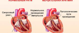
Myogenic bradycardia is associated with cardiac diseases and is the first manifestation of sick sinus syndrome. Normally, the sinus node (the structure of the heart responsible for producing electrical impulses) functions at a frequency of 60–90 impulses per minute, and the heart muscle (myocardium) contracts at the same frequency. With pathology of the sinus node, the production of impulses slows down and bradycardia occurs. The same thing happens when there is a disruption in the transmission of impulses from the sinus node to the underlying structures of the heart (impulse block).
- Pharmacological, or medicinal, bradycardia is possible with abuse and overdose of certain medications (cardiac glycosides, beta-blockers, calcium channel blockers, antidepressants), narcotic substances, etc.
According to clinical manifestations, the following forms of bradycardia are distinguished:
- latent form, characterized by the absence of clinical and ECG manifestations, and sinus node dysfunction is determined by electrophysiological study;
- compensated form with mild clinical manifestations (complaints of dizziness and weakness), ECG changes are present;
- decompensated form, in which persistent sinus bradycardia is determined, manifested by dizziness, fainting, transient paresis, and heart failure.
Possible causes of slow heart rate
In order to understand the cause of bradycardia, it is necessary to know the mechanisms that regulate heart rate.
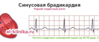
There is a system of fibers in the heart - the cardiac conduction system, which is responsible for the generation of electrical impulses, their conduction through the heart muscle and the correct contraction of the myocardium. Normally, impulses are produced in the sinus node (also called the sinoatrial node, or SA node) at a rate of 60–90 per minute. sinus bradycardia
occurs .
Next, the electrical impulse travels to the atrioventricular node through a system of conduction bundles. If fiber damage occurs at this level, non-sinus bradycardia
. The types of bradycardia are no different in their manifestations, but this division is important for making a diagnosis and choosing the right treatment tactics.
Reasons that disrupt the structure of the conduction system of the heart, i.e. cause organic damage to the myocardium are:
- myocardial infarction with subsequent replacement by connective tissue of destroyed heart structures, including important bundles that conduct electrical impulses from the sinus node;
- coronary heart disease, when, due to atherosclerosis of the coronary arteries, adequate blood supply to the myocardial areas in the area of the conduction bundles is disrupted and sclerodegenerative fibrosis (replacement with connective tissue) of the affected areas develops;
- myocarditis - inflammatory damage to the heart muscle;
- congenital anomalies of the structure of the conduction system of the heart.
During intoxication during infectious diseases, with severe sweating, vomiting and diarrhea, a significant loss of electrolytes (potassium, sodium and magnesium ions) occurs, which leads to disruption of the water-salt balance in the body and the occurrence of various heart rhythm disturbances.
Some thyroid diseases, such as hypothyroidism, are characterized by a decrease in the synthesis and function of thyroid hormones, as a result of which metabolism is disrupted and heart rate is reduced.
Possible causes of bradycardia include general hypothermia, increased intracranial pressure (with cerebral edema, tumor process, meningitis, cerebral hemorrhage), lead poisoning, nicotine, etc.
What diseases cause bradycardia?
The list of diseases and conditions for which bradycardia is diagnosed is quite extensive:
- sick sinus syndrome;
- atrioventricular block;
- myocardial infarction;
- cardiac ischemia;
- myocarditis;
- infective endocarditis;
- heart defects;
- hypothyroidism;
- some infectious diseases (typhoid fever, viral infections);
- poisoning with lead, organophosphorus compounds, including pesticides, narcotic substances;
- general hypothermia;
- heart tumors.
Which doctors should you contact if you have bradycardia?
Treatment of diseases in which bradycardia is one of the symptoms is carried out by cardiologists, therapists, and endocrinologists.
Diagnosis and examinations for bradycardia
First of all, to identify cardiac arrhythmias, examination techniques such as counting pulse and heart rate are used.
When examining a patient, the doctor uses auscultation to listen to the heart rhythm and detect other accompanying symptoms to diagnose the disease that has caused the change in heart rate.
When making a diagnosis, instrumental and laboratory diagnostic methods must be used:
- electrocardiographic study (ECG), echocardiography (EchoCG), and if dynamic monitoring is necessary, 24-hour Holter monitoring;
Treatment of pathology
Bradycardia in the fetus during pregnancy is treated by normalizing the woman’s lifestyle and changing her diet, as well as prescribing vitamin complexes.
Even in case of slight deviations from the norm, the expectant mother should be constantly under the supervision of a doctor. If the fetal heart rate is too low, it is important to follow all its instructions.
In most cases, hospitalization and treatment in a hospital are necessary.
If the problem arose at a later stage, and the intrauterine development of the embryo proceeds without deviations, then cesarean section is recommended as a method of delivery.
If an ultrasound showed a deterioration in the fetus’s heart function, slowing or stopping of movements, the presence of sudden respiratory movements and convulsions, the woman should:
- Completely give up bad habits.
- Normalize your lifestyle.
- Use vitamin and mineral complexes selected by your doctor.
- Drink teas and tinctures based on medicinal plants.
- Take selected medications.
In each case, separate methods for solving the problem are prescribed.
Treatment of bradycardia in children and adolescents
Treatment of mild or moderate bradycardia and arrhythmia in a child of any age is aimed at normalizing the heart rate, as well as eliminating the cause of the pathological condition.
Depending on the situation, emergency medications and ongoing medications may be used to stabilize the heart. The choice of treatment method for bradycardia is made by a cardiologist. If a child develops severe bradycardia, accompanied by frequent fainting, Morgagni-Adams-Stokes attacks and other conditions that disrupt the usual rhythm of life, surgical treatment is used. Experienced cardiac surgeons implant an electrical pacemaker that controls the functioning of the heart. This treatment is also relevant if the condition arose against the background of severe defects and blockades.
Prevention measures
To prevent any disturbances in the development of the embryo, more attention should be paid to:
- Planning pregnancy.
- Treatment of chronic diseases before conception.
- Healthy lifestyle.
- Walking in the fresh air.
- Consumption of vitamins and minerals.
- Regular rest.
- Eliminate stress and physical activity.
- Passing preventive examinations.
It is much easier to try to avoid bradycardia than to eliminate it. By following simple recommendations, you can successfully carry and give birth to a completely healthy child.
Preventive measures help avoid the development of bradycardia and eliminate the pathological process in the initial stages of formation. This can also help reduce the likelihood of abnormalities in fetal development.