Sinus bradycardia - a decrease in heart rate to 60 beats per minute. This condition is not a pathology. It can be either a variant of the norm or a sign of a serious illness. Bradycardia is often observed in professional athletes. With regular exercise, the heart pumps blood more actively and provides the necessary blood flow through fewer contractions. In swimmers, skiers and track and field athletes, the resting heart rate can reach 30 beats per minute. In this case, this is not considered a deviation. However, if the heart rate is less than normal and other disturbing symptoms are observed, there is a reason to seek advice from a cardiologist.
Symptoms of bradycardia
- Semi-fainting, attacks of loss of consciousness or dizziness during a decrease in pulse.
- Arterial hypertension, unstable blood pressure.
- Increased fatigue even after minor physical activity.
- Chronic circulatory failure.
- Angina pectoris of exertion and rest in combination with a decrease in heart rate.
- Weakness.
- Cold sweat.
- Pain in the heart area.
- Shortness of breath (uncharacteristic symptom).
Severe bradycardia
(less than 40 beats per minute) is a very dangerous condition that can cause the development of heart failure, and therefore the patient may require surgery to implant a pacemaker.
Normal or pathological?
Bradycardia (heart rate less than 60 beats per minute) is not uncommon. Especially for those who have been involved in sports for a long time. At the end of intense physical activity, the “athlete’s heart” (there is even such a term in cardiology) is programmed to ensure that the pulse quickly returns to normal values.
The heart rate slows down in some patients simultaneously with an increase in blood pressure. And in this sense, bradycardia can be considered a tool of self-regulation of our body, which by nature is conceived very intelligently.
Article on the topic Under the beat of the heart. How to choose a tonometer
It’s another matter if bradycardia occurs suddenly and is accompanied by a deterioration in well-being. In this way, for example, atrial fibrillation can manifest itself (in which the so-called “tachy-brady” syndrome is often diagnosed, when frequent contractions are replaced by rare ones), fraught with stagnation of blood, as a result of which blood clots form in the chambers of the heart (usually in the left atrium). But the worst thing happens when small fragments of a blood clot (embolus) break off and, along with the blood flow, enter the coronary vessels, causing a heart attack. Or a cardioembolic stroke if the deadly “bullet” reaches the carotid artery.
An embolus can “fly” into the system of mesenteric vessels supplying blood to the intestines, causing necrosis of one of its sections followed by peritonitis, or into the vessels of the lower extremities, causing their critical ischemia (impaired blood supply) with the development of gangrene.
Factors in the development of bradycardia
- Sclerotic changes in the myocardium - the muscular layer of the heart, affecting the sinus node.
- Increased tone of the parasympathetic nervous system.
- Increased intracranial pressure (with cerebral edema, tumor, meningitis, cerebral hemorrhage).
- The effect of certain medications for the treatment of cardiovascular diseases (beta blockers, antiarrhythmics, etc.).
- Toxic poisoning with lead, nicotine.
- Hypothyroidism is a decrease in thyroid function.
- Starvation.
- Age-related changes in the heart.
- Diseases of the cardiovascular system (atherosclerosis of the coronary vessels, ischemic heart disease, cardiosclerosis, heart attack, endocarditis, myocarditis, post-infarction scars, etc.).
- Disorders of the metabolism of minerals and electrolytes involved in conducting electrical impulses in the heart.
- Obstructive apnea (stopping breathing during sleep).
- Infectious and inflammatory diseases (rheumatism, systemic lupus erythematosus).
- Hemochromatosis (accumulation of iron in organs and tissues).
- Some diseases not related to the functioning of the cardiovascular system (typhoid fever, jaundice, etc.).
Causes of bradycardia
There are many reasons that can cause bradycardia. Most often, such a rhythm disturbance occurs as a result of damage to the sinus node, the so-called weakness syndrome in the sinus node, or when the conduction system is damaged - heart block. Accordingly, any reasons that can lead to node weakness or heart block automatically lead to bradycardia. Such reasons may be:
- Deficiency of thyroid hormones.
- Increased bile acids in the blood.
- Dystrophic or sclerotic changes in the sinus node as a result of inflammatory processes or after myocardial infarction with appropriate localization.
- Increased sensitivity or irritation of the vagus nerve. In this case, bradycardia is paroxysmal in nature, which can occur with the slightest impact on the carotid sinus.
- Autonomic-vascular nervous dysfunctions. For example, high intracranial pressure, swelling or bleeding in the brain, brain tumors.
- Physiological reasons: resting state in athletes, sleep, symptom of cholemia, and so on.
- Toxic causes: typhoid fever, uremia, sepsis, hepatitis or poisoning with various organophosphorus compounds, β-blockers, quinidine, glycosides, morphine, sympatholytic drugs or calcium channel blockers.
- With cardiosclerosis, myocardial dystrophy or myocarditis, tachy-brady syndrome is often observed, when rapid heartbeat (tachycardia) alternates with bradycardia. This condition occurs as a result of the alternation of pacemakers. If complications occur, chronic bradycardia may develop.
- Myocardial infarction, when the lesion is localized in the sinus node. With extensive myocardial infarction, acute bradycardia, both sinus and heterotopic, can develop.
Symptoms Moderate bradycardia usually does not have pronounced symptoms. Weakness, slight dizziness, increased fatigue, slowness and other general symptoms may occur. With chronic bradycardia, symptoms of heart failure are added, which develops against the background of decreased blood circulation in both circles. In acute bradycardia or tachy-brady syndrome, general weakness is accompanied by: - Morgagni-Adams-Stokes syndrome (dizziness, loss of function and cerebral ischemia). - Half-consciousness or fainting. - Development of angina pectoris with all its symptoms. - Collapse. Also, symptoms of bradycardia may be accompanied by manifestations of the underlying disease. Diagnosis of bradycardia First of all, it is necessary to identify the underlying disease that caused bradycardia. For example, determining the level of thyroid hormones, determining intracranial pressure, cerebral edema, and so on. To diagnose bradycardia directly, ECG, Holter monitoring, EchoCG, bicycle ergometry and, if necessary, X-ray examination or transesophageal examination of the myocardium, in particular the conduction system, are prescribed.
Types of bradycardia
- Absolute bradycardia
- a decrease in heart rate (heart rate) is constant and does not depend on the conditions in which the patient is located. - Relative bradycardia
is a decrease in heart rate under certain conditions (after physical activity, against the background of diseases - fever, traumatic brain injury, typhus, meningitis), a lag between the increase in heart rate and the increase in body temperature. This group also includes “athletes’ bradycardia” – a decrease in heart rate in well-trained people when they perform certain physical exercises. - Moderate bradycardia
- most often observed in children suffering from respiratory arrhythmia during deep inspiration, during sleep, or in the cold. Bradysphygmia, a rare pulse with a normal heart rate, should be distinguished from bradycardia. - Extracardiac vagal bradycardia
– observed in patients suffering from neurological diseases, diseases of internal organs, kidney diseases (nephritis), myxedema and other diseases. - Depending on the reasons that caused bradycardia, physiological and pathological bradycardia are distinguished. Pathological bradycardia is represented by two main forms - sinus bradycardia and bradycardia due to heart block.
Diagnosis of bradycardia in athletes
To identify cardiac bradycardia during sports, it is necessary to undergo a comprehensive medical examination; it is not possible to independently identify the disease during physical activity.
To determine the degree and severity of bradycardia during physical activity, it is necessary to undergo the following studies:
- Electrocardiography.
- Echocardiography.
- Comprehensive clinical assessment.
- Stress Testing.
Typically, athletes only need to undergo an ECG. Other research methods are necessary if there are symptoms of pathological bradycardia in order to exclude other diseases.
Complications of bradycardia
Rarely bradycardia
can also occur in healthy people.
A moderate degree of bradycardia
is not dangerous and does not cause serious disturbances in the functioning of the cardiovascular system.
However, if attacks of bradycardia
are repeated, this can lead to a lack of blood supply and oxygen starvation of organs and tissues, as a result of which their full functioning is disrupted. Acute oxygen deficiency can cause Morgagni–Adams–Stokes syndrome, in which a sudden loss of consciousness occurs.
A more serious complication of bradycardia
is the depletion of the myocardium - the heart muscle, which occurs as a result of fibrillation of the ventricles of the heart. Over time, rupture of the heart muscle (myocardial infarction) and death are possible.
“Catch” a pause
Bradycardia can also be a symptom of myocardial infarction, which can occur mildly, without classic pain, but with the development of disorders of the conduction system of the heart. In this case, there cannot be two opinions: such a person must be hospitalized immediately.
Article on the topic
Rules for a healthy heart: what to eat, how to exercise and what to avoid
With persistent, pronounced bradycardia, blood pressure also decreases (which should not be normal), which may require emergency installation of a pacemaker: in this mode, the work of the heart to supply blood to vital organs becomes ineffective.
The need to install a pacemaker may also arise in elderly patients who, against the background of atherosclerotic changes in the myocardium, develop the so-called sick sinus syndrome, in which dangerous pauses in the heart rhythm occur.
True, such arrhythmia can only be “caught” with the help of 24-hour Holter ECG monitoring. In some cases, doctors resort to another diagnostic procedure - transesophageal cardiac stimulation, during which a sensor inserted into the esophagus transmits impulses to the heart muscle and, against the background of excessive impulses, problems are identified that are not visible under normal conditions.
Article on the topic
All about the circulatory system: numbers and facts
In all other cases, bradycardia can be a normal tool of self-regulation of your body, which in this way can react, for example, to an increase in blood pressure. If this happens regularly (for example, in patients with hypertension), it is enough to lower blood pressure and the heart rate will immediately increase. For some patients with bradycardia, doctors prescribe dihydrodipyridine calcium antagonists. But this does not help all patients.
Treatment of bradycardia
Treatment of bradycardia
at GUTA CLINIC it begins with a correct diagnosis - after all, the successful outcome of treatment depends on establishing the exact cause of the disease.
If, against the background of a decrease in heart rate, no diseases that could cause bradycardia
are diagnosed, and there are no symptoms of the disease, then
bradycardia
is not treated
- If a slow heart rate is caused by a medical condition, such as hypothyroidism or electrolyte imbalance, correcting the problem is usually enough to stabilize your heart rate.
- If bradycardia
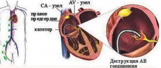
is a consequence of taking any medications, the attending physician will recommend reducing the dose or prescribing an analogue of the drug being taken that does not cause changes in heart rate. If for some reason it is not possible to take these measures, the patient may be recommended to have a pacemaker implanted.
Regardless of the cause of the disease, treatment of bradycardia
is to bring the heart rate to a level at which the blood supply to the body is sufficient.
Untreated bradycardia
or its incorrect treatment can lead to quite serious consequences - from fainting and general deterioration in health to paroxysms and death.
Diagnosis of bradycardia
at GUTA CLINIC it is carried out using the most modern expert-level equipment, and treatment is carried out by highly qualified cardiologists, candidates of medical sciences and doctors of the highest qualification category, who regularly improve their knowledge and exchange experience with foreign colleagues.
Many of our specialists have completed internships and training in the largest specialized medical centers in Europe and the USA, and have a number of scientific publications on the diagnosis and treatment of cardiovascular diseases, incl. and bradycardia
.
Treatment of bradycardia
at GUTA CLINIC it is carried out individually depending on the patient’s age, symptoms, characteristics of the course of the disease, the form of bradycardia and other individual indicators.
The cardiologists at GUTA CLINIC have hundreds of successfully treated patients with various forms of bradycardia
, including complicated ones.
Some features of the ECG in children involved in sports
S.A. IVYANSKY, L.A. BALYKOVA, A.N. Urzyaeva, L.S. ZAGRYADSKAYA, Yu.O. SOLDATOV, A.V. SAMARIN
Mordovian State University named after. N.P. Ogareva, Saransk
Children's Republican Clinical Hospital of the Ministry of Health of the Republic of Moldova, Saransk
Republican medical and physical education clinic of the Ministry of Health of the Republic of Moldova, Saransk
Ivyansky Stanislav Alexandrovich
Art. Lecturer at the Department of Pediatrics, Medical Institute, Mordovian State University. N.P. Ogareva
430032, Saransk, st. Rosa Luxemburg, 15, tel. (8342) 35-30-02, e-mail
The article presents the results of an ECG examination of 198 children aged 10-15 years playing football and tennis, with a sports experience of 2-5 years (initial training group) and 5-7 years (special sports training group).
It has been established that the level of average heart rate in athletes differs from the corresponding indicator in peers who do not engage in sports after 5 years of regular exercise, while the level of the minimum heart rate of athletes remains at a comparable value. Limits have been established for the duration of the QTc (460-480 ms), requiring additional diagnostic measures.
ECG changes requiring additional examination (bradycardia less than 2%, AV block of the second degree, ST-T changes, heart rhythm disturbances, prolongation of the QTc interval above 460-480 ms) were recorded in 4.9% of cases in the initial training group and in 9% of cases in the special sports training group. Key words:
child athletes ECG, heart rate, QT interval, signs of maladaptation.
SA IVYANSKY, LA BALYKOVA, AN URZYAEVA, LS ZAGRYADSKAYA, Yu.O. SOLDATOV, AV SAMARIN
Mordovian State University named after NP Ogarev, Saransk
Children Republican Clinical Hospital of the Ministry of Health of the Republic of Mordovia, Saransk
Republican medical exercises dispensary of the Ministry of Health of the Republic of Mordovia, Saransk
Some features of electrocardiography in children going in for sports
The article gives the results of electrocardiography of 198 children 10-15 years old, playing football and tennis with sports experience of 2-5 years (group of initial training) and 5-7 years (group of special athletic training).
It was found that the level of the average heart rate of the athletes is different from the corresponding figure of those not involved in sports after 5 years of regular exercise; the level of the minimum heart rate of athletes is at a comparable value. We set the limits of the interval length QTc (460-480 ms), requiring additional diagnostic procedures.
ECG changes that require further investigation (bradycardia less than 2%, AV-block II degree, ST-T changes, arrhythmias, QTc interval prolongation above 460-480 msec) were recorded in 4.9% of cases in the group of initial training and 9 % in the group of special sports training. Key words:
young athletes, ECG, heart rate, QT duration, desadaptation sings.
Currently, the attention of sports specialists continues to be drawn to the state of the cardiovascular (CVS) system as it determines the level of sportsmanship and health status of young athletes. The most accessible and informative method for examining the cardiovascular system in athletes continues to be standard electrocardiography (ECG). Despite its simplicity and accessibility, the economic feasibility of widespread implementation of ECG screening in sports practice continues to be actively discussed in America [1], although European specialists have been using this approach since the late 80s [2-4]. In our country, an ECG performed at rest and during exercise is included (according to Order of the Ministry of Health of the Russian Federation No. 613) in the list of required examination methods for admission to mass and professional sports and is carried out at least 2 times a year during sports activities [5] .
Interpretation of ECG data from examinations of young athletes can cause certain difficulties, since the available regulatory documents are based mainly on the results of examinations of adult athletes [2, 4-6]. An additional number of problems arise due to the fact that even in practically healthy children and adolescents who do not engage in sports, many ECG parameters vary within very wide limits [7, 8]. In addition, gender, age, living conditions, constitutional features, psycho-vegetative status and the nature of training loads have a significant impact on the electrophysiological parameters of the myocardium in young athletes (as well as athletes over 18 years of age).
The only large-scale study in this direction was carried out by V.N. Komolyatova et al. (2013) and concerned “elite” athletes (members of youth Olympic teams) aged 15-18 years [8]. However, in real everyday practice, doctors are much more likely to encounter children involved in sports at the initial preparation stage (1-3 years of classes) and the educational and training stage (4-6 years of classes), and untimely detection of cardiac pathology in them can lead to the most serious consequences .
That is why our study set the goal of identifying the features of a standard ECG in child athletes with no more than 6 years of training experience, in comparison with healthy peers leading a normal lifestyle, and determining the interpretation of the results obtained.
Materials and methods
Using standard 12-channel electrocardiography (Schiller AT-1), during a mandatory examination, 192 children and youth sports school students aged 11-14 years (among them 158 boys and 40 girls) involved in football and tennis were examined. The criteria for inclusion in the study were: availability of access to sports activities based on the results of a previous in-depth medical examination (IME) and the absence of acute and chronic diseases in the acute stage at the time of examination. Athletes with clinically significant somatic pathology (bronchial asthma, Gilbert's disease, atopic dermatitis, etc.), including those taking certain medications for medical reasons, were excluded from the study.
The examination was carried out during the inter-competition period. All child athletes were at the stage of initial or educational training and were divided into groups according to sports affiliation and level of sports skill (Table 1). Thus, 4 study groups were formed, with group No. 1 in each sport including children training for 2-3 years, and group No. 2 - for 4-6 years, with a load intensity of at least 6-6 years. 7 hours a week.
Table 1.
Age and sex characteristics of the study and control groups
| boys | girls | duration of classes | average age, years | |
| Children playing football, No. 1 | 36 | 0 | 2,3+0,68 | 11,3+1,16 |
| Children playing football, No. 2 | 72 | 0 | 5,4+0,71 | 11,4+1,09 |
| Children playing tennis, No. 1 | 24 | 22 | 2,4+0,84 | 12,1+1,32 |
| Children playing tennis, No. 2 | 26 | 18 | 4,7+0,57 | 13,1+1,03 |
| Control group No. 1 | 10 | 0 | 12,4+1,25 | |
| Control group No. 2 | 7 | 7 | 0 | 12,6+1,09 |
Control groups were selected on the “experience-control” principle for young football players from healthy untrained boys of the same age, and for tennis players from children comparable to them in gender and age.
ECG data were analyzed manually at a recording speed of 50 mm/s, measuring the duration of the RR, PQ, QT intervals, the polarity and height of the main ECG waves, assessing the nature of the basic rhythm, the presence of cardiac rhythm and conduction disorders, as well as disorders of the final part of the ventricular complex (ST segment and T waves). The corrected QT interval was calculated using the Bazett formula. The data obtained were interpreted according to the recommendations of L.M. Makarov [7].
The research results were subjected to statistical processing using the statistical software package Statistica7.
Results and discussion
Analysis of the results of a standard resting ECG indicated a lower level of average heart rate in group of football players No. 2, who had been training for more than 3 years (p<0.05), relative to the corresponding control group (Table 2). In the group of tennis players with similar experience in sports, the average heart rate values, although they were significantly lower than the corresponding indicator in control group No. 2, did not reach statistically significant differences (p>0.05). This fact is probably due to the peculiarities of the training process in a sport such as tennis, in the form of a significant proportion of special exercises and technical training and relatively small volumes of speed-strength load, which probably somewhat slows down the processes of cardiovascular remodeling in the initial stages of training.
It is worth noting that the values of the minimum heart rate in the groups of athletes, regardless of the length of experience in sports, did not reach statistically significant differences (p>0.05) with the same indicator in the control groups (Table 2). This fact is in good agreement with the results of observations of highly trained young athletes conducted at the Center for Syncope and Cardiac Arrhythmias in Children of the Federal Medical and Biological Agency of Russia [8] and makes it possible to interpret the changes in heart rate detected in athletes from the perspective of population norms. Thus, we believe that heart rate values that are in the range of 2-5% for a population of children of the same sex who do not engage in sports should be considered unphysiological for child athletes, and pathological - below 2%. It is bradycardia of this level that should be considered as potentially dangerous and requiring additional in-depth examination.
Table 2.
Some ECG indicators in child athletes
| Football No. 1 | Football No. 2 | Tennis No. 1 | Tennis No. 2 | Control No. 1 | Control No. 2 | |
| Wed. Heart rate, beats/min | 74,1+6,42 | 70,8+2,21* | 75,5+4,37 | 72,8+4,92 | 80,9+6,85 | 82,6+7,38 |
| Min heart rate, beats/min | 58,3+5,97 | 56,2+4,89 | 59,3+4,77 | 57,1+5,3 | 60,8+6,45 | 58,2+6,28 |
| QT, ms | 343+17,3 | 354+14,3* | 338+15,9 | 351+12,8* | 340+14,8 | 347+18,6 |
| QTс, ms | 371+8,6 | 399+10,3* | 379+7,2 | 393+9,1 | 368+7,9 | 376+8,2 |
Note: * - differences in the corresponding values of the group of children playing football are significant at p<0.05
When analyzing the duration of the QT interval in young athletes, its increase was revealed, which was adequate to the slowdown in heart rate and at the same time reached significant (p<0.05) differences from the corresponding control indicators in both groups of athletes at the educational and training stage of preparation (training for over 3 years). As for the duration of the QTc interval, its differences from the indicator of untrained peers were recorded only in highly trained football players (group No. 2), which obviously reflects the process of formation of myocardial hypertrophy and confirms our previous statement about the powerful impact of speed-strength “ragged” loads and psycho-emotional stress (in such an aggressive sport as football) on the processes of myocardial remodeling. At the same time, a clear relationship between the duration of QTc and the duration of regular training was revealed both in the group of football players (r = 0.52) and in the group of tennis players (r = 0.58). This makes it advisable to monitor this indicator in young people during sports activities.
We identified 3 football players (1.6%) involved in a special sports training group with a QTc interval duration of more than 460 ms. Moreover, in one athlete, the extension of the electrical systole was combined with a single ventricular extrasystole. According to current recommendations, this required an in-depth examination and, in one case, withdrawal from sports activities. It is worth noting that recently the possibility of increasing the permissible duration of the QTc interval for adult highly trained athletes to 480 ms and even 500 ms (especially for women) has been actively discussed in the literature. However, in our opinion, for children under 18 years of age, a slowdown in the QTc interval over 460 ms should already be considered pathological and require the exclusion of congenital and acquired long QT interval syndrome.
In a large-scale study by V.N. Komolyatova revealed parity indicators of the average duration of the QTc interval and higher values of the average absolute QT in athletes compared to the population [8]. Although, of course, the proportion of people with a longer QTc interval among athletes will be slightly higher than among untrained people. Obviously, this fact is due to an increase in myocardial mass and neurohumoral changes as a result of regular and intense sports activities.
The average duration of the PQ interval in our study did not differ between athletes and practically healthy children leading a normal lifestyle, although the proportion of people with slowed atrioventricular conduction was slightly higher among athletes of group No. 2 (1.8-2.1%), relative to the control (0%). The average duration of the QRS complex was slightly higher among athletes, which is obviously due to the fairly high prevalence of incomplete blockade of the right bundle branch (12.8%) among the examined athletes.
Analysis of the frequency of various ECG syndromes in athletes confirmed the well-known opinion about the high prevalence of bradycardia among people involved in sports. The greatest prevalence of sinus bradyarrhythmia was recorded among athletes of special sports training, especially in the group of football players. In addition, in the same group, sinus node dysfunction was detected significantly more often than the control group (p<0.05): pacemaker migration (MPM) and sinoatrial block, as well as pre-excitation syndromes.
Other researchers demonstrate similar results [8,9]. Conduction disturbances such as atrioventricular (AV) block of the first degree, incomplete blockade of the right bundle branch of His, which are a classic “ECG attribute” of an athletic heart and regarded as physiological, were somewhat more common among athletes (p>0.05), predominantly in groups of special sports training (both among football players and among tennis players).
Second degree AV block and partial block of the left leg of the Gis were recorded only in the group of football players of special sports training and, according to international recommendations, were regarded as potentially dangerous and, of course, required additional examination. A single extrasystole (ventricular and supraventricular) was recorded significantly more often (p<0.05) in the group of football players of special sports training and was not found in the control and groups of athletes of initial training.
Changes in the ST-T segment in the group of football players at the training stage were recorded more often (p<0.05) than in the control group, where they were not recorded in any child. It is known that repolarization disorders in athletes may reflect the formation of working hypertrophy and/or electrolyte and neurohumoral changes. The nature and severity of these changes can vary widely among athletes, reflecting both adaptive changes, which should be regarded as benign, and the formation of potentially dangerous disorders, which are a sign of pronounced myocardial remodeling due to stress and physical overexertion [10].
It is probably not possible within the framework of this work to discuss in more detail the possible causes and mechanisms of the formation of repolarization disorders in athletes, but it should be said that: 1) T wave inversion of more than 1 mm in two or more leads except III, AVR and V1 (excluding V2 and V3 in women under 25 years of age), not disappearing or appearing after FN; 2) depression of the ST segment by 0.5 mm below the isoelectric line between point j and the beginning of the T wave in leads V4, V5, V4, I and AVL or by 1 mm in any leads; 3) a pathological Q wave with a depth of more than 3 mm or a duration of more than 40 ms in any leads, excluding AVR, AVL and V1, should be a reason to exclude organic pathology.
An analysis of the standard ECG findings showed that among athletes at the initial level of training, 4.9% (4 people) of children had pathological ECG results in the form of severe bradycardia in the region of 2-5%) and conduction disturbances such as AV block of the second degree. Among athletes in the sports training group, pathological and borderline ECG results in the form of changes in the ST segment, single extrasystole, conduction disturbances such as AV block of the second degree and pre-excitation phenomena (WPW) were recorded in 7% (8 people) of cases, mainly among football players.
conclusions
Thus, according to the results of the study, the average level of heart rate in athletes differs from the corresponding indicators of their healthy untrained peers (downwards) only after 3 years of regular exercise, while the minimum heart rate in all groups remains comparable. This fact allows us to speak about the possibility of assessing the criticality of bradycardia from the standpoint of ECG standards proposed for healthy peers leading a normal lifestyle.
The average values of the absolute QT interval in trained young athletes were higher than those in the control group, and in terms of the QTc interval, they did not differ for either the average or threshold values. The prevalence of potentially dangerous ECG changes requiring additional examination (bradycardia less than 2%, second degree atrioventricular block, ST-T disturbances, phenomena of ventricular preexcitation, extrasystole, prolongation of the QTc interval above 460-480 ms), among athletes in the special sports training group was 9%, while among athletes in the initial training group it was only 4.9%.
LITERATURE
1. Maron BJ, Doerer JJ, Haas TS et. al. Sudden deaths in young competitive athletes: analysis of 1866 deaths in the United States, 1980-2006. - 2009. - Vol. 119, No. 8. - R. 1085-92.
2. Corrado D., Basso C., Pavei A., Michieli P., Schiavon M., Thiene G. Trends in sudden cardiovascular death in young competitive athletes after implementation of a preparticipation screening program // JAMA. - 2006. - Vol. 296. - R. 1593-601.
3. Hevia AC, Fernández MM, Palacio JM, Martín EH, Castro MG, Reguero JJ ECG as a part of the preparticipation screening program: an old and still present international dilemma // Br J Sports Med. - 2011. - Vol. 45, No. 10. - P. 776-779.
4. Steinvil A., Chundadze T., Zeltser D., Rogowski O., Halkin A., Galily Y., Perluk H., Viskin S. Mandatory electrocardiographic screening of athletes to reduce their risk for sudden death proven fact or wishful thinking ? // J Am Coll Cardiol. - 2011. - Vol. 57, No. 11. - P. 1291-1296.
5. Order of the Ministry of Health and Social Development of Russia No. 613n. — Access mode https://www.rg.ru/2010/10/01/sport-dok.html, free.
6. Corrado D., Pelliccia A., Heidbuchel H. et al. Recommendations for interpretation of 12-lead electrocardiogram in the athlete // European Heart Journal. - 2010. - Vol. 31. - R. 243-259.
7. Makarov L.M. ECG in pediatrics. - M.: Medpraktika-M, 2006. - 544 p.
8. Standard ECG parameters in children and adolescents / ed. M.A. Shkolnikova. - Moscow, 2010. - 232 p.
9. Papadakis M., Sharma S. Electrocardiographic screening in athletes: the time is now for universal screening // Br J Sports Med. - 2009. - Vol. 43. - R. 663-8.
10. Uberoi A., Stein R., Perez MV et al. Interpretation of the Electrocardiogram of Young Athletes Circulation. - 2011. - Vol. 124. - R. 746-757.
REFERENCES
1. Maron BJ, Doerer JJ, Haas TS et. al. Sudden deaths in young competitive athletes: analysis of 1866 deaths in the United States, 1980-2006. - 2009. - Vol. 119, No. 8. - R. 1085-92.
2. Corrado D., Basso C., Pavei A., Michieli P., Schiavon M., Thiene G. Trends in sudden cardiovascular death in young competitive athletes after implementation of a preparticipation screening program // JAMA. - 2006. - Vol. 296. - R. 1593-601.
3. Hevia AC, Fernández MM, Palacio JM, Martín EH, Castro MG, Reguero JJ ECG as a part of the preparticipation screening program: an old and still present international dilemma // Br J Sports Med. - 2011. - Vol. 45, No. 10. - P. 776-779.
4. Steinvil A., Chundadze T., Zeltser D., Rogowski O., Halkin A., Galily Y., Perluk H., Viskin S. Mandatory electrocardiographic screening of athletes to reduce their risk for sudden death proven fact or wishful thinking ? // J Am Coll Cardiol. - 2011. - Vol. 57, No. 11. - P. 1291-1296.
5. Prikaz Minzdravsocrazvitija Rossii No. 613n. — Rezhim dostupa https://www.rg.ru/2010/10/01/sport-dok.html, svobodnyj.
6. Corrado D., Pelliccia A., Heidbuchel H. et al. Recommendations for interpretation of 12-lead electrocardiogram in the athlete // European Heart Journal. - 2010. - Vol. 31. - R. 243-259.
7. Makarov LM JeKG v pediatrii. - M.: Medpraktika-M, 2006. - 544 s.
8. Normativnye parametry JeKG u detej i podrostkov / pod red. MA Shkol'nikovoj. - Moskva, 2010. - 232 s.
9. Papadakis M., Sharma S. Electrocardiographic screening in athletes: the time is now for universal screening // Br J Sports Med. - 2009. - Vol. 43. - R. 663-8.
10. Uberoi A., Stein R., Perez MV et al. Interpretation of the Electrocardiogram of Young Athletes Circulation. - 2011. - Vol. 124. - R. 746-757.
Physiological sinus bradycardia - causes:
1. Sleep
In sleep, the whole body rests, including the heart. It, of course, does not stop completely, but it slows down and this is important for proper rest of the whole body and the heart itself. After all, the heart is an organ that works around the clock. With different activity, depending on the time of day.
The night is called the “kingdom of the vagus”, since it is at night that less adrenaline is produced, and the hormone norepinephrine mainly works and is characterized by a pulse-suppressing effect. This is an adaptive (protective mechanism) given to us by nature and it allows the “eternal worker” - the heart - to rest.
2. Clamping of large vascular-nerve bundles
(on the shoulder, hip) with a long-term non-physiological position (sitting at an angle, prolonged awkward position of the arm)
And in the event of a damaging effect, a reflex decrease in heart rate occurs. This goes away without a trace when the influence of the provoking factor ceases. For example, compression of the neurovascular bundle by a hematoma, mechanical compression in a forced position (working at an angle, sitting at an angle).
3. Systematic exercise (compensatory or adaptive bradycardia)
Any sport is an adrenaline rush. To protect the body from excessive exposure to this hormone, norepinephrine (anti-adrenal hormone) begins to be produced compensatoryly over time.
This is accompanied by a number of signs, including the formation of a rare pulse - bradycardia. In this case, the heart is ready for sports stress, works smoothly and economically, and wears out less. The last position is relative, big sport always means heart problems.
The fact is that all large vessels in the body (femoral, brachial artery) are intertwined with small thin branches of the autonomic (peripheral) nervous system.
4. Excessive tone of the parasympathetic nervous system (so-called vagotonia)
In some people, norepinephrine dominates in the body from birth and this condition is called vagotonia.
A person has a central and peripheral nervous system. The peripheral nervous system is a “braid” of all internal organs and blood vessels. And normally, two nerves approach each vessel and internal organ - adrenal and noradrenal.
If the adrenal system dominates, then this is usually manifested by an increased pulse (tachycardia), excessive sweating, and excessive excitability. If the norepinephrine system prevails, the patient has a rare pulse and is prone to sweating and chilliness. Often suffers from pathology of the gastrointestinal tract (typical signs of vagotonia).
5. Eating too much food.
The mechanism of development of bradycardia in this case is similar to all the positions listed above.
6. Carotid sinus syndrome
– reflex bradycardia due to compression of the carotid artery and the so-called carotid sinus located next to it. This reflex is used for tachyarrhythmias, when massage of the area where the carotid artery is located reflexively reduces the pulse.

