Nina Andreeva-Ross Health June 03, 2020
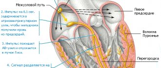
Every third inhabitant of the planet suffers from arrhythmia, that is, a violation of the heart rhythm. Many do not consider this a disease at all and do not receive any treatment - until they suddenly lose consciousness on the street, and this often happens to young people. Moreover, 25% of ischemic strokes are associated with arrhythmia. If your heart beats out of sync, how can you restore its rhythm? The director of the National Medical Research Center for Surgery named after A. A. V. Vishnevsky, President of the Russian Society of Arrhythmology, Academician of the Russian Academy of Sciences Amiran REVISHVILI.
Photo: Pixabay/Pexels
— Amiran Shotaevich, what are the causes of arrhythmia? Why does the heart, which for the time being had a clear rhythm, suddenly begin to lose its rhythm?
— This could be due to many circumstances: heart disease, disorders of the autonomic nervous system, arterial hypertension, changes in hormonal status (the risk increases in menopausal women), thyroid disease, bad habits (smoking, alcohol), hypothermia and much more...
Including the same stress. Their constant “presence” triggers inflammatory processes in the body that destroy cardiac tissue and cause heart rhythm disturbances. Or emotional burnout syndrome. That is, a feeling of dissatisfaction with life combined with overwork and irritability - the heart rate can also suffer from it...
Therefore, I advise everyone to try to be optimistic, lead an active lifestyle, and walk in the fresh air for at least 45 minutes every day. Do not smoke and be sure to monitor your blood pressure.
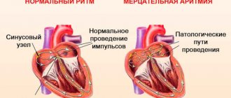
— People are mainly familiar with three types of arrhythmias: tachyarrhythmia (with a heart rate of up to 120 - 150 beats per minute), bradyarrhythmia (with a heart rate of less than 55 beats per minute) and atrial fibrillation. But cardiologists say that there are many more varieties and each type is treated differently.
- This is true! In addition to tachycardia and bradycardia, arrhythmias are divided into supraventricular and ventricular, depending on what part of the heart they originated in. According to another classification, arrhythmia can be paroxysmal (paroxysmal) and constant (chronic).
There are also many arrhythmias, in which the heart beats unevenly. The most common of them are extrasystolic (premature contraction of the entire heart or just the ventricles) and atrial fibrillation (chaotic contraction of individual groups of muscle fibers, the pulse reaching 100 - 150 beats per minute).
Atrial fibrillation is one of those sudden disturbances in heart rhythm in which you should immediately call an ambulance. However, she is already one of the most frequent reasons for her arrival.
— How dangerous is this or that arrhythmia?
— With the same atrial fibrillation, there is a high risk of the formation of blood clots, which can move through the vessels and clog them, creating the threat of a heart attack or stroke. If rhythm disturbances are associated with a rare pulse, then there is a threat of short-term cardiac arrest, stopping blood circulation and leading to loss of consciousness.
Related article:
Scientists from St. Petersburg are creating a vaccine against coronavirus in the form of “yogurt”
But there are types of arrhythmias that are completely harmless and do not lead to significant consequences. The insidiousness of this disease is that arrhythmia can be asymptomatic. Therefore, even if you feel well, an irregular rhythm detected by a home blood pressure monitor is a reason to contact a cardiologist.
It is worth consulting with him, even if no further treatment is required. For example, sometimes patients complain of heart failure, freezing followed by a sharp jolt. This condition often occurs during physical activity or from excitement, and quite often occurs at rest. With such extrasystole, it is useful to take a sedative.
And, for example, during magnetic storms, sometimes this happens: an arrhythmia occurs, but after some time it goes away safely. But you need to know that at this time there was a slowdown in blood flow, which worsened the nutrition of vital organs, and even the blood became thicker. This means that you need to keep medications against the formation of clots in your blood vessels on hand. But a doctor must prescribe them.
There are also a few very simple tips that can somehow calm down a sudden strong heartbeat. If a person is simply very worried or afraid of something. Sit in a chair or lie on the bed, breathe slowly, deeply, measuredly - this will quickly normalize your rapid pulse. And one more thing: take a deep breath, pinch your nose, tense your stomach - your pulse will quickly return to normal.
You can use this psychological technique. Feeling an attack approaching, a person usually intuitively grabs his heart, as if trying to block the pain - he seems to calm the heart, giving it the command to beat slower. Give the same command to your heart: “Beat slower... Even slower...”.
— Atrial fibrillation, as you said, is one of the common causes of strokes. Is it possible to somehow get rid of it?
— Treatment must begin with the elimination of the underlying disease that led to the arrhythmia (for example, high blood pressure). There is also drug treatment. If the doctor has already accurately diagnosed “atrial fibrillation,” then he will prescribe it. If therapy does not help, then you should consider surgery - it is called ablation.
Until recently, arrhythmia was treated by open surgery with the connection of a cardiopulmonary bypass system. Although the focus of the disease is tiny, only a few millimeters, it is difficult to find. We have developed a new method for searching for this very source - an original procedure for computer surface mapping.
Electrodes are placed on the patient's body and they simultaneously record electrocardiograms. A lot - up to 240 cardiograms. The patient then undergoes an MRI of the chest. After processing the tomography results, the doctor receives a 3D model of the heart, on which the source of the arrhythmia is indicated with maximum accuracy. Today our technology is used in many countries around the world.
Once the source of the arrhythmia is identified, it is eliminated using a catheter procedure. Catheters are inserted through the femoral artery or vein, which, moving through the vessels, reach the heart and “cauterize” the arrhythmogenic zone. Treatment is carried out under X-ray control. Moreover, with the help of X-ray surgery, diagnosis and treatment are easier and faster. The procedure is performed under local anesthesia and takes no more than an hour and a half.
Today in Russia, electrocardiographic heart mapping technology is used in many clinics. Tens of thousands of procedures are performed, but the need is one and a half times greater.
Another procedure, the installation of a pacemaker, was worked out in the same way. It is required for patients with bradycardia. Every year more than 40 thousand operations are performed in the country, which is fifteen times more than was performed ten years ago, but three times less than necessary.
— What prevents all those in need from undergoing the procedure? Are there not enough medical centers?
— There are not enough highly qualified specialists! Each patient has a different type of arrhythmia, so there cannot be one standard operation for everyone.
In general, an arrhythmologist is a rather rare specialty. This is a multidisciplinary doctor. He must be equally proficient in all treatment methods (electrophysical diagnostics, catheter, thoracoscopic and surgical methods) in order to choose the treatment method that is optimal for the patient. To work independently, he needs to study for at least ten years and perform at least a thousand procedures during this period.
BY THE WAY
Experts give the following advice on preventing arrhythmia:
- To keep your heart working smoothly, include oranges, pears, raspberries, and currants in your diet. In winter, let it be frozen berries, it’s still good. The diet should also include red sweet peppers, tomatoes, beets, parsley, apples, and corn;
- a very healthy mixture of honey, walnuts, lemon, dried apricots and raisins. These products contain a lot of potassium, magnesium, calcium, which are needed for normal heart function;
— for arrhythmia and cardiovascular diseases, it is better to drink mint tea. Mint relieves irritability and calms. In this case, you should keep the liquid in your mouth longer and drink it in small sips. You could also take a mint candy before bed;
- An infusion of viburnum berries is also useful. It contains a large amount of vitamin K, which has a beneficial effect on the heart and blood vessels and normalizes cardiac activity. Viburnum is also rich in vitamin C, which strengthens the immune system, and vitamin P, which is necessary for the body to absorb ascorbic acid. This is why viburnum is indispensable for high blood pressure. The recipe is simple:
Pour 1 cup of fruit with 1 liter of hot water, put on low heat and let boil for 10 minutes. Then strain and cool. Drink half a glass three times a day;
— what about coffee? Scientists have come to the following conclusion: if a strong coffee overdose can lead to arrhythmia, then a couple of cups of good grain coffee a day, as it turns out, for some reason stabilize the heart rhythm. This mainly concerns atrial fibrillation.
#diseases #heart #health
The material was published in the newspaper “St. Petersburg Vedomosti” No. 094 (6692) dated 06/03/2020 under the title “If the heart beats out of sync.”
Share on VKontakte Facebook
Cool
Treatment methods for arrhythmia
Treatment of arrhythmia depends on its type, the cause of the arrhythmia, and the individual condition of the patient.
For any arrhythmia, actions aimed at normalizing lifestyle and general health are useful, including proper nutrition, healthy sleep, quitting smoking and other bad habits, dosed physical activity, such as therapeutic walking, swimming, cycling.
Drug treatment
The basis of drug treatment is antiarrhythmic drugs. In some cases, the doctor may decide that drug treatment is not required. Under no circumstances should you use medications at your own discretion, without consulting a doctor.
Specialist consultation
To treat arrhythmia, you should contact a cardiologist. Cardiologists at Family Doctor JSC have extensive experience in diagnosing and treating arrhythmias. If you need an in-depth examination of the condition of your heart, contact the high-tech divisions of our network - the Rehabilitation Center (Polyclinic No. 5) and the Hospital Center.
Make an appointment Do not self-medicate. Contact our specialists who will correctly diagnose and prescribe treatment.
Rate how useful the material was
thank you for rating
Section materials
December 23, 10:55
Faster than delta. Doctors warned about the high contagiousness of omicron
December 15, 12:54
Don't let go. The doctor talks about the appearance of blood clots in the body
December 01, 18:58
Over the year in St. Petersburg, the number of residents with HIV infection increased by 17%
November 30, 12:10
In St. Petersburg, the majority of HIV-positive mothers give birth to healthy children
November 26, 17:21
The WHO Healthy Cities Conference will be held in St. Petersburg
Resulting
Why does the heart, which for the time being had been beating a clear rhythm, suddenly begin to lose track of it? This could be due to many circumstances:
- congenital or acquired heart disease;
- arterial hypertension;
Article on the topicHalfway to a stroke? Dispelling the main myths about hypertension
- electrolyte imbalance (deficiency or excess of potassium, calcium, magnesium in the body);
- hormonal imbalances (therefore, in menopausal women, the risk of arrhythmia increases);
- taking certain medications (diuretics and sympathomimetics, for example);
- concomitant diseases (thyroid pathologies, in particular);
- bad habits (smoking, alcohol, drug addiction).
Modern tactics of drug rhythm-slowing therapy for atrial fibrillation
M
atrial fibrillation (AF) is one of the most severe and common heart rhythm disorders, occurring in 0.4% of the general population (M.S. Kushakovsky, 1999) and in more than 5% of people over 69 years of age (Braunwald, 1996). In the vast majority, AF leads to the appearance (or worsening) of heart failure, which, in addition to the disappearance of atrial systole, is caused by tachyarrhythmia itself, i.e. two terms: tachycardia and arrhythmia of the heart (unproductive - too long and too short - diastolic pauses). When treating AF, it is generally accepted that it is necessary to reduce the work of the heart (the second component - the arrhythmia itself - is usually, unfortunately, not taken into account). It is known that a decrease in heart rate is indeed accompanied by a noticeable decrease in signs of heart failure. There are even studies (PIAF, 2000; AFFIRM, 2002) that show that, taking into account such end points as the incidence of strokes, heart attacks, thromboembolic and hemorrhagic complications, mortality, as well as quality of life, effective conservative treatment of MA (and this, first of all, rhythm-slowing therapy) is not inferior to the tactics of restoring and maintaining sinus rhythm. However, these data were obtained on a fairly specific group of patients (elderly people with decreased contractile function of the heart, with obviously poor chances of stable maintenance of sinus rhythm and an increased risk of developing complications of the underlying disease - widespread atherosclerosis and antiarrhythmic therapy), and they cannot be extended to the entire population of patients suffering from MA.
To slow down the rhythm in MA, first of all, digitalis preparations are used, as they are the most effective and, it would seem, have been sufficiently studied. In addition, b-blockers, calcium channel blockers, and, less commonly, class III antiarrhythmic drugs (amiodarone, d,l-sotalol) are used. Currently, some authors believe that even monotherapy with b-blockers or calcium antagonists is possible, as the most severe means of slowing down the rhythm. More often, they are still recommended to be added to digitalis drugs if the reducing effect of the latter is insufficient. This takes into account the better ability of b-blockers and calcium antagonists compared to digitalis to slow down the rhythm not only at rest, but also during physical activity, as well as the possibility of using such combination therapy to prescribe smaller doses of cardiac glycosides with a correspondingly lower risk of digitalis intoxication.
The selection of drugs is carried out empirically, based on the current ideas that the only pathogenetic mechanism of the reducing effect of these drugs is the inhibition of the conduction of ff waves through the atrioventricular (AV) system (the effect on the degree of arrhythmia of the heart rhythm, as mentioned above, is not taken into account at all ).
To date, we have obtained data that allows us to supplement existing ideas about the mechanisms of the reducing effect of digitalis and other drugs. These works were started by us (A.V. Nedostup, E.A. Bogdanova, A.A. Platonova) in the mid-70s [1–4] and continued (in collaboration with O.V. Blagova) in recent years [5–9]. In accordance with this, a more objective approach to the tactics of rhythm-slowing therapy for MA has become possible. Below is a list of new provisions
,
which should be taken into account when rhythm-reducing therapy in patients with AF
.
1. When exposed to the heart of a patient with AF, cardiac glycosides have a significant effect not only on conduction in the AV system, but also on the process of atrial fibrillation itself, which is characterized by a certain wave period ff and its inverse frequency. At the same time, in the vast majority of patients (about 80%), at the first stage of digitalization, “shredding” of ff waves occurs, i.e. reducing their period from the initial 0.15–0.20 s. up to 0.12–0.14 s. with a corresponding increase in flicker frequency. This process precedes in time the influence of digitalis on the AV system and is the leading slowdown mechanism at the average rate of digitalization: the flow of more frequent flickering waves, according to the law of parabiosis, passes through the AV system to the ventricles with great difficulty, resulting in a slowdown in the heart rate, directly proportional to degrees to the original wave period ff.
2. Slowing of AV conduction under the influence of glycosides is achieved in most patients only at the second (later) stage and coincides with the appearance of a characteristic “digitalis” curve on the ECG, and then ventricular extrasystole and other manifestations of hyperdigitalization. Only in 20% of patients who, as a rule, have initially small ff waves (with a period of 0.12–0.14 s), their further “grinding” does not occur and a direct slowdown of AV conduction develops in the early stages of digitalization.
3. The decrease in ventricular rate in patients with AF under the influence of b-blockers and calcium channel blockers is achieved only due to their inhibitory effect on AV conduction (stronger for b-blockers, which additionally enhance latent conduction through the AV junction) - and a significant increase is noted the minimum RR interval (RRmin), which directly reflects the refractoriness of the AV node during MA. These drugs do not affect atrial activity.
4. Class III antiarrhythmics (amiodarone, d,l-sotalol) reduce ff waves, up to the transformation of atrial fibrillation into flutter, which facilitates their conduction through the AV system, and on the other hand, inhibit AV conduction. Thanks to these two counter-directional processes, the decrease in ventricular rhythm under the influence of these drugs is moderately pronounced (more pronounced when prescribing d,l-sotalol due to its b-blocking effect, leading not only to an increase in the refractoriness of the AV junction, but also to an increase in latent conduction in it) .
5. In MA, monotherapy with b-blockers or verapamil leads, along with a slowdown in the rhythm, to negative changes in its structure (an increase in arrhythmia, alternation of short and long RR intervals, which is subjectively poorly tolerated by patients). These changes are somewhat less pronounced when verapamil is prescribed due to its lack of influence on latent conduction in the AV node. Due to a more uniform redistribution of RR intervals as a result of the conduction of “crushed” flicker waves to the ventricles, the administration of digitalis preparations is accompanied by a smaller spread of RR values, while the structure of the ventricular rhythm is close to optimal (symmetrical monomodal histogram of RR intervals).
6. In a number of patients with persistent tachyarrhythmia, which is difficult to respond to digitalis therapy, tachycardia is caused by the superposition of periods of frequent, fairly regular rhythm), which on the histogram of RR intervals corresponds to a peak in the zone of short RR distributions (“early” RR peak). Probably, in this case, there is periodic abnormally fast conduction of ff waves to the ventricles or the presence of periods of frequent supraventricular rhythm (ectopic or due to re-entry
in the AV node and other myocardial structures). In such cases, an effective reduction in rhythm is achieved by amiodarone, which affects all possible mechanisms of the formation of the “early” peak; d,l-sotalol and b-blockers may also be effective.
From the above, when starting MA therapy, it is obvious that it is necessary to take into account the initial data not only on the frequency (and structure) of the ventricular rhythm, but also on the frequency of ff waves. Approximate data about it can be obtained by analyzing a regular ECG in lead V1, where flicker waves are clearly visible. When recording at a speed of 25 mm/s, the wave period ff 0.15–0.20 s corresponds to a wave width of 3.5–5.0 mm. Accurate data can be obtained using a special computer program created to work in the Matlab 5.3 environment and allows for high-resolution ECG analysis in Frank orthogonal leads (X, Y, Z). Using this program, you can obtain a periodogram of ff waves, construct an autocorrelation function of ff wave periods, a histogram of RR intervals, a cardiointervalogram and a scatterogram of RR intervals, and calculate statistical parameters of ventricular rate variability.
For practical work (prescribing therapy), a periodogram of ff
and histograms of RR intervals. We repeat that an indicative analysis of atrial ff waves can be done using a conventional ECG; it can also be used to determine the refractoriness of the AV node, corresponding to the minimum RR interval, and to evaluate the structure of the RR intervals and the nature of their alternation, identifying periods of frequent, relatively regular rhythm (preferably according to Holter monitoring). Of course, when selecting therapy, well-known clinical criteria remain important - the nature of the underlying disease, concomitant pathology, experience of previous therapy, etc. However, without analyzing the characteristics of MA itself, therapy cannot be truly individual.
The most general principles
for prescribing rhythm-lowering drugs,
depending on the nature of atrial activity and ventricular rhythm, are as follows:
1. The most rational is the combined prescription of drugs with various mechanisms of reducing action
: reducing the period of digoxin ff waves and only later inhibiting AV conduction - with drugs that exclusively block AV conduction (b-blockers, verapamil), in some cases - with suppressing ventricular ectopy, normal and abnormally fast AV conduction and ectopic supraventricular rhythms , amiodarone and d,l-sotalol, although their simultaneous undesirable “enlargement” effect on ff waves should be taken into account. It is also possible to combine amiodarone with beta-blockers, which deepen the slowing of AV conduction and reduce the risk of developing ventricular arrhythmias.
2. In the presence (or appearance at any stage of therapy) of ff waves of large and medium periods, it is not possible to achieve a stable reducing effect with obtaining the optimal structure of ff
using digitalis preparations.
3. In the presence of periods of frequent relatively regular ri, which correspond to an “early” peak on the histogram of RR intervals), the reducing effect is achieved only with the help of combination therapy, which usually includes digoxin and a class III antiarrhythmic or b-blocker.
Figure 1 shows an algorithm for individual selection of reducing therapy for a permanent form of MA, depending on its initial parameters. Let's comment on this algorithm.
Rice. 1. Algorithm for selecting rhythm-slowing therapy depending on the initial parameters of atrial fibrillation
As can be seen, when selecting therapy for patients with the tachysystolic form of AF, first of all, it is necessary
to determine the initial value of the wave period ff
.
With its values from 0.15 s and above (medium and large waves ff)
All patients are prescribed cardiac glycosides, which, depending on the presence or absence of ventricular extrasystole, is carried out immediately or after its suppression. In this case, an important factor determining the success of glycoside monotherapy is (along with the degree of initial tachycardia) the presence of an “early” RR peak and its resistance to digoxin.
A. In the presence of large and medium waves ff (from 0.15 s) and the absence of frequent ventricular extrasystole
Digoxin is prescribed (average rate of digitalization - 0.5 mg per day). When normosystole is achieved (which is accompanied by a decrease in the period of ff waves, i.e., their frequency) and a good subjective state, the goal of treatment is achieved. If (which happens more often) the feeling of palpitations persists during exercise, a b-blocker is added to digoxin (having previously reduced the dose of digoxin), monitoring the refractoriness of the AV node using the dynamics of the minimum RR interval (RRmin should be within 50 ms or less).
If, during treatment with digoxin, the period of ff waves decreases to 0.14 s and below, but tachysystole persists, a b-blocker or verapamil is also added to digoxin (without reducing its dose), after which a good result can be obtained.
If after this tachysystole persists and against this background there are no zones of relatively rhythmic tachycardia (“tachycardic chains” that give an “early” peak in the histogram of RR intervals), the dose of a b-blocker (not digoxin) should be increased. If the effect is good (normosystole), but the feeling of heartbeat during exercise is preserved, you can once again carefully increase the dose of the b-blocker. In this case, however, you should pay attention to the patient’s feeling of “arrhythmic” heartbeat, which is characteristic of therapy with fairly high doses of b-blockers. If such a feeling exists (it is better to confirm this by analyzing the histogram of RR intervals, which reveals a large “scatter” of RR values), carefully increase the dose of digoxin, counting on the slowdown of AV conduction at this stage of its administration (and, accordingly, controlling the value of RRmin , an increase to 55–60 ms signals deep inhibition of AV conduction with the threat of developing complete AV block against the background of AF, i.e. Friederik's syndrome). As a rule, in this way it is possible to achieve a fairly regular normosystolic rhythm.
If, during therapy with digoxin and a b-blocker, tachyarrhythmia persists and areas of “tachycardic circuits” are visible
with a corresponding “early” peak in the histogram of RR intervals, without canceling digoxin, replace the b-blocker with amiodarone or d,l-sotalol, which usually eliminate areas of rhythmic tachycardia (if the rhythm slowdown is insufficient during treatment with digoxin and amiodarone, a b-blocker is added again) . This is usually not required with digoxin and amiodarone; if the patient suffers from broncho-obstructive syndrome, then verapamil or diltiazem is added instead of d,l-sotalol or b-blocker.
B. In the case of initial frequent ventricular extrasystole in patients with initial tachysystole and wave period ff 0.15 s or higher
d,l-sotalol or amiodarone (the latter in combination with b-blockers) is prescribed. Only after achieving the disappearance of extrasystole, digoxin should be added, which is prescribed without fail, given its ability to “grind” ff waves and optimize the rhythm structure. In the presence of frequent ventricular extrasystole against the background of already ongoing digitalization, which does not lead to normosystole, digitalis preparations are discontinued, d,l-sotalol or amiodarone is prescribed and, having suppressed ventricular extrasystole, digitalis preparations are carefully added to the treatment.
These options usually exhaust the situations faced by a doctor who begins therapy with initial ff waves of large and medium (0.15–0.20 s.) periods (and, accordingly, small and medium frequencies of atrial activity).
With tachysystole and the initial period of ff waves 0.12–0.14 s (“crushed” ff waves)
, which is sometimes initially characteristic of a number of patients with AF, but is more often noted during treatment with digoxin, it is obvious that it is possible to achieve the necessary slowdown of the rhythm by prescribing digoxin (or increasing digitalization) only by slowing down AV conduction, because Digitalis preparations no longer act on flickering waves of this frequency. Nevertheless, the use of digoxin in most of these patients is advisable:
1) to maintain a high frequency of ff waves (especially with the simultaneous administration of class III antiarrhythmics);
2) to optimize the rhythm structure;
3) to achieve a cardiotonic effect.
A. In the presence of initially “small” ff waves (0.12–0.14 s) in combination with the absence of an “early” RR peak (“tachycardic chains” on the ECG) and ventricular ectopy
Digoxin monotherapy may be used with caution. It should be remembered that only significant doses of digitalis, close to toxic, affect AV conduction, and therefore a combination of digoxin with b-blockers or verapamil or monotherapy with the latter is preferable. Having obtained normosystole with an optimal rhythm structure, you can proceed to maintenance therapy. If the rhythm structure is not optimal (a large “scatter” of RR values remains against the background of normosystole), carefully increase the dose of digoxin (monitoring the RRmin value and, if necessary, reducing the dose of b-blockers or verapamil).
B. If patients with an initial ff wave period of 0.12–0.14 s do not have an “early” RR peak, but have ventricular ectopy
, it is best to prescribe d,l-sotalol, which both eliminates ventricular ectopy and slows down the rhythm. Digoxin can be added only after eliminating ventricular ectopy (if the patient is already receiving digoxin, it should be discontinued). If d,l-sotalol is ineffective in this regard, “cordaronization” should be performed (with the possible addition of a b-blocker, remembering that amiodarone itself does not slow down the rhythm well) and then, having eliminated the ventricular ectopy, add digoxin. Carrying out constant monotherapy with class III antiarrhythmics (especially amiodarone) will lead to “enlargement” of ff waves with loss or weakening of the reducing effect, as well as deterioration of the rhythm structure. Let us remind you once again that in patients with bronchial obstruction it is necessary to “sacrifice” d,l-sotalol and b-blockers, replacing them with amiodarone in combination with verapamil.
B. With tachysystole with an initial wave period ff of 0.12–0.14 s and an “early” peak of RR in the absence of frequent ventricular ectopy
You can try to carefully increase the dose of digoxin in combination with a b-blocker. If after this the zone of rhythmic tachycardia (“chain”) remains, digoxin is left, replacing the b-blocker with d, l-sotalol or amiodarone, which, as a rule, gives a good effect. If the “early” peak disappears, but the tachycardia persists, as already mentioned, a b-blocker (verapamil) is added to the digoxin-amiodarone combination.
D. With the simultaneous presence of an “early” peak and ventricular ectopy in patients with small waves ff
There are even stronger reasons for prescribing amiodarone or d,l-sotalol. At the same time, all of the above is true about the undesirability of monotherapy with these drugs (if it is impossible to prescribe digoxin due to incomplete suppression of ventricular ectopy, b-blockers can be used).
As can be seen from the above comments, despite the apparent complexity of the algorithm, the number of possible initial variants of MA and accompanying rhythm disturbances is not so large and can be treated within the framework of more or less standard rules.
Among internists (even before cardiology was identified as a separate discipline), it was always believed that treatment with digitalis drugs was one of the most difficult therapeutic tasks. There was even an aphorism: “Whoever knows how to treat with digitalis can be considered a doctor.” It is clear that this applied, in particular, to digitalis therapy for patients with MA. Gradually, with the advent of ECG and clinical pharmacology as an independent discipline, the “secrets” of digitalis therapy began to be revealed. The task of slowing down the rhythm in atrial fibrillation was further simplified with the introduction into clinical use of b-blockers, calcium and potassium channel blockers, although their appearance posed new problems - preferable therapy for certain patients, combinations of medications, etc. And yet, therapy for atrial fibrillation is still quite empirical.
We hope that the new data we have presented on the mechanism of action of digitalis in MA, as well as other observations underlying the above recommendations, will serve as another step towards optimization and a scientific approach to therapeutic tactics for rhythm-slowing therapy in patients with MA.
Literature:
1. Egorov D.F., Leshchinsky A.A., Nedostup A.V., Tyulkina E.E. Atrial fibrillation (strategy and tactics of treatment on the threshold of the 21st century). – Izhevsk: Alphabet, 1998. – 413 p.
2. Nedostup A.V., Bogdanova E.A., Mikhnovsky E.I. Analysis of the structure of the heart rhythm during atrial fibrillation using a specialized computer. Cardiology, 1975, No. 1: pp. 64–69.
3. Nedostup A.V., Bogdanova E.A., Platonova A.A. et al.: Analysis of the structure of the heart rhythm during digitalis therapy of patients with atrial fibrillation. Cardiology, 1977, No. 4: p. 85–90.
4. Nedostup A.V., Bogdanova E.A., Platonova A.A. Study of the process of atrial fibrillation in clinical practice using statistical methods of analysis. Cardiology, 1980, No. 10: p. 73–78.
5. Nedostup A.V., Blagova O.V., Bogdanova E.A., Platonova A.A. The importance of analyzing the electrical activity of the atria during atrial fibrillation for the selection of rhythm-slowing therapy // Abstracts of the Congress “Cardiostim-2000”. “Bulletin of Arrhythmology”, 2000, No. 15: p. 53
6. A.V.Nedostup, O.V.Blagova, E.A.Bogdanova, A.A.Platonova. Possibilities of using a combined analysis of the electrical activity of the atria and the structure of the ventricular response in patients with a permanent form of atrial fibrillation to control the decreasing ri, 2001, December, T.6, No. 1, Suppl. A: s. 26–35
7. A.V.Nedostup, O.V.Blagova, E.A.Bogdanova, A.A.Platonova. Drug reduction therapy for atrial fibrillation: a new approach to an old problem // “Therapeutic Archive”, 2002, No. 8: p. 35–42
8. A.V.Nedostup, O.V.Blagova. New pathogenetic approach to rhythm-reducing therapy for atrial fibrillation, 2002, No. 12: pp. 12–16
9. A.V.Nedostup, O.V.Blagova, E.A.Bogdanova, A.A.Platonova. Correction of the frequency and structure of the ventricular rhythm in a permanent form of atrial fibrillation: a comprehensive pathogenetic approach. "Cardiology", 2003, No. 12, p.
Symptoms of cardiac arrhythmia
Many people believe that it is impossible not to pay attention to heart rhythm disturbances. But most often, cardiac arrhythmia is asymptomatic. All signs are attributed to stress, overload or fatigue. But nevertheless, it is very important to diagnose heart pathology in a timely manner. Contact a specialist immediately if you notice at least one of the symptoms of arrhythmia:
- cardiopalmus;
- chest pain, a feeling that someone is pressing on the chest;
- daily dizziness and headaches;
- sensations of interruptions in the rhythm of the heart or simply the feeling of your own heartbeat;
- shortness of breath even during everyday simple activities;
- loss of consciousness or lightheadedness.
Please note that these symptoms are common to many other diseases. Often this is the reason why people do not contact a specialist on time. For example, fainting is attributed to overwork or hunger, and headaches are attributed to frequent stress. Therefore, we would like to draw your attention once again to the fact that if any of the symptoms are present, consult a doctor immediately! To which? We'll tell you a little later.
Tachycardia. Diagnosis and treatment
With this type of arrhythmia, the number of heartbeats per minute exceeds 90 beats. Tachycardia is of two types: pathological or physiological.
Pathological tachycardia
Characterized by disorders of the cardiovascular and other systems. This type of tachycardia can cause myocardial infarction, acute heart failure and sudden cardiac arrest.
Physiological tachycardia
It differs from pathological in that it is caused by exposure outside the body. Often physiological tachycardia is a normal reaction of the body to any changes. For example, during physical exertion, during excitement or anger and similar situations, physiological tachycardia is the norm.
General rules for first aid
The rules for providing first aid for the treatment of paroxysm of atrial fibrillation are carried out differently depending on several characteristics of the attack:
- blood pressure level;
- shortness of breath at rest;
- duration of the attack;
- heart rate;
- primary or repeated paroxysm.
Depending on this, emergency doctors either try to restore sinus rhythm or reduce the heart rate, while simultaneously preventing the formation of blood clots. For this purpose, medications are used, and, if necessary and conditions exist, electropulse therapy is used.
What you can and cannot do at home during an attack
If an attack of irregular heartbeat develops, you must immediately call an ambulance.
Before the medical team arrives, you can:
- give the patient a semi-sitting position;
- unbutton tight clothes;
- provide fresh air access to the room;
- invite the patient to breathe with his stomach, wipe his face with a handkerchief dipped in cold water;
- give 20 - 30 drops of Corvalol in half a glass of water;
- prepare for the arrival of the team: organize its meeting, prepare medical documents, previous ECGs, think about transporting the patient to the ambulance (such a need may arise, but the duties of the ambulance personnel do not include carrying the patient);
- reassure the patient, tell him to call the doctors.
When detecting an attack of MA before the arrival of the ambulance, you cannot:
- Diet for atrial fibrillation: permitted and prohibited foods, sample menu
- give the patient medications before the ambulance arrives, including nitroglycerin;
- massage the eyeballs or the area of the carotid arteries;
- waste time measuring blood pressure without preparing for the arrival of medical personnel;
- collect things for hospitalization (this will be the time while the doctor examines the patient, relieves the attack, etc.; hospitalization is not always required);
- worry and panic.
How to stop an attack of MA on your own (a pill in your pocket)
Some patients whose diagnosis of “paroxysmal atrial fibrillation” has been established for a long time, and attacks occur less than once a month, can learn to independently stop such paroxysms. This tactic is called “pill in the pocket.”
It is used in intellectually intact patients who can adequately assess their condition. The “pill in your pocket” strategy should not be used if the next attack of arrhythmia caused any new symptoms:
- chest pain;
- dizziness;
- weakness in the limbs;
- facial asymmetry and so on.
In such cases, you should not stop the paroxysm on your own, since these symptoms may be a sign of the development of a heart attack or stroke.
If paroxysmal fibrillation proceeds as usual, the patient can take the drug propanorm at a dosage of 450–600 mg.
The patient should consult his cardiologist in advance about in what cases and in what dose to take this medicine. It is better if the first dose of propanorm is taken in a hospital, under the supervision of medical professionals.
Extrasystole. Diagnosis and treatment
Characterized by additional (extraordinary) contractions. Extrasystole is of two types according to the type of occurrence: ventricular or atrial. According to medical data, every person had episodes of extrasystole, even those who had never complained of heart problems. On average, every healthy person is allowed 4% extrasystoles of the total number of heart beats per day. In this case, frequently recurring episodes of extrasystole should become a cause for concern, since cerebral and coronary blood flow decreases, and there is a high probability of developing angina pectoris (a form of coronary heart disease).
Symptoms of extrasystole
There is a feeling of anxiety, a feeling of an extra blow with a feeling of a push, a feeling as if the heart is stopping.