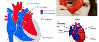
The configuration of the heart is its outline, a kind of projection onto the front surface of the body. Normally, on an x-ray, it has smooth, rounded contours, there is a waist, the upper part is smaller than the lower, 2 arches are visible along the right contour, 4 along the left.
If the waist is smoothed, and there is an expansion of the cardiac shadow to the left and partially to the right, then this configuration is called mitral. It appears in diseases of the valve (congenital and acquired) between the left parts of the heart, as well as diseases of the lungs.
With aortic waist, the waist is emphasized; this configuration is a sign of aortic valve defects, hypertension, pathologies of the heart muscle (heart attack, cardiomyopathy). The trapezoidal shape occurs with pericarditis, myocardial inflammation, and defects. There are also cor pulmonale with expansion of the right ventricle and “bovine” with enlarged chambers.
What is the configuration of the heart, its indicators are normal
The configuration of the heart is the shape, contours, showing the expansion or contraction of the atria, ventricles, and vascular bundle. It includes the borders (upper, left and right), waist (narrowing at the junction of the bundle of blood vessels to the left ventricle), angles between the diaphragm (reflecting the inclination of the axis) and the arcs of the chambers of the heart. If the configuration is normal, this means that they correspond to the parameters (see table) of healthy people:
- relative dullness of the heart (its actual size) in the chest and absolute (the part that is not covered by the lungs and is directly adjacent to the ribs);
- length (length) and width (diameter);
- waist (retraction of the contour, narrowing of the diameter);
- there are angles between arcs.
| Contour type and main parameters | How was she educated? | Upper | Left | Right |
| Relative Dullness | Real contours | Bottom of 3rd ribs on the left edge of the sternum | The fifth intercostal space is 1.5 cm to the right from the conditional line drawn from the middle of the left clavicle | The fourth intercostal space is 1 cm to the right from the edge of the sternum |
| Absolute | Right ventricle | Bottom of 4th ribs on the left edge of the sternum | 2.5-3.2 cm to the right of the line from the middle of the left clavicle | Fourth intercostal space along the left edge of the sternum |
| Width of the bundle of large vessels (vascular) | Aorta, pulmonary artery, veins | The space between the 2nd and 3rd ribs does not extend beyond the borders of the sternum (approximately 4.5-5.5 cm) | ||
| Right contour (arcs) | Superior vena cava, right atrium | The edge of the sternum to the 3rd rib and the third or fourth space between the ribs 1 cm to the right | ||
| Right atriovasal angle | Between the superior vena cava and the atrium | 3-4 intercostal space | ||
| Cardio-phrenic angle right | Diaphragm, right atrium | 5th intercostal space | ||
| Left contour (arches) near the left edge of the sternum | Aorta | 1st intercostal space | ||
| Pulmonary artery | 2nd intercostal space | |||
| Left atrium | Level 3 ribs | |||
| Left ventricle | Inferior to the 3rd rib, just behind the left atrial appendage | |||
| Waist | Bundle of blood vessels, left ventricle (left), right atrium (right) | Level 3 ribs | ||
| Length | Heart length | From the apex to the right cardiophrenic angle the length should be between 11.5-12.7 cm | ||
| Diameter | Width of relative cardiac dullness | From the right border to the midline and from the left (the most protruding part) to the middle, normally 11-13 cm | ||
If the patient has diseases of the heart and/or large vessels, especially valve defects, then the load on its parts changes. Then the atria and ventricles enlarge, which is manifested by a shift in the boundaries of relative and absolute dullness, changes in size, smoothing of angles, and disappearance of the waist.
Determination of heart configuration by percussion
To determine the configuration of the heart, you first need to find the boundaries of dullness. They are so named because they can be examined during a medical examination. To do this, use the percussion (tapping) method. Above the lungs, the sound is clear, since they are filled with air, and as soon as the finger reaches the contour of the heart, dullness appears (relative dullness), and then a completely dull sound (absolute).
First, the true border of the heart is found in the chest. To do this, the finger of the left hand moves from the outside to the center. Since the organ’s entire surface is not adjacent to the chest, part of it is covered by the lungs, and a small area remains open, when tapping it can be identified by a dull (dull) sound. This zone (absolute dullness) reflects the size of the right ventricle. It is found by moving from the center to the edges.
When the boundaries of cardiac dullness change
An increase in the relative dullness of the heart is a ghost of expansion of its cavities (ventricles and atria). Occurs with valve defects, high blood pressure, weakening of the heart muscle or its hypertension (thickening). The absolute dullness of the heart can also change normally - for example, with bloating or pregnancy, the diaphragm rises upward and brings the heart closer to the ribs.
Expansion may be affected by:
- compaction of lung tissue (pneumosclerosis);
- adhesions in the chest;
- dilatation of the right ventricle.
Emphysema (increased airiness), an asthma attack, a low position of the diaphragm with weakness of the respiratory muscles can reduce the area.
X-ray configuration
If the boundaries of dullness (relative and absolute) can be found only by tapping, then measurements of length, width, finding the waist, angles and arcs of the configuration occur only on a chest x-ray. In the conclusion, the radiologist indicates the conformity of the configuration to a certain type and measures the angles, length and diameter.
It is important to understand that X-ray diagnostics will only show an expansion of the heart shadow or its displacement, an unusual shape, but to determine the cause, an ultrasound (echocardiography) will often be required. Only the latter method will help to study the movement of blood through the valves, the contractility of the heart muscle and its thickness.
Expert opinion
Alena Ariko
Expert in Cardiology
An atypical configuration of the heart is not always a sign of illness. For example, young women may have a cardiac shadow shape that is close to the mitral shadow, while older overweight patients may have an aortic shadow shape. Therefore, a diagnosis is never made based on X-ray findings alone.
Types of heart configuration
The following types of heart configurations have been identified:
- normal - the right angle between the vascular bundle is approximately in the middle, the second and third arches are almost equal (2 cm each);
- aortic – the waist is rarely emphasized;
- mitral - expansion mainly to the left, partially to the right, no waist;
- close to geometric figures: trapezoid, ball, triangle;
- “bullish” with a significant increase in all cameras.
Acquired heart disease - symptoms and treatment
There is no medicine that can reverse the process and restore the valve to its original state. With the help of medication, it is only possible to influence the cardiovascular system and reduce the risk of complications. In severe cases, surgical treatment methods are used.
Drug treatment
The goal of therapy is to eliminate the causes of circulatory failure, improve the functional state of the myocardium, restore normal blood circulation, microcirculation (transport of blood cells and substances to and from tissues) and prevent recurrent circulatory disorders. Treatment for chronic circulatory failure includes a complete balanced diet and drug therapy.
The main groups of drugs used for valve dysfunction:
1. ACE inhibitors (angiotensin-converting enzyme) - enalapril, lisinopril, ramipril, perindopril, fosinopril. The drugs block the conversion of the hormone angiotensin I to angiotensin II. Angiotensin II has a vasoconstrictor effect and causes a rapid increase in blood pressure. ACE inhibitors are used to treat hypertension and treat or prevent heart failure.
2. Beta blockers - carvedilol, bisoprolol, metoprolol. These drugs lower blood pressure and normalize heart rate. The action is caused by blocking beta-adrenergic receptors, which are responsible for the body's response to stress.
3. Mineralocorticoid receptor antagonists - spironolactone, eplerenone. The drugs lower blood pressure and have a diuretic effect, reducing fluid content in tissues.
There are different types of heart defects, and different combinations of drugs are used to treat each of them. Concomitant pathologies and individual characteristics of the patient are also taken into account.
For example, when congestion predominates, diuretics (furosemide, torsemide) are additionally prescribed, which reduce the volume of circulating blood, reducing congestion in the pulmonary and systemic circulation. If blood clots are present or there is a high risk of their occurrence, anticoagulants are used - drugs that reduce blood clotting (warfarin, rivaroxaban, dabigatran, apixaban).
Relatively recently, a new class of drugs was developed - angiotensin-neprilysin receptor inhibitors (sacubitril). Their main effect is to increase the amount of peptides cleaved by neprilysin. The drugs increase diuresis (urine volume), natriuresis (sodium excretion in the urine), cause relaxation of the myocardium and prevent processes of disruption of the structure and function of the heart.
If it is impossible to use beta blockers, it is recommended to replace them with If channel inhibitors (ivabradine), which also reduce the heart rate. If it is impossible to use ACE inhibitors, angiotensin receptor blockers (losartan, valsartan, candesartan, telmisartan, irbesartan, olmesartan) are used. They have properties similar to ACE inhibitors, but do not reduce the synthesis of angiotensin II, but block angiotensin receptors [6].
Surgery
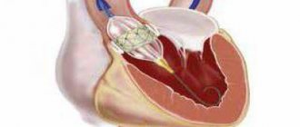
If there are indications, the effectiveness of drug treatment is insufficient and there are no contraindications, surgical methods are used. Many patients are afraid of the need for heart surgery. In some cases, surgical methods do carry some risk, but there are fairly safe operations that are not open heart and without large incisions. Such operations include balloon commissurotomy for mitral valve stenosis. The method consists of widening the valve using a catheter passed through the artery.
Before surgery, the necessary laboratory and instrumental studies are carried out and the patient’s condition is stabilized with medications. When preparing for surgery, it is important to reduce shortness of breath, swelling, and normalize pulse and blood pressure.
A common surgical method is artificial or biological valve replacement. There are also valve-sparing operations, which involve plastic surgery of a damaged valve.
Surgical treatment significantly improves the quality of life, however, after surgery, medication continues, but is adjusted if necessary. In addition, after certain operations, such as artificial valve replacement, anticoagulant therapy is constantly taken. The drugs are necessary because the risk of thromboembolic complications (blockage of arteries with blood clots) increases [5].
Waist Heart
The cardiac waist is the junction of the vascular bundle (arteries and veins entering and exiting the heart) into the left ventricle. Its apex is the left atrium, so when it enlarges, the waist disappears. With the expansion of the left ventricle, the waist will be well defined, “emphasized”.
Diameter of the heart
To measure the diameter of the heart, the midline of the body is visually determined, and from it the distances to the right and left edges of the relative dullness of the heart are measured. In a healthy person of average build, the right size does not exceed 4 cm, and the left - 9 cm; for thin people, a decrease of 1 cm is allowed.
The right diameter is larger than normal when:
- expansion of the right chambers (atrium and/or ventricle);
- accumulation of fluid in the pericardial sac (exudative pericarditis);
- displacement of the heart shadow to the right due to the large left ventricle.
The left size increases with diseases of the left chambers of the heart, less often it is shifted by the dilated right ventricle.
Normal dimensions of the vascular bundle
The vascular bundle is formed by the aorta or superior vena cava on the right and the pulmonary artery on the left; its size normally does not exceed 5-6 cm. It is covered by the sternum and does not protrude beyond its edges. If its width is greater, then this may be a sign of an aortic aneurysm or atherosclerosis.
Right heart contour
The right contour of the heart includes only 2 arches. The first is the superior vena cava or the ascending aorta (they are very close), and the second is always represented by the right atrium. Between these arches there is an angle called the right atriovasal.
Its location determines the configuration of the heart:
- normally it is approximately in the middle (between equal arcs);
- with mitral it is displaced upward;
- with aortic it moves down;
- with trapezoidal, triangular, spherical it disappears.
Left outline
The left contour includes the arcs:
- the first is part of the descending aorta;
- the second is the pulmonary artery;
- third - left atrium;
- the fourth is the left ventricle.
With a mitral configuration, they are all present, but shifted to the left. With aortic, the first and second arches, as well as the third and fourth, merge, the angle (left atriovasal) between the second and third is sharply expressed. If the patient has a trapezoid or a ball, then there is only the first arch, the angle, and the other three are connected into one large line .
Special pathological forms of the heart
Special pathological forms of the heart include pulmonary and bovine.
Pulmonary heart
With cor pulmonale, the load on the right side increases, which leads to dilation of the atrium and ventricle. The X-ray shows an expansion of the shadow to the right (2nd arc) and a displacement of the left contour due to the large right ventricle, a mitral configuration appears.
Chronic diseases of the bronchi and lungs, which last for several years, are accompanied by cor pulmonale in 25% of cases. It can also occur acutely when the chest and diaphragm are damaged within a few hours.
If the patient is severely obese, then excess fat raises the diaphragmatic dome upward, which makes breathing difficult and causes changes in the heart shadow. Subacute cor pulmonale (develops up to 3-7 days) occurs when a pulmonary artery is blocked by a blood clot, severe pneumonia, tumor metastases, poliomyelitis, muscle weakness (myasthenia gravis).
Bull's heart
Enlargement of the chambers of the heart (dilated cardiomyopathy) or thickening of the heart muscle (hypertrophic cardiomegaly) leads to the fact that the cardiac shadow increases in all directions and a “large” or “bull” heart is formed.
Its configuration will be close to trapezoidal or spherical, less often the waist will be preserved.
The causes of large heart syndrome may be:
- atherosclerosis, angina pectoris, heart attack and its complications: aneurysm, post-infarction cardiosclerosis;
- vices;
- hypertonic disease;
- inflammation of the myocardium, immune or due to rheumatism, bacterial, viral, fungal infections;
- cardiomyopathy due to alcoholism, contact with chemicals, metals, industrial dust;
- severe hypovitaminosis, nutritional deficiency, especially lack of protein, when the heart muscle is weakened and stretched.
The expansion of the chambers of the heart leads to poor circulation, stagnation of blood in the lungs, edema, and enlargement of the liver.
We recommend reading the article about x-rays of the heart. From it you will learn what an x-ray of the heart shows, why x-rays are needed in three projections, with contrast of the esophagus, as well as how to prepare for x-rays of the heart and what the results will tell you. And here is more information about the structure of the human heart.
The configuration of the heart can be normal: 2 arches on the right, 4 on the left, there is a waist. With defects of the heart, large vessels, diseases of the lungs, myocardium, a mitral, aortic, trapezoidal shape appears, and pulmonary and “bull” hearts can be detected on an x-ray. Just a change in configuration does not indicate the presence of a disease.
Aortic insufficiency
Aortic insufficiency is a dysfunction of the aortic valve, in which the valves do not close completely and blood returns from the main vessel of the body - the aorta - back to the heart.
What is aortic insufficiency?
Aortic insufficiency can be congenital (due to impaired formation of the aortic valve during intrauterine development) and acquired (due to the effects of pathological processes, that is, diseases).
Why does it arise?
Normally, the aortic valve has 3 thin cusps, which, when opening, allow blood from the heart to the internal organs, and when closing, prevent blood from getting back into the heart and overloading it.
Quite often, as a result of a violation of intrauterine development, the aortic valve from birth may have only 2 leaflets, this leads to accelerated “aging” of the valve with the formation of a narrowing of its lumen and/or unevenness of the leaflets.
The second common cause of aortic insufficiency is infective endocarditis. This extremely dangerous disease is characterized by inflammation and destruction of vital structures of the heart with the formation of heart failure and vegetations (accumulations of colonies of microorganisms on destroyed heart structures). An absolutely healthy person can develop infective endocarditis against the background of complete well-being. It is worth noting that aortic valves with congenital malformations are most often affected by infection.
Everyone knows such a disease as atherosclerosis; it affects the vessels of all organs, as well as some structures of the heart. The aortic valve is an intravascular formation as it is located in the lumen of the main artery of the body, the aorta. When atherosclerotic plaques develop on the aortic valve, thickening of the leaflets may occur. Thickened valves lose elasticity and become covered with calcium salts, which leads to disruption of closure and backflow of blood.
Who can get sick
The disease is typical for patients of any age. Children are born with a bicuspid aortic valve, which makes them sick from birth; often the disease is detected when a dangerous complication occurs - infective endocarditis or dissection (dissection) of the aorta. ( Any age )
Atherosclerosis progresses with age in all people, causing the gradual formation of aortic insufficiency in a certain proportion of the population. ( Most typically from 45 to 70 years old )
A special group consists of patients with infective endocarditis. ( Any age )
What are the symptoms of the disease?
Aortic insufficiency is a disease in which no symptoms at all may occur for a long time; this is due to the compensatory capabilities of the body. In acute aortic insufficiency, symptoms appear at lightning speed and often lead to death in the first days of the disease.
The following complaints are typical for this disease:
- Shortness of breath during moderate physical activity (people around you may notice that you get tired more quickly)
- Palpitations, sensation of heart pulsation in the left side of the chest
- Interruptions in heart function
- Feeling of pulsation in the vessels of the whole body, head, fingertips. The pulsation of the vessels of the neck is especially noticeable.
- Dizziness when changing body position, or physical effort, stress.
- Late stages of the disease are characterized by sudden attacks of loss of consciousness
- Edema of the lower extremities
How is the diagnosis made?
Most often, when undergoing a professional examination, medical examination, or fluorography, doctors may notice significant deviations in the shape of the heart or changes in the electrocardiogram. An experienced cardiologist will always be able to hear a heart murmur characteristic of aortic insufficiency and will prescribe a number of studies:
- Electrocardiograms
- X-ray of the chest organs
- ECHO-cardiography (ultrasound of the heart)
- In difficult cases, multislice computed tomography with the introduction of a contrast agent is required.
This scope of research allows you to confirm or exclude the diagnosis, determine the stage of the disease and prognosis.
What are the stages of the disease?
Aortic insufficiency occurs:
- Easy
- Moderate
- Expressed
- Heavy
Who is treating?
It all depends on the cause, severity and presence of symptoms. Mild to moderate aortic insufficiency, not associated with infective endocarditis, and not accompanied by symptoms of heart failure, is observed by a cardiologist with periodic consultations with a cardiac surgeon.
If there are signs of infective endocarditis, treatment is carried out by a cardiac surgeon, regardless of the degree of damage and the presence of symptoms. Severe aortic insufficiency is treated by a cardiac surgeon. Treatment method: surgical replacement of the aortic valve, plastic surgery of the aortic valve, replacement of the ascending aorta.
What are the approaches to surgical treatment?
Depending on the stage of the disease, the patient’s age and many other factors, there are open surgical techniques and minimally invasive methods.
Each method has its own advantages and disadvantages.
Aortic valve replacement using an open technique under artificial circulation.
During open surgery, a longitudinal incision of the sternum and complete visualization of the heart are performed under artificial circulation. Then a heart-lung machine is connected, the heart stops, the altered heart valve is excised, and a prosthetic heart valve is securely sewn in its place.
Advantages of the method:
- Wide use
- This approach is considered excellent
- It is easy to influence complications that arise during surgery
Disadvantages of the method:
- Significant surgical trauma (scar length 20-25 cm).
- The need to sleep strictly on your back for 3 to 6 months.
Duration of the operation: from 3 to 6 hours.
In the intensive care unit: usually about 36 hours.
Total duration of hospitalization: 12-15 days.
Risk of intervention: about 1.5%
Aortic valve replacement from a mini-access under artificial circulation
This surgical correction option is characterized by a smaller incision, which is less traumatic for the patient. The course of the operation is similar to open surgery.
Advantages of the method:
- Less traumatic method
- Cut about 10 cm long
- Early rehabilitation
- There is no need to sleep on your back for long periods of time
Disadvantages of the method:
- Great technical complexity
- Limited ability to influence complications during surgery, or requires switching to a larger incision
Duration of the operation: from 2 to 8 hours.
In the intensive care unit: usually about 24 hours.
Total duration of hospitalization: 10-15 days.
Risk of intervention: more than 2%
Aortic valve plastic surgery
In rare cases in adults and quite often in children, the valve can be preserved by modifying its weak points surgically.
Advantages of the method:
- There is no need for lifelong use of anticoagulants, you continue to live with your own valve
Disadvantages of the method:
- It is difficult to predict the duration of the effective correction period
- Repeat surgery may be required if the valve ruptures again
Duration of the operation: from 3 to 8 hours.
In the intensive care unit: usually about 36 hours.
Total duration of hospitalization: 12-15 days.
Risk of intervention: moderate
What's the prognosis?
If you seek medical help in a timely manner, most patients, after successful surgical treatment, fully return to normal life within 3-6 months. People who work physically can return to work after 4-6 months; if the work does not involve physical effort, 6-8 weeks is often enough for rehabilitation. It should be understood that in each specific case the pace of rehabilitation is individual and depends on the patient’s age, the degree of “neglect” of the case, the presence of concomitant diseases, and complications during treatment.
I need to get examined! What are the stages?
1. Consultation with a cardiac surgeon.
After an in-person examination and a standard physical examination, it is possible with a high degree of probability to confirm or refute aortic stenosis.
Research will be conducted:
- Electrocardiography
- ECHO-cardiography (ultrasound of the heart)
If the diagnosis is confirmed, further examination is indicated before hospitalization in a specialized hospital.
- Determination of blood group, Rh factor
- Diagnosis of HIV, hepatitis B, C, syphilis
- General urine analysis
- General blood analysis
- Standard biochemical blood test
- X-ray of the chest organs, or fluorography
- Consultation with a urologist for men, a gynecologist for women
- Fibrogastroscopy
The main purpose of the examination before planned hospitalization is to exclude possible contraindications.
2. Hospitalization to a specialized hospital for further examination
In a hospital setting the following will be performed:
- Coronary angiography (men over 40 years old, women in menopause)
- Control ECHO-cardiography
- Examination with the participation of the head of the department or a professor as part of a council in order to determine the indications for surgical treatment and the choice of treatment tactics.
3. Discharge from the hospital with the exact date of hospitalization for surgical treatment if consent and indications are available.
4. Registration of a quota of high-tech medical care for surgical intervention.
5. Hospitalization for surgical treatment
6. Postoperative observation.
If you find yourself with similar symptoms, or your cardiologist suggests this diagnosis, do not hesitate, seek qualified medical help, we will conduct an examination on an outpatient basis, accompany hospitalization in a specialized hospital for high-tech additional examination and surgical treatment, and ensure proper monitoring after the operation.
Without surgical treatment of severe forms, the long-term prognosis for life is unfavorable. Patients die from acute myocardial infarction, sudden arrhythmic death, rupture of aortic aneurysm, aortic dissection, and progressive congestive heart failure.
Remember: aortic valve insufficiency is a structural change in the heart, cannot be cured by any drug, and if the cause is infective endocarditis, emergency surgery is often required!!!
Make an appointment now
