The human heart is a kind of trigger for the productive work of the whole organism. Thanks to the impulses of this organ, which are issued on a regular basis, blood is able to circulate throughout the body, saturating the body with vital substances. If the heart is normal, then the whole body works as productively as possible, but sometimes you still have to face certain health problems.
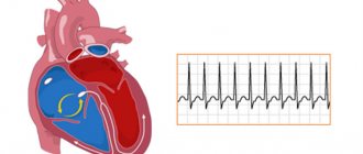
If a person comes for an examination to a doctor and the specialist suspects that something is wrong with his heart, he will send the patient for an ECG. Sinus rhythm on an ECG is a very important indicator and clearly provides data on the real state of the human heart muscle. What exactly can be determined by looking at the cardiogram is worth considering in more detail.
Peculiarities
ECG sinus rhythm what is it? Sinus rhythm detected on the ECG indicates good activity of the heart muscle, in which there are no pathologies. This rhythm characterizes oscillations that arise from impulses in a certain node and diverge throughout the atrium and ventricle. As a result, the heart muscle contracts. For the examination to show the correct result, the patient should not worry, he needs to be in a calm state.
What does sinus rhythm ECG mean? If the doctor notes on the transcript that sinus rhythm is present, then this means that the P peaks manifest themselves on a homogeneous basis, the pulse is 60-80 beats per minute, the distances between P-P and RR are similar.
Compliance of features is checked as follows:
- The elevations P are equal in height;
- Before the QRS complex, the presence of P armholes is mandatory;
- The PQ distance remains stable;
- The P notch in the second lead is positive.
ECG results - sinus rhythm
If all the parameters reflected on the cardiogram correspond to sinus rhythm, this means that the innervation impulses correctly follow from top to bottom. Otherwise, the impulses come from secondary parts of the heart.
What does a vertical position mean when there is sinus rhythm on an ECG? This is the normal location of the heart in the thoracic region on the line of the conventional location of the central axis. The location of the organ is permissible at different angles of inclination and in different planes, both vertical and horizontal, as well as in intermediate ones. This is not a pathology, but only indicates the distinctive characteristics of the structure of the patient’s body and is detected as a result of an ECG examination.
How to make an appointment with a specialist
Don't waste time - sign up for a consultation in just a few clicks. Leave your details in the feedback form or contact us at the contact number. If you need to call an ambulance at JSC "Medicine" (clinic of Academician Roitberg), you can call.
Geographically, the clinic is located in the Central District of Moscow, not far from the Mayakovskaya metro station, a 5-minute walk. Address: 2nd Tverskoy-Yamskoy lane, building 10.
Among our specialists there are candidates and doctors of science, highly experienced doctors who successfully solve complex clinical problems.
Do not delay visiting a specialist, even if your child is already feeling better. The sooner you seek treatment for sinus arrhythmia in children, the higher the chances of taking control of the possible disease and curing it forever.
Pathologies
Tests of the heart muscle may reveal some abnormalities.
When deciphering an ECG, a discrepancy between heart activity and sinus rhythm indicates an arrhythmia or blockade. The blockade occurs as a result of the transmission of impulses by the central nervous system to the heart. An increased heart rate means that the vibrations are faster. If we talk about rhythm disturbances, then in total there is a discrepancy between the frequency of contractions of the heart muscle and the sequence.
Irregular cyclicity of sinus rhythm can be observed on the ECG by the difference in the distances between the peaks. This basically indicates a weak node. To verify arrhythmia, it is necessary to conduct Holter monitoring and a drug test. This way it is possible to identify disturbances in the self-regulation of the autonomic system and the source of oscillations.
Functional arrhythmias
This is also a fairly large group, including:
- Rhythm disturbances of neurogenic origin It is known that the heart is under the influence of the autonomic nervous system, which controls the activity of all internal organs. It consists of sympathetic and parasympathetic nerves. Their effect on the heart is opposite. An increase in the tone of the vagus nerve (this is a parasympathetic nerve) inhibits the work of the heart, and an increase in the tone of the sympathetic nervous system, on the contrary, stimulates its activity. Usually the influence of the vagus nerve and the sympathetic nerves are in a state of balance. However, during the day the activity of the sympathetic division of the autonomic nervous system prevails, and at night - the parasympathetic one. Excessive activation of sympathetic tone is promoted by stress, strong emotions, intense mental or physical work, smoking, drinking alcohol, strong tea and coffee, and spicy foods. The arrhythmias that arise at these moments are called sympathodependent. Often such rhythm disturbances occur in patients with neuroses. Activation of sympathetic tone also occurs in diseases of the thyroid gland, intoxication, feverish conditions, and blood diseases.
- Another group consists of vagus-dependent arrhythmias (from the Latin nervus vagus - vagus nerve). In such patients, interruptions in heart function occur at night. Diseases of the gastrointestinal tract: intestines, gallbladder, peptic ulcer of the stomach and duodenum, and diseases of the bladder can lead to an increase in the parasympathetic effect on the heart and, accordingly, to the appearance of gastrointestinal rhythm disturbances. Reflexes are formed in diseased organs, as a result of which the activity of the vagus nerve increases.
Signs of violations
Cardiac weakness syndrome is detected on the basis of clinical and ECG studies. To make sure of the diagnosis of arrhythmia, you need to compare the current results of the cardiogram with the transcript with normal data on the patient’s heart condition. Uniform and positive P waves in one lead, as well as uniform location at a distance of 0.11-0.20 s in front of the QRS complex.
In one minute, the number of blows should not exceed 90. This indicator is determined by the method of dividing 60 seconds. for the duration of the R – R segment. Or the number of complexes that occurred in 3 seconds. Multiply by 20 (this is approximately 15 cm of tape).
Conclusion ECG sinus rhythm may reflect pathologies such as:
- Arrhythmia. The R – R intervals on the cardiogram vary by values exceeding 0.15 seconds. Here there is a direct connection between the number of heart beats and respiratory activity (inhalation - exhalation);
- Tachycardia. Contractions of the heart muscle increase to 90 beats per minute. Other rhythm parameters remain normal. In such cases, oblique descending depression of PQ and ascending ST are often found. The image in this “picture of the disease” resembles an anchor. If the heart rate exceeds 150 beats per minute, there is a risk of second degree block;
- Bradycardia. The main indicators of sinus rhythm are present in the ECG, but the number of heart beats is reduced. Therefore, the P-P interval increases to 0.21 seconds;
- Rigid sinus rhythm. The frequency of contractions of the heart muscle is increased. The P-P interval has a difference of up to 0.05 seconds. In this case, there is damage to the node or pathology of neurovegetative regulation.
Diagnostics
During the appointment, the doctor examines the little patient. If a child has poor contact with strangers, in particular with doctors, it is worth asking him at home in more detail what exactly is bothering him. During the interview, the doctor analyzes the child’s complaints and collects anamnesis. Most likely, clinical blood tests will be required. Of course, an ECG for sinus arrhythmia in a child will be performed immediately. You may also need the results of Holter monitoring and echocardiography. Perhaps an orthostatic test will be informative.
All analyzes and instrumental studies are carried out in one place - you do not need to travel and waste time. We are located in the Central Administrative District, not far from the Mayakovskaya metro station.
Reasons for violations
Cardiac dysfunction in the human body occurs for the following reasons:
- Regular consumption of drinks containing strong alcohol;
- Constant smoking;
- Heart disease;
- Heart failure;
- Excess thyroid hormones;
- Mitral valve protrusion;
- Uncontrolled use of glycosides or drugs against arrhythmia.
An increase in heart rate eliminates disturbances in a person’s respiratory activity.
An electrocardiogram (ECG) is not a new and time-tested method of identifying heart pathologies. This procedure requires very little time and does not require any preparatory steps. However, in order to get the correct result, transcript and doctor’s conclusion, sometimes you need to undergo such an examination several times. Based on the data obtained and clinical examinations, the cardiologist will diagnose the patient and prescribe treatment.
Why ECG readings may deviate from the norm
Irregular heart rhythm can be provoked not only by serious pathological abnormalities, but also by factors more familiar to a person’s daily life.
If the result of the electrocardiogram does not always correspond to the norm, this means that this state of the body could be provoked by the following factors:
- the person regularly drinks alcoholic beverages;
- the patient has been smoking cigarettes on a regular basis for quite a long time;
- a person is regularly exposed to various types of stressful situations;
- the patient often uses antiarrhythmic drugs;
- a person has problems with the functioning of the thyroid gland.
Of course, an accelerated heart rate or too slow may indicate problems of a more serious nature. If the results of the cardiogram are not normal, this may indicate acute heart failure, valve displacement, or congenital heart defects.
Our doctors
Kedrinskaya Larisa Ivanovna
Experience 18 years / Doctor of the highest category, Candidate of Medical Sciences / General practitioner, cardiologist
Çà ïèñà òüñÿ ê âðà ÷ó
Vershuta Elena Vasilievna
More than 23 years of experience / Highest category, Candidate of Medical Sciences / Pediatric cardiologist, cardiologist, therapist
Çà ïèñà òüñÿ ê âðà ÷ó
+
What to do?
If your baby feels unwell, but you have not previously consulted a doctor and do not have a reliable diagnosis, do not give your child any medicine, even if you are firmly convinced that the medicine will help.
Do not under any circumstances resort to the methods of so-called “traditional” medicine.
When to see a doctor
First, make an appointment with your pediatrician, and then, if necessary, your pediatrician will make a referral to a pediatric cardiologist.
Of course, it is impossible to independently detect sinus arrhythmia in a child. You can suspect the development of a cardiac disease in a child based on an increased heart rate. Often, sinus arrhythmia practically does not manifest itself, and it is discovered during a routine examination.
Our pediatrician will refer you to a pediatric cardiologist, the doctor will carefully study your problem and will definitely help. The doctor sees you in the center of Moscow, a 5-minute walk from the Mayakovskaya metro station.