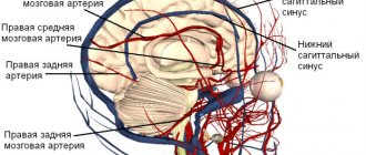
Brain atrophy: causes, symptoms, diagnosis
Atrophic changes in the cerebral cortex lead to the destruction of neural connections and a decrease in the activity of functional centers.
The condition leads to disruption of intracerebral metabolism, dementia, and the formation of a number of mental diseases (Alzheimer's, amyotrophic lateral sclerosis, dementia). Clinical symptoms depend on the type, stage, and degree of the disease. The multisystem form is accompanied by diffuse death of neurons and gradual loss of body functions.
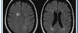
Brain atrophy on MRI
Causes of the disease
Understanding the topic of what atrophy that occurs in the brain is, it should be noted that this is always a secondary diagnosis that develops against the background of long-term damaging effects on the central nervous system. Doctors name several reasons why brain cells die:
- Genetic predisposition. The most important factor.
- Intoxication of the body, repeated with high frequency, associated with the use of alcoholic beverages and drugs.
- Injuries to the skull and soft tissues inside the skull.
- Insufficient blood supply to tissues, cerebral ischemia.
- Chronic anemia – insufficient oxygen supply. The condition occurs as a result of low concentrations of hemoglobin protein and red blood cells in the blood, which deliver oxygen to the tissues.
- Infections that affect the nervous system - polio, meningitis, Kuru disease, leptospirosis, brain tissue abscess.
- Diseases of the cardiovascular system - ischemia of the heart muscle, heart failure, atherosclerotic vascular pathologies.
- Decortication due to coma.
- Intracranial pressure. It is often the cause of cerebellar atrophy in newborns.
- Large tumors that compress surrounding tissues and interfere with normal blood supply to parts of the brain.
- Cerebrovascular disease is destructive changes in the vessels located in the brain.
If a person avoids mental activity, the risk of developing atrophic diseases occurring in the brain increases. Among the factors that increase the likelihood of the death of neurons located in the brain are smoking, low mental stress, chronic arterial hypertension, hydrocephalus, and long-term use of drugs that constrict blood vessels.
Causes of brain atrophy
After age 50, the risk of neurodegenerative conditions increases. Provoking factors increase the likelihood of the occurrence of a nosological form:
- Decreased kidney function (failure);
- Long-term increase in intracranial pressure (hydrocephalus);
- Frequent use of alcohol, drugs;
- Infectious damage to the cerebral cortex (retroviruses, poliomyelitis, encephalitis);
- Traumatic brain injury;
- Vascular diseases (thrombosis, atherosclerosis, aneurysm);
- Metabolic conditions;
- Mental illnesses - Alzheimer's, Itsenko-Cushing's syndrome, Parkinson's, Whipple, Gellervorden-Spatz.
Increases the likelihood of nosology - metabolic disorders, birth injuries, sexually transmitted infections, lack of B vitamins, folic acid.
The main causes of atrophy of the cerebral cortex
Scientific studies show a high probability of damage to cortical and subcortical structures in people 50-55 years old due to genetic predisposition. Cortical atrophy develops in patients suffering from hereditary Huntington's chorea.
Other reasons:
- Traumatic brain injuries accompanied by hematoma, neuronal death, and cyst formation;
- Chronic alcoholism, drug addiction, and the use of certain medications reduce the thickness of the cerebral hemispheres and subcortical sphere. Long-term alcohol intoxication disrupts intracellular metabolism and ensures the gradual death of neurons;
- Chronic cerebral (brain) ischemia is caused by vascular diseases (atherosclerosis, hypertension). Lack of oxygen contributes to irreversible tissue death;
- Congenital hydrocephalus in newborns leads to increased intracranial pressure and atrophy of the brain matter;
- More than seventy percent of cases of the disease in people over 55 years of age are due to neurodegenerative diseases - Pick, Lewy, Alzheimer's, Parkinson's. Nosologies form senile dementia.
Less common etiological factors of nosology are hypoxia of newborns, hydrocephalus, multiple congenital cysts in a child.
Causes of cerebral atrophy in newborns
The main etiological factor in reducing the thickness of the hemispheres of newborns is intrauterine hypoxia and problems during childbirth. Damage to the baby's head when passing through the birth canal provokes a traumatic brain injury and contributes to the appearance of hydrocephalus (dropsy).
Causes of atrophic cerebral changes in newborns:
- Damage to the bones of the skull;
- Increased amount of cerebrospinal fluid (hydrocephalus);
- Intrauterine infections (cytomegaly, herpes, meningitis).
There are no effective treatments for neonatal atrophy. Timely detection using MRI allows you to prescribe maintenance therapy and reduce the progression of the disease. Moderate changes are correlated with drug therapy. The child will be able to attend kindergarten and study in a special school.
Reasons provoking the violation
In the development of brain atrophy, and as a result of senile dementia, there is a complex of reasons that cause atrophic processes in the brain. The formation of the disease is influenced by the following factors:
- blood oxygen saturation deteriorates;
- the body's ability to regenerate deteriorates;
- as a result of atherosclerosis, there is a disruption in the blood supply to brain tissue;
- processes of genetic predisposition to atrophic pathologies are activated;
- reduction of mental load and thought processes.
What is typical, even if a person has a predisposition to brain atrophy (genetic), but in the process of life he studied foreign languages, was interested in science, art, read a variety of literature, developed intellectually in every possible way and used the work of the intellect in practice, that is, made the most of his potential brain, he is less susceptible to developing dementia.
The development of congenital cortical cerebral atrophy is essentially brain hypoplasia, since its normal formation does not occur. Despite this, the process is also called atrophic.
Also in adulthood, the destructive process can develop for a number of reasons:
- atrophy can occur as a result of toxic poisoning (alcoholism); with frequent consumption of alcoholic beverages, neurons die off, and due to ongoing intoxication, their restoration does not occur, which leads to this problem;
- constant low blood pressure;
- use of vasoconstrictor drugs;
- lack of mental stress;
- injuries resulting in compression of blood vessels and development of cerebral edema;
- cysts and tumors lead to compression of blood vessels; neoplasms that have stopped growing are especially dangerous; they influence the development of atrophy; growing neoplasms have a less negative impact;
- in rare cases, neurosurgery may be the cause.
Subatrophy of the brain - the first stage of senile dementia
Before clinical symptoms appear, subatrophic changes develop. There are no external symptoms. The condition is accompanied by a partial decrease in the function of a segment of the hemispheres.
Morphological types of subatrophy:
- Frontal;
- Frontotemporal;
- Parieto-occipital.
The first type is characterized by a decrease in mental activity, loss of speech and motor functions.
Damage to the frontotemporal areas leads to a decrease in a person’s hearing ability, communication functions are lost (difficulty communicating with other people), and the functioning of the cardiovascular system is disrupted.
Subatrophy reduces the volume of gray and white matter. Disturbances in conduction and motor function and fine motor activity occur.
Features of cortical atrophy
The death of cortical cells begins in the frontal lobes, where the functional centers for controlling movement and speech are located. Gradually, atrophy spreads to surrounding structures. In older people, the pathology leads to senile dementia.
Diffuse cortical changes are accompanied by microcirculation disorders and progressive clinical symptoms. Fine motor skills of the upper limbs and coordination of movements are impaired. The pathological complex leads to Alzheimer's disease and senile dementia.
MRI of cortical atrophy shows a decrease in the size of the frontal lobes. If there are changes on both sides, the functioning of the internal organs controlled by the frontal lobes is disrupted.
Congenital cortical atrophy of newborns is localized on one side. Symptoms are mild. With the help of rehabilitation procedures, it is possible to socialize the child.
Types of pathology
The generalized form of cerebral atrophy involves multiple areas of nerve cells in the brain tissue. Diffuse brain atrophy is the uniform death of neurons in all areas of the brain structures. It develops as a result of arterial hypertension, which is characterized by damage to small vessels located in each part of the brain.
The initial symptoms of diffuse atrophy resemble dysfunction of the cerebellum. The progressive course leads to a rapid increase in symptoms, which makes it possible to differentiate the pathology at later stages. Unlike the cortical type, with diffuse atrophy the symptoms of damage to the controlling, dominant hemisphere are clearly expressed. With cortical subatrophy occurring in the brain, destruction and tissue destruction are only just beginning.
Subatrophy, which occurs in the brain, is a condition that precedes the stage of neuronal death. The mechanism of the disease has already been launched, destructive processes have begun, but the body independently compensates for the violations that have arisen. Subatrophic changes are accompanied by mild symptoms. Bihemispheric cortical atrophy occurs in the tissues of both hemispheres. Manifested by Alzheimer's syndrome.
Alcohol atrophy developing in the brain
Organic damage to the structures of the brain matter, which develops against the background of constant exposure to ethanol, is called toxic encephalopathy. Affects all parts of the brain. The cortical layers and cerebellum are especially sensitive to the negative effects of alcohol. Often leads to cranial nerve palsy. The frontal lobes are responsible for behavior, intelligence, emotions and moral qualities - properties that characterize a conscious personality.
Developing pathology causes atrophic changes in tissues and is one of the main causes of dementia. Dementia, as a consequence of alcoholism, is diagnosed in 10-30% of patients who abuse alcoholic beverages. A person becomes infantile and loses the ability for abstract logical thinking. As the disease progresses, the patient loses basic skills - the ability to brush teeth, tie shoelaces, and hold cutlery.
Clinical symptoms of multiple system atrophy
Diffuse neurodegeneration is accompanied by problems in the reproductive and urinary areas. Necrosis of many parts of the brain is simultaneously accompanied by a variety of clinical symptoms:
- Muscle tremors in Parkinsonism;
- Impaired gait and mobility coordination;
- Loss of erection;
- Vegetative-vascular disorders.
Before the advent of magnetic resonance imaging, early diagnosis of the disease was problematic. Only nuclear magnetic resonance verifies a decrease in the thickness of the brain parenchyma.
Clinical symptoms of brain atrophy
The manifestations of pathology are largely determined by the causes and provoking factors. Most older people have dementia, frontal lobe syndrome, and internal multiple organ pathology.
How does frontal lobe syndrome manifest?
- Lack of appetite;
- Loss of memory, intellectual activity;
- Frequent emotional breakdowns;
- Lack of communication with surrounding people;
- Irritability;
- Lack of self-criticism.
Psychoorganic syndrome is accompanied by cerebroasthenic disorders, affective disorders, and amnesia.
The patient lacks an adequate assessment of surrounding events and self-criticism. Primitive thinking appears, a one-sided representation of the essence of the detail. Speech reserve decreases, paramnesia appears.
Concomitant affective disorders lead to depressive syndrome and inadequate mental state. Tearfulness, touchiness, irritability, unreasonable aggression are typical manifestations of pathology.
Symptoms
Initial signs of atrophy affecting the tissues and structures of the brain usually appear in people over 45 years of age. Pathology is more often diagnosed in female patients. Characteristic symptoms:
- Changing personality type. Apathy, indifference, narrowing of interests.
- Psycho-emotional disorder. Mood swings, depression, increased irritability.
- Impaired memory function.
- Reducing vocabulary.
- Motor dysfunction, impaired coordination of movements and fine motor skills.
- Deterioration of mental activity.
- Decreased performance.
- Epileptic seizures.
The body's regenerative reactions weaken. Reflexes are depressed. Symptoms become brighter and more expressive. Atrophic changes are manifested by Parkinson's and Alzheimer's syndrome. The following signs indicate a specific affected area:
- Medulla. Deviations in the functioning of the respiratory, digestive, and cardiovascular systems. Defense reflexes are suppressed.
- Cerebellum. Weakness of skeletal muscles, malfunctions of the musculoskeletal system.
- Midbrain. Inhibited or absent reactions to external stimuli.
- Diencephalon. Pathological deviations in the functioning of the thermoregulation system, disruption of the hemostatic and metabolic systems.
- Frontal lobes. Secrecy, aggression, demonstrative behavior.
Signs such as impulsiveness, previously unusual rudeness, increased sexuality, decreased self-control, and apathy indicate malfunctions in the functioning of the main organ of the central nervous system.
Types and classification of brain atrophy
According to the degree of danger, there are two types of atrophic changes in the brain:
- Physiological;
- Pathological.
The first type is natural. Throughout human development, the death of the umbilical arteries and ductus arteriosus (in newborns) initially accompanies it. After puberty, the tissue of the thymus gland is lost.
In old age, degenerative changes in the genital area occur. In elderly people, cortical destruction and involution of the frontal part appear. The condition is physiological.
Types of pathological atrophy:
- Dysfunctional – develops with a decrease in the functional activity of the brain;
- Compression – provoked by increased pressure on brain tissue (hydrocephalus, hematoma, copious accumulation of blood);
- Ischemic (dyscirculatory) occurs due to narrowing of the lumen of the arteries due to atherosclerosis, blood clots, and increased neurogenic activity. Generalized cerebral hypoxia is accompanied not only by mental dementia and sclerotic intracerebral changes;
- Neurotic (neurogenic) is formed due to a decrease in the flow of nerve impulses to the internal organ. The condition is formed due to gradual hemorrhages, the presence of intracerebral tumors, atrophy of the optic or trigeminal nerve. Occurs during chronic intoxication, exposure to physical factors, radiation therapy, long-term treatment with non-steroidal anti-inflammatory drugs;
- Dishormonal - occurs against the background of endocrine imbalance in the ovaries, testes, thyroid gland, mammary glands.
Morphological types of brain atrophy:
- Smooth – the surface of the brain is smoothed;
- lumpy - uneven distribution of areas of necrosis forms a special structure;
- Mixed.
Classification according to the extent of damage:
- Focal - only isolated areas of atrophic damage to the cerebral cortex can be traced;
- Diffuse - spreads over the entire surface of the parenchyma;
- Partial – necrosis of a limited part of the brain;
- Complete – atrophic changes in white and gray matter, degeneration of the trigeminal and optic nerves.
The nature of morphological changes in the brain is revealed by magnetic resonance scanning. Scanning should be done after the first clinical symptoms appear.
How to help a patient
Cortical cerebral atrophy cannot be completely cured. The main task is to prescribe comprehensive treatment aimed at slowing the development of symptoms. Atrophies that appear at a young age can be effectively corrected if the etiological factor is excluded.
For the treatment of cortical atrophy, patients are prescribed drugs from the following groups:
- To improve blood microcirculation . The most popular drug is Trental. A drug with a vasodilating effect, increasing the lumen of capillaries, improving gas exchange through the walls of blood vessels and blood microcirculation.
- Nootropics . Drugs in this group improve blood circulation and brain metabolism. Highly effective drugs: Piracetam, Ceraxon, Cerepro, Cerebrolysin. Medicines in this group have a beneficial effect on the patient’s thinking abilities.
- Antioxidants . Drugs in this group stimulate regeneration processes, increase metabolic rate, slow down atrophy, and reduce the impact of oxygen free radicals.
- In order to relieve associated symptoms such as headache, the use of non-steroidal anti-inflammatory drugs .
An important factor in the treatment of cortical atrophy is monitoring the patient’s neuropsychic state. This diagnosis should be accepted adequately by family members, as should their attitude towards the patient:
- the patient needs walks in the fresh air daily;
- moderate physical activity;
- the patient must be entrusted with all possible self-care procedures;
- in case of a neurasthenic state, the use of mild sedatives is permissible.
The disease progresses very quickly and the result of this process is personality degradation. The manner of communication, speech, and behavior takes on an ornate connotation. The vocabulary becomes significantly scarcer, which leads to the use of monosyllabic phrases in speech.
The prognosis for patients with cortical atrophy of the brain is always unfavorable: whether the process progresses slowly or quickly, it always leads to degradation and, ultimately, death.
There are currently no effective methods for preventing the destructive process. The only thing that can slow down the process is:
- timely treatment of all existing diseases;
- active lifestyle;
- working on your memory;
- development of intelligence from youth;
- positive attitude.
Brain atrophy is destructive changes that provoke depletion of organ tissue, deterioration of vitality, and loss of function. Accompanied by necrosis of nerve cells and severance of neural connections within chemically or functionally related groups. The volume of brain tissue decreases. Destructive processes spread to different parts - the cortex and subcortical (subcortical) areas. Often occurs in patients over 50 years of age. Diagnosed in newborn infants and children under one year of age.
The death of the cells that make up the brain provokes serious consequences. There is a violation of cognitive abilities, which include speech, spatial orientation, understanding, logical thinking, reasoning, calculation and learning. The disease causes neurological disorders and motor dysfunction.
Doctors give a negative answer to the question of whether cerebral atrophy, which occurs in the brain, affects life expectancy. Neurons die gradually. From the initial signs of pathology to a state where a large area of the brain atrophies with the subsequent development of dementia, more than 20 years can pass. The death of a patient is usually caused by other diseases that cause disruptions in the functioning of the body that are incompatible with life.
Discussions on how long patients with atrophic lesions live do not correctly reflect the characteristics and influence of the pathology. Cerebral atrophy does not reduce life expectancy, but significantly impairs its quality. Leads to dementia and disability. The person is not capable of self-care and needs constant medical supervision and care. Often forced to spend the rest of his life in a specialized dispensary.
Forms of multiple system atrophy
The danger of multiple lesions of brain structures is determined by a complex of pathological damage from the hemispheres, subcortical formations, cerebellum, spinal trunk, and white matter. Concomitant changes in the optic nerve lead to blindness, and in the trigeminal nerve – disruption of the innervation of the face.
Forms of multiple system atrophy:
- Olivopontocerebellar – damage to the cerebellum with impaired mobility;
- Striatonigral degeneration – muscle tremors with manifestations of Parkinsonism;
- Shy-Drager syndrome – vegetative-vascular dystonia, decreased blood pressure;
- Kugelberg-Welander amyotrophy is brain atrophy with muscle wasting and hyperplasia of connective tissue fibers.
Symptoms are determined by the predominant form of the lesion.
Prevention
Pathology is often a consequence of arterial hypertension and atherosclerosis. In order to prevent negative consequences, it is recommended to promptly treat diseases that provoke atrophic processes in the tissues of the brain matter. Doctors advise giving up bad habits, leading a healthy lifestyle, loading the brain with logical tasks, and stimulating intellectual activity.
Brain atrophy is a long-term pathological process that, in the absence of correct therapy, leads to dementia, disability, and complete dependence on staff. Often the patient requires hospitalization. In order to promptly identify and stop the development of the disease, at the first alarming symptoms it is better to consult a neurologist.
The main stages of atrophic brain changes
The disease has five degrees of progression. Based on clinical symptoms, it is possible to verify nosologies starting from the second or third stage.
Degrees of cortical atrophy:
- There are no clinical symptoms, but the pathology progresses rapidly;
- 2nd degree – characterized by a decrease in communication skills, lack of an adequate response to critical remarks, and an increase in the number of conflicts with other people;
- Lack of behavior control, causeless anger;
- Loss of adequate perception of the situation;
- Elimination of the psycho-emotional component of behavioral reactions.
Identifying any symptom requires additional study of the structure of the brain.
Symptoms of each stage of the destructive process
Cortical cerebral atrophy goes through five stages of development, each of which has its own symptoms and manifestations:
- The initial stage is characterized by a complete absence of manifestations, but its development occurs rapidly and quickly moves into the next stage.
- At the second stage, communication with others deteriorates very quickly. A person cannot adequately perceive criticism, becomes irritable, conflicts involving him are a normal phenomenon that recurs regularly. Very often the thread of the conversation gets lost.
- The third stage - the patient loses control over his behavior in several stages, which becomes outrageous. Frequent manifestations of despondency or the occurrence of causeless outbursts of anger.
- four : awareness of the essence of events is completely lost. The patient cannot adequately respond to the demands of others.
- Fifth stage - the patient does not take pictures of the events taking place at all, and, accordingly, this does not cause any emotions in him.
Depending on who and what lobe of the brain was affected by atrophy, some speech disorders arise in the early stages, unreasonable euphoria or indifference, lethargy, sexual hyperactivity, and some types of mania appear.
Principles of diagnosing atrophy
The initial stage involves taking an anamnesis, examination, and physical examination. The second stage is clinical and instrumental methods (ultrasound, CT, MRI of the brain, scintigraphy, PET/CT). Damage to the optic nerve is confirmed by ophthalmoscopy, tonometry, contrast CT or MRI angiography.
The best way to detect pathology of the soft tissues of the brain is MRI. The procedure must be performed several times (a month apart) to identify atrophy of varying depth and extent.
Magnetic resonance examination reveals the smallest local lesions and helps to correctly determine the degree of disease progression.