02 Sep 2021 at 14:47 MRI of the head in Tushino 9441
According to current statistics, cerebral ischemia is diagnosed in every fourth resident of our country. As a rule, this phenomenon is age-related, however, its symptoms are increasingly observed among representatives of the younger generation (up to 40 - 45 years). In order to minimize the negative consequences of this dangerous disease, it is necessary to promptly contact specialists and listen to their professional recommendations.
There are three stages of chronic cerebral ischemia:
- Stage 1 (initial) is characterized by a large number of various complaints of headache, irritability, insomnia, a feeling of “heaviness” in the head, decreased memory for immediate events, fatigue, poor coordination of movements, and instability when walking. These changes are not noticeable to others and patients remain adapted to society. A neurological examination reveals minimal symptoms, a clear neurological deficit has not yet formed, and adequate therapy can reduce or eliminate individual manifestations of the disease.
- Stage 2 (subcompensation) the frequency of complaints about loss of memory and performance, instability, and dizziness increases. A leading clinical syndrome is formed, and a neurological deficit becomes obvious. Cognitive, personality, and emotional-volitional disorders worsen. Adaptation to society and professional skills fade, but the possibility of self-care remains.
- Stage 3 (decompensation) Objective neurological disorders come to the fore. The number of complaints decreases due to decreased criticism of one’s condition. On examination, several well-defined neurological syndromes are noticeable. Cognitive impairments at the advanced stage of chronic cerebral ischemia are very often the leading cause of disability, their everyday adaptation suffers, patients are unable to work, require constant outside care, and vascular dementia develops.
In the clinic of chronic cerebral ischemia, several main neurological syndromes are distinguished. These include cephalgic, vestibulo-atactic, psychopathological, paroxysmal syndrome, pyramidal insufficiency, pseudobulbar syndrome.
Cephalgic syndrome is characterized by pain in various parts of the head. Often there is no connection between headaches and hemodynamic disorders. As chronic cerebral ischemia progresses, headache attacks become less frequent.
Vestibulo-atactic syndrome is one of the most common in patients with chronic cerebral ischemia. It includes vertigo, loss of coordination, instability, and noise in the head. These complaints arise from circulatory failure in the vertebrobasilar system, as well as age-related changes in the vestibular apparatus, ischemia of the vestibulocochlear nerve.
Paroxysmal disorders in chronic cerebral ischemia are varied and include episodes of transient global amnesia, syncope, drop attacks, and epileptic seizures. The severity of such conditions increases as ischemia and concomitant diseases progress.
Mental disorders are often present at all stages of chronic cerebral ischemia. At first, they are of the nature of asthenoneurotic, depressive, and anxiety disorders; at stages II-III, they are joined by cognitive impairment, and dementia occurs.
Symptoms of focal brain damage
Any interruptions in brain function affect the daily natural functions of human activity, and the localization of lesions affects muscle functions and the normal functioning of internal organs. Pathologies of vascular origin often lead to mental changes and can cause hypertension, stroke, etc. Subcortical lesions, as a rule, do not manifest themselves in any way and are detected by chance.
The main signs of the presence of lesions in the brain are:
- hypertension - accelerated blood circulation caused by insufficient oxygen flow in the brain, which is caused by dystrophic changes in the blood vessels;
- epilepsy;
- mental abnormalities arising from pathological processes in the subrachnoid spaces, which may be accompanied by hemorrhage, congestion in the fundus with the formation of dark spots, burst vessels, and retinal tears;
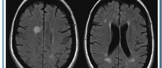
- strokes, which can be prevented with timely MRI examination, since pre-stroke changes are very clearly visualized on images;
- attacks of headaches, dizziness, in which a general examination is required, incl. using MRI;
- uncontrolled muscle contractions.
Signs of single lesions of the discircular medulla on the tomograph screen always mean certain disturbances in the functions of the vascular system, which is associated mainly with hypertension.
Basic examinations in patients with chronic cerebral ischemia:
- MRI of the brain, preferably with angiography
- blood pressure and ECG monitoring.
- general blood test, general urinalysis, biochemical blood test, coagulogram, blood lipid spectrum.
- fundus examination
- examination by an otoneurologist
- neuropsychological study.
- CDK BCS, TKDG, ECHO-KG
- examination by a psychotherapist to identify anxiety-depressive conditions and cognitive deficits).
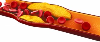
Treatment and prevention of chronic cerebral ischemia should be aimed at correcting the current vascular disease. The focus is on antihypertensive therapy, prevention of thrombosis, and lowering blood cholesterol levels. Also an important component of treatment is the correction of concomitant diseases (diabetes mellitus, heart rhythm disturbances, heart failure, obesity), sleep disorders and anxiety-depressive disorders. Vascular therapy, neuroprotection, and improvement of neurometabolism play a significant role. A comprehensive treatment is recommended, including non-drug methods of influence: psychotherapy, exercise therapy, massage, physiotherapeutic treatment.
The First Neurology clinic presents a variety of hardware techniques that help improve cerebral circulation, such as mesodiencephalic modulation (MDM therapy), brain micropolarization, and electrosleep. Using the modern CEFALY neurostimulation technique, the intensity of headaches decreases, memory and concentration improve.
What to do if MRI shows chronic ischemia?
If, when decoding the results of a head scan, the presence of ischemic brain disease is detected, it is recommended to consult a neurologist. You may need to consult a therapist or cardiologist. To slow the progression of ischemic damage, it is necessary to achieve target levels of blood pressure, blood glucose and cholesterol levels. Doctors will evaluate metabolic changes and select treatment to compensate for chronic diseases.
The diagnostic department performs MRI of the brain from 2,690 rubles. Contrasting is carried out from the age of 12. You can ask the administrator any questions you may have by calling +7 (812) 407-32-31.
Our specialists
Tarasova Svetlana Vitalievna
Expert No. 1 in the treatment of headaches and migraines. Head of the Center for the Treatment of Pain and Multiple Sclerosis.
Somnologist.
Epileptologist. Botulinum therapist. The doctor is a neurologist of the highest category. Physiotherapist. Doctor of Medical Sciences.
Experience: 23 years.Derevianko Leonid Sergeevich
Head of the Center for Diagnostics and Treatment of Sleep Disorders.
The doctor is a neurologist of the highest category. Vertebrologist. Somnologist. Epileptologist. Botulinum therapist. Physiotherapist. Experience: 23 years.
Palagin Maxim Anatolievich
The doctor is a neurologist. Somnologist. Epileptologist. Botulinum therapist. Physiotherapist. Experience: 6 years.
Romanova Tatyana Alexandrovna
Pediatric neurologist. Experience: 24 years.
Temina Lyudmila Borisovna
Pediatric neurologist of the highest category. Candidate of Medical Sciences.
Experience: 46 years.
Zhuravleva Nadezhda Vladimirovna
Head of the center for diagnosis and treatment of myasthenia gravis.
The doctor is a neurologist of the highest category. Physiotherapist. Experience: 16 years.
Mizonov Sergey Vladimirovich
The doctor is a neurologist. Chiropractor. Osteopath. Physiotherapist. Experience: 8 years.
Bezgina Elena Vladimirovna
The doctor is a neurologist of the highest category. Botulinum therapist. Physiotherapist. Experience: 24 years.
Drozdova Lyubov Vladimirovna
The doctor is a neurologist. Vertebroneurologist. Ozone therapist. Physiotherapist. Experience: 17 years.
Read also
Transient ischemic attack (TIA)
What it is?
Why is this happening? Is this condition dangerous? What to do if doctors make such a diagnosis? These questions are always asked by patients who come to see a neurologist. According to the classification... Read more
Encephalopathy
These can range from minor abnormalities in brain function to serious neurological disorders that require urgent treatment. Encephalopathy can be congenital or acquired. Congenital...
More details
Swelling of the legs
Causes of Swelling in the Legs The legs are common sites for swelling due to the effect of gravity on the fluids in the human body. However, fluid retention is not the only cause of leg swelling. Injuries...
More details
Alzheimer's disease
47,000,000 people suffer from dementia in the world. Doctors call Alzheimer's disease the most common cause of dementia. Alzheimer's disease is a progressive neurodegenerative disease accompanied by...
More details
Vegetative-vascular dystonia
Vegetative-vascular dystonia is a dysregulation of the autonomic nervous system, which manifests itself in the form of various clinical symptoms. This disease is diagnosed at different times...
More details
Diffuse and focal changes - what are they?
Diffuse and focal changes - what is it, what is the difference
At the conclusion of the ultrasound examination, when pathology is detected, the doctor may indicate the presence of focal or diffuse changes. These are diagnostic criteria that describe the degree of organ damage in various diseases.
general information
With an ultrasound, the doctor must determine the condition of the organ. After carefully examining it, the specialist makes a conclusion - pathology or normal. If the organ is normal, then it is simply described, where it all ends.
If the examination shows a deviation, then the degree of damage to the organ is determined. The doctor must answer the question of what changes are present - focal or diffuse.
Diffuse and focal changes - what is the difference
It should be clarified that the division into focal and diffuse changes is conditional. It helps in the work of specialists to better understand the picture of the disease.
Diffuse is damage to the entire organ. Whatever part the doctor examines, he sees pathological changes. The organ is completely different from a healthy one. It is impossible to determine which area is normal.
Focal changes are a pathological process affecting part of an organ. There are areas that differ from healthy ones. However, all other parts look normal.
Examples of diffuse and focal changes
With advanced hepatitis, an inflammatory disease of the liver, the entire organ is affected. The doctor will see diffuse changes. When the pathology is still at an early stage, healthy areas will also be identified, that is, focal changes.
With a malignant liver tumor at the initial stage, the doctor sees focal changes. When cancer affects the entire organ, it is no longer possible to determine its healthy boundaries - this is a diffuse pathology.
Relationship between changes and disease
What changes have occurred is of great importance. Diffuse damage is always a sign of severe pathology that has already affected the entire organ. However, the presence of focal changes does not mean that the disease is still at an early stage. It depends on the disease.
Ultrasound examination cannot show a specific disease. The doctor can only see anatomical changes. The same diffuse and focal processes can be observed with completely different deviations. Usually, after an ultrasound, when the affected area is identified, other, more accurate research options are prescribed.