Causes
Pinching of cervical vessels can occur for a number of reasons, including the following:
- hernias,
- injuries,
- spondyloarthrosis,
- tumors.
But most often the cause of infringement is osteochondrosis. This pathology is so common that doctors call it a kind of payment for a person for walking on two legs, and not on four legs. That is, the human muscular corset is forced to constantly maintain the spine in a straight, anatomically correct position, and if the muscles are poorly developed, osteochondrosis will certainly make itself felt. At the same time, the muscles in the cervical spine are naturally weakly developed, and they bear a large load.
The psycho-emotional state of a person also has an impact: with chronic stress, the tone of the neck muscles increases, which significantly increases the risk of pinched blood vessels, the formation of a local inflammatory process and swelling that impedes blood circulation.
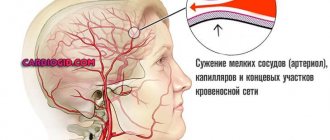
The most common cause of pinched vertebral artery is degenerative-dystrophic lesions of the cervical spine
Symptomatic picture
The intensity and localization of pain depends on the cause. People with weak ligaments often complain of regular pain, usually dull, aching, localized in a small area of the neck, which occurs in the morning, after sleeping on an uncomfortable pillow, and after prolonged work in one position. Often, the unpleasant sensations go away on their own during the day, but they can also intensify. Characterized by headache, dizziness, numbness of the shoulder girdle, back of the head, tingling sensation, goosebumps. Sudden changes in blood pressure are possible: there may be a sudden decrease in pressure, manifested by unexplained drowsiness, weakness, darkening of the eyes, or a significant increase.
If a ligament is sprained or torn, most often acute pain, muscle spasm and swelling immediately occur, but there are rare cases where the first symptoms appear only several hours after the injury.
With osteochondrosis, the pain intensifies with movement and is practically not felt at rest; it can radiate to the ear, back of the head, lower jaw, and less often to the suprascapular region.
In the presence of intervertebral disc herniation, pain occurs suddenly after some kind of physical activity or even after a careless, too sudden movement, it is highly intense, and intensifies not only with movement, but even with coughing or sneezing. May radiate to the arm or shoulder. The patient is forced to keep his head tilted in the direction opposite to the location of the pain.
With infectious myopathies, pain usually occurs on the right or left and affects not only the neck, but also the shoulder, arm, even fingertips, and swelling of the hand may be observed.
Myofascial syndrome of the trapezius muscle is characterized by acute pain between the shoulder blades.
Symptoms
Clinical symptoms may vary depending on whether the process is acute or chronic.
In case of acute pinching, the main symptom is severe pain in the neck. It may intensify when turning the head, or the person will complain of a feeling of numbness when it is physically impossible to turn the head. This happens when a person has suffered a minor injury (strained neck muscles) or slept in an uncomfortable position.
Arteries pass through the cervical vertebrae, feeding brain tissue and carrying out venous drainage. When pinched, this process is disrupted. As a rule, this process develops gradually; the body is able to compensate for a long time for the lack of oxygen and other substances entering the brain with the bloodstream. Therefore, the clinical picture increases progressively.
Symptoms of chronic pinching of the vertebral artery from the side of the brain are expressed as follows:
Cervical gymnastics
- headache;
- fast fatiguability;
- memory loss;
- deterioration of attention;
- visual disturbances (floaters or spots before the eyes);
- hearing loss, ringing in the ears;
- fainting or dizziness.
When such symptoms are constantly present, a person’s quality of life decreases: sleep is disturbed, depression develops, and motivation for any activity disappears. Therefore, if you notice any of the described signs, you should consult a doctor as soon as possible.
Headache and dizziness are the most common symptoms of pinched vessels in the cervical spine
If the pinching is caused not by osteochondrosis, but by another disease (problems with the skeletal system of the body, pathologies of vascular origin, tumor) or injury, the clinical picture may differ. Its intensity will depend on how quickly the underlying disease develops.
Diagnosis and treatment of pain in the neck and upper extremities
Professor O.S. Levin
RMAPO
Neck pain is one of the most common manifestations in clinical practice [3,9,17]. Neck pain affects almost half of the adult population , with persistent neck pain affecting approximately 23% of women and 17% of men [15]. The cervical spine is the most mobile part of the spinal column, which makes it particularly vulnerable to injury and degenerative changes. However, degenerative-dystrophic changes in the spine are far from the only cause of neck pain, which thus requires careful differential diagnosis [7,17,21]. Pain in the neck and arm is usually divided into vertebrogenic (caused by spinal pathology) and non-vertebrogenic, and the causes of both types of pain are very numerous (Table 1). Each variant of vertebrogenic pathology can lead to the development of 4 main clinical syndromes: • local pain syndrome (cervicalgia), often accompanied by local vertebral syndrome in the form of pain, deformity, limited mobility or instability of one or more adjacent segments of the spine; • reflex (irritative) syndromes include referred pain (for example, cervicobrachialgia, cervicocranialgia), muscular-tonic syndromes, neurodystrophic manifestations, repercussion autonomic disorders with a wide range of secondary manifestations: enthesopathies, periatropathies, myofascial syndrome, tunnel syndromes, etc. d.; • radicular syndrome (radiculopathy), caused by irritation or compression of the spinal roots; • syndromes of compression (ischemia) of the spinal cord (due to herniated intervertebral discs, spinal canal stenosis, etc.).
From a pragmatic point of view, back pain, regardless of its origin, is useful to subdivide according to its course and location (Table 2). Back pain can be nociceptive, neuropathic or psychogenic in origin. Nociceptive pain is associated with the activation of pain receptors - nociceptors, and generally corresponds to the degree of tissue damage and the duration of action of damaging factors. It is felt at the site of origin (local pain) or at a distance (referred pain). Neuropathic pain is associated with dysfunction or damage to the nervous system. Neuropathic back pain is usually associated with root or dorsal root ganglion involvement [1,12].
Acute neck pain in most cases is a consequence of protrusion or herniation of the intervertebral disc, less often - trauma or myofascial syndrome. Chronic (persistent or constantly recurring) pain, which has a particularly important medical and social significance, most often occurs against the background of degenerative-dystrophic changes, although the latter is not always its direct cause. Degenerative changes, as a rule, affect intervertebral discs, joints, ligaments and other tissues that form the spinal motion segment (SMS). An important role in the development of degenerative-dystrophic changes is played by heredity, age, physical activity on the elements of the PDS, and injuries. At different stages of the degenerative cascade, clinical manifestations of spinal osteochondrosis can be predominantly associated with intervertebral disc herniation, in others - with arthrosis of the intervertebral (facet) joints (spondyloarthrosis), the formation of osteophytes and other changes [6].
Herniated intervertebral discs Characteristic clinical features of pain syndrome with a herniated cervical intervertebral disc: - acute onset after physical activity, awkward movement or injury; increased pain in the neck and arm with increased pressure in the epidural space (with coughing, sneezing, straining, compression of the jugular veins);
Table 2. Classification of back pain
| With the flow | By localization |
| Acute - less than 6 weeks Subacute 6-12 weeks Chronic more than 12 weeks | Cervicalgia - neck pain Cervicobrachialgia - neck pain spreading to the arm Cervicocranialgia - neck pain spreading to the head area Cercicothoracalgia - neck pain spreading to the interscapular area |
Table 1. The main causes of pain in the neck and upper extremities Vertebrogenic pain Herniated intervertebral disc Cervical spondylosis Painful dysfunction of the SMS Instability of the SDS Rheumatoid arthritis Seronegative spondyloarthropathy Trauma Tumors Spondylitis Osteoporosis Non-vertebrogenic pain Brachial plexopathy Peripheral nerve lesions Reflex sympathetic dystrophy ia Myofascial syndrome Fibromyalgia Damage to joints and periarticular tissues Damage bones (osteomyelitis, osteoporosis) Damage to blood vessels (thromboembolism, vasculitis, Raynaud's syndrome) Viscerogenic pain (diseases of the pleura, heart) Psychogenic pain
- increased pain in the neck and arm when bending the neck, tilting the head towards the pain with an axial load on it, rotating the head towards the pain with throwing it back; - forced position of the head with a slight tilt forward and the side opposite to the localization of pain, a sharp limitation of the mobility of the cervical spine and tension of the neck muscles; - reduction of pain when traction of the head or placing a hand behind the head (due to the expansion of the intervertebral foramen).
Depending on the level of the lesion, pain can radiate to the medial parts of the scapula, interscapular region, back of the head, shoulder girdle and arm. With radiculopathy, pain, sensory disturbances (paresthesia, dysesthesia), loss of reflexes, and sometimes muscle weakness are detected in the area of the corresponding root (Table 3). The cause of radiculopathy is most often lateral disc herniations C6-C7 and C5~C6, much less often - C7-T1 and C4-C5. Root damage may be associated with its mechanical compression or tension, compression of its arteries and veins with the development of ischemia and edema, inflammation, demyelination, axonal degeneration, fibrosis, but it is difficult to clinically differentiate these types of root damage. An important role in symptom formation may be played by the irritation of the spinal ganglion cells and the sympathetic fibers that follow the root [11].
The cause of spinal cord compression is usually a median hernia, which is most often localized at the C5-C6 or C3-C4 levels. Dysfunction of the spinal cord can be associated with its direct mechanical compression (static or dynamic - during movement) or compression of the vessels supplying the spinal cord. A factor that increases the likelihood of compression is congenital or acquired spinal canal stenosis. At the level of the cervical spine, spinal canal stenosis is diagnosed if the ratio of the anteroposterior diameter of the spinal canal and the vertebral body is less than 0.8. Compression of the spinal cord is manifested by weakness and impaired sensitivity in the arms and legs, hyperreflexia and spasticity in the legs, changes in gait, pathological foot reflexes, pelvic disorders, Lhermitte’s symptom (the sensation of current passing through the spine and legs when bending it) [9,12].
Cervical spondylosis Spondylosis is understood as a set of degenerative changes, which include the formation of osteophytes, degenerative changes and hypertrophy of intervertebral joints (spondyloarthrosis), hypertrophy of the ligamentous apparatus, sclerotic changes in uncovertebral “joints”, inflammatory damage to periarticular tissues, etc. These changes can be a source of pain, and the narrowing of the intervertebral foramina and spinal canal they cause can lead to radiculopathy or myelopathy. In contrast to disc herniation, spondylosis often affects the upper cervical (C2-C4) roots, pain usually increases with extension rather than with flexion, and the prognosis is less favorable [12,17].
Damage to the facet joints can be not only a consequence of increased load on these joints (including disc herniation) or trauma, but also a manifestation of systemic arthropathy (polyosteoarthrosis, rheumatoid arthritis, seronegative spondyloarthropathy). It manifests itself as acute unilateral or bilateral neck pain, often regressing within 7-10 days. When the upper cervical joints are affected, the pain radiates to the back of the head and forehead, when the middle cervical joints are affected - to the area of the shoulder girdle and shoulder, the lower cervical joints - to the scapula and interscapular area. Torticollis may develop as a result of protective muscle spasm (8).
Exacerbations of pain syndrome with spondyloarthrosis occur with varying frequency. They are often caused by minor trauma, unsuccessful movement, hypothermia, or prolonged stay in an uncomfortable position (including during sleep). In some patients, the pain becomes permanent over time. Upon examination, limited mobility of the cervical spine in the corresponding SMS is revealed, especially during extension - while flexion and rotation are preserved; pain on palpation of the facet joints is usually on both sides (approximately 1-2 cm from the midline). Pain is provoked by straightening the neck or bending towards the more affected joint. The diagnosis is confirmed by a decrease in pain after joint blockade. Osteophytes, whose sanogenetic function is to stabilize the SDS, sometimes compress the vertebral artery and esophagus, causing vertebrobasilar insufficiency and swallowing disorders (dysphagia).
| Table 3. Signs of damage to the cervical and upper thoracic roots | |||||
| Signs / roots | Localization of pain | Decreased sensitivity | Decreased reflex | Paresis | Possible location disc herniation |
| C5 | Outer surface of the shoulder, medial part of the scapula | Upper outer shoulder (above the deltoid) | Biceps reflex | Abduction and external rotation of the shoulder, partially flexion of the forearm | C4-C5 |
| C6 | Lateral surface of the forearm and hand, 1st and 2nd fingers |
| Biceps reflex | Flexion and internal rotation of the forearm, partially extension of the hand | C5-C6 |
| C7 | The back of the shoulder and forearm up to 2-3 fingers | 2nd and 3rd fingers, back of the hand and forearm | Triceps reflex | Shoulder extension, wrist and finger extension, partial wrist flexion | C6-C7 |
| C8 | Inner surface of the forearm, hand up to fingers IV-V | IV-V fingers, inner surface of the hand and forearm | No | Flexion and extension of fingers | S7-T1 T T1-T2 |
| T1 | Inner surface of the shoulder and forearm, axillary area | Inner surface of the shoulder and upper forearm, axilla | No | 4 Spreading fingers | |
As a result of narrowing of the spinal canal and intervertebral foramina due to the proliferation of osteophytes, hypertrophy of the articular processes and the yellow ligament, compression of the anterior spinal artery, as well as the vertebral or medullary arteries feeding it, is possible. Compression of the anterior spinal artery at the cervical level can cause damage to the cervical spinal cord. When the vertebral artery is compressed, the following may also be observed: posterior cervical sympathetic syndrome (see below), repeated episodes of vertebrobasilar insufficiency, episodes of drop attacks.
Painful dysfunction of the spinal motion segment Disc degeneration can lead not only to a hernia, but also to uniform moderate protrusion of the disc, a decrease in its height and a change in the relative position of the main elements of the spinal motion segment. This can cause instability or pathological fixation of the segment, leading to chronic pain syndrome.
Myofascial syndrome Myofascial pain is localized primarily in the proximal limbs, muscles of the shoulder girdle (trapezius, levator scapulae, multifidus, erector spinae), suboccipital muscles, facial and masticatory muscles. Referred pain is noted in the head, shoulder, and eye area. The pain syndrome may resemble radicular pain (“pseudoradicular syndrome”), but is sometimes observed simultaneously with radicular pain, which causes difficulties in diagnosis. Spasm of the scalene or pectoralis minor muscles can cause compression of the brachial plexus (myogenic thoracic outlet syndrome).
Cervicocranialgia Cervicocranialgia is characterized by the presence of pain in the cervical region, spreading to the occipital, sometimes frontotemporal region. The pain can be bilateral or unilateral, and the side of the pain usually does not change. The pain often intensifies when moving the head, staying in an uncomfortable position for a long time, palpation of the cervical-occipital muscles, and is accompanied by limited mobility of the cervical spine. Cervicocranialgia may be associated with pathology of the cervical osteoarticular or muscular structures innervated by the roots of the superior cervical spinal nerves (C2, C3, less often - irritation of the sympathetic plexus of the vertebral arteries (with the development of posterior cervical sympathetic syndrome). Constant dull pain in the cervico-occipital region usually occurs caused by myofascial syndrome or pathology of the facet joints [4].
Posterior cervical sympathetic syndrome is a condition associated with irritation or compression of the sympathetic plexus of the vertebral artery and manifested by a combination of neck pain, unilateral migraine-like headache with signs of autonomic dysfunction on the same side (pupil dilation, facial hyperhidrosis, less often - ptosis or miosis), transient dizziness, ringing in the ears, blurred vision, neck pain, dysesthesia in the scalp, which often develop against the background of anxiety-depressive syndrome [12].
In some patients, it is caused by neurodystrophic syndrome of the nuchal ligament (in this case, maximum pain and induration are detected at the site of attachment of the nuchal ligament to the occipital bone). In addition, the cause of cervicocranialgia can be neuralgia of the occipital nerve, damage to the upper cervical roots or the cervical part of the nuclear apparatus of the trigeminal nerve due to cervical spondylosis, as well as the consequences of a whiplash injury to the neck. craniovertebral anomalies or tumors. Neuralgia of the occipital nerve is manifested by short-term paroxysms in the zone of innervation of the medially located greater or laterally located lesser occipital nerves, accompanied by pain upon percussion along the course of the nerve and impaired sensitivity in the zone of its innervation [2,7].
Cervicocranialgia may be accompanied by cervicogenic dizziness, provoked or aggravated by movements in the cervical spine (especially when turning to the sides and throwing back the head) and caused by pathological proprioceptive impulses from the neck muscles, ligaments, joints, less often - irritation or compression by bone osteophytes of the vertebral arteries passing through holes in the transverse processes of the cervical vertebrae. In the latter case, dizziness occurs in the context of other manifestations of vertebrobasilar insufficiency and is more often observed in elderly patients with stenosing atherosclerosis of the main arteries of the head [12].
Psychogenic back pain Chronic back pain is rarely purely psychogenic in nature. More often it occurs against the background of another psychogenic disorder (anxiety-phobic, hypochondriacal syndromes, depression, hysteria, etc.) and potentiates the manifestations of vertebrogenic pathology, contributing to the chronicity of the pain syndrome. The main diagnostic signs of psychogenic back pain can be: discrepancy between the pain zone and traditional topography; the course of the pain syndrome, determined by fluctuations in the psychological state of the patient; unusual localization of neurological symptoms (for example, weakness or numbness is felt not in the characteristic zones of innervation, but in the entire limb): pain of a superficial nature (painful skin); discrepancy between the severity of pain and the absence of limitation of spinal mobility, the appearance or intensification of pain with axial load on the spine (with pressure on the vertex area) [6,20].
Other causes of neck pain Neck pain, especially long-term or increasing pain, should always be the basis for a thorough somatic and oncological search. It is important first of all to exclude infectious diseases (nonspecific or tuberculous spondylitis, epidural abscess, discitis) and tumors. They are characterized by persistent, increasing pain of a non-mechanical nature (intensified rather than relieved at rest and at night), accompanied by systemic manifestations (loss of body weight, increased fatigue, fever, increased ESR, leukocytosis, anemia), severe local pain of the spinous processes . Characteristically, there are signs of compression of the roots and spinal cord. Most cervical spine tumors are metastases from lung, breast, or prostate cancer. Especially often, the metastatic process also affects the bodies of the C7 and T1 vertebrae. X-rays at an early stage may not reveal changes. Radioisotope scintigraphy and MRI are more sensitive.
Idiopathic diffuse skeletal hyperostosis (Forestier disease) affects mainly men after 50 years of age. The clinical picture includes pain, stiffness, and tenderness of the spine to palpation. Cervical spine involvement is often accompanied by dysphagia. X-rays reveal numerous osteophytes and defoliation of the anterior longitudinal ligament. Often the disease is asymptomatic and is accidentally detected during radiography.
Tendinitis of the longus colli tendon causes progressive pain along the anterior neck and dysphagia that worsens with head movement. Upon examination, severe pain is revealed on palpation of the upper cervical vertebrae from the front.
Styloid process syndrome (Eagle syndrome) is characterized by elongation of the styloid process and is manifested by pain along the anterior surface of the neck, radiating to the ear, as well as persistent sore throat. Patients often undergo tonsillectomy by mistake. The diagnosis is made by radiography. Treatment includes analgesics, physical therapy, and sometimes surgery. Inflammatory or neoplastic diseases of the thyroid gland can cause pain along the anterior surface of the neck, radiating to the ear, lower jaw, and back of the head.
Diagnosis When examining a patient with pain in the neck and arm, first of all it is necessary to exclude serious diseases that require emergency intervention: tumor, osteomyelitis, epidural abscess, meningitis, retropharyngeal abscess, fracture or subluxation in the cervical spine, subarachnoid hemorrhage, thrombosis or carotid or vertebral dissection arteries [4,17].
Having ruled out the possibility of a so-called “serious pathology” based on the anamnesis, one should differentiate between radiculopathy and reflex pain syndrome, as well as disc herniation and cervical spondylosis. When trying to identify the source of pain, you need to palpate the muscles, identifying painful and trigger points, spinous processes and facet joints.
Pain in the arm, occurring separately from pain in the neck (brachialgia), combined with sensory disturbances, paresis, amyotrophy and/or vegetative-trophic disorders, may be associated not with damage to the spine, but with involvement of the brachial plexus, compression neuropathies, and reflex sympathetic dystrophy. Pain in the arm, not accompanied by neurological symptoms, is more often caused by damage to soft tissues (arthrosis, enthesopathies, glenohumeral periarthropathy, myofascial syndrome, etc.), vessels of the upper extremities, as well as somatic diseases that cause referred pain (for example, angina pectoris).
Particularly significant diagnostic difficulties are caused by the diagnosis of glenohumeral periarthropathy, mainly associated with the pathology of soft periarticular tissues. There are several variants of glenohumeral periarthropathy: 1) with rotator cuff tendinitis, the pain is diffuse or limited to the lateral surface of the shoulder; Abduction of the shoulder is the most painful, but other movements, such as raising the shoulder, can also be painful. On palpation, pain is detected in the subacromial region; 2) when inflammation spreads from the cuff to the subacromial bursa, subacromial bursitis occurs; 3) biceps tendinitis is manifested by pain in the shoulder and tenderness of the muscle tendon, which is palpated during external rotation of the shoulder along its anterior surface. Flexion of the forearm and supination against resistance are also painful; 4) arthritis of the acromioclavicular joint is manifested by diffuse pain, which intensifies when raising the arm, as well as pain in the joint area; 5) adhesive capsulitis - the final stage of any pathology of the glenohumeral tissues, as well as neurological diseases that limit movement in the shoulder joint. It manifests itself as diffuse pain in the shoulder, limitation of both active and passive movements in the shoulder joint [4,8]. Local pain in the elbow area, as a rule, does not have a vertebrogenic nature and is more often caused by myofascial syndrome, tunnel neuropathy (for example, damage to the superficial branch of the radial nerve), epicondylitis, arthrosis of the elbow joint, or olecranon bursitis [12].
Additional research methods can help in correct diagnosis. However, their results should always be assessed with caution, analyzed in the context of clinical data. In patients with chronic neck pain radiography of the cervical spine most often reveals a decrease in the height of the intervertebral discs, sclerosis of the end plates, hypertrophy of the articular processes, osteophytes, and uneven narrowing of the spinal canal. Oblique films may reveal narrowing of the intervertebral foramina. However, the main purpose of cervical x-rays, which are performed in most patients, is to rule out causes of pain such as tumor, spondylitis, or osteoporosis. Detection of radiological signs of osteochondrosis is not of great clinical importance, since they can be found in the vast majority of mature and elderly people. On the other hand, young individuals with a herniated disc may not have any radiographic changes. To identify spinal instability, functional radiography , taking pictures in extreme flexion and extension. A disc herniation can be verified using CT, MRI or myelog raffia CT and MRI are especially important when signs of spinal cord compression appear . The diagnostic value of myelography has sharply decreased in recent years, but nevertheless it is sometimes performed (as part of preoperative diagnosis). Functional diagnostic methods include ENMG, a method for studying evoked potentials [17].
Treatment In the absence of “serious pathology,” the prognosis is generally favorable - in most cases, complete recovery occurs, which sometimes (especially with radiculopathy) lasts for several weeks or months. Treatment should be aimed at accelerating the regression of symptoms, preventing chronic pain syndrome and further exacerbations.
In the acute period, with severe pain, short-term immobilization of the neck using a soft collar is indicated. It is possible to temporarily wear a cervical collar for 1-2 weeks (primarily at night). Individual selection of the collar is important, since if the collar is too wide, excessive extension of the neck may occur. As with lumbosacral pain, chronic pain can be prevented by returning to normal levels of daily activity as quickly as possible. In turn, this requires quick and adequate relief of pain. To control pain, analgesics and nonsteroidal anti-inflammatory drugs (NSAIDs) are usually used [1,5].
In recent years, NSAIDs have been especially widely used to relieve pain in patients with vertebrogenic syndromes. The main mechanism of their action is associated with inhibition of cyclooxygenases (COX) - enzymes that ensure the biotransformation of arachinodonic acid into prostaglandins. Two isoforms of COX have been identified: COX™ 1 and COX-2. COX-1 plays an important role in the functioning of the gastric mucosa, kidneys, platelets, and vascular endothelium. Its suppression significantly mediates the development of side effects of NSAIDs. COX-2 is induced directly in the area of inflammation and is involved in the development of the inflammatory process and pain syndrome. Inhibition of COX-2 underlies the analgesic and anti-inflammatory effect of NSAIDs. In addition, blockade of COX-2 in the central nervous system appears to be able to further block the transmission of pain impulses at the level of the spinal cord [1,9,16]. Based on their effect on various COX isoenzymes, NSAIDs are usually divided into three main groups: non-selective COX inhibitors - suppress the activity of both COX isoforms approximately equally (diclofenac, indomethacin, ibuprofen, ketoprofen, lornoxicam); agents predominantly acting on COX-2 (nimesulide, meloxicam); • selective COX-2 inhibitors (celecoxib) [1.19]. When using selective COX-2 inhibitors, the risk of developing complications, primarily from the gastrointestinal tract, is lower than with traditional NSAIDs that act on both COX-1 and COX-2. Selective COX-2 inhibitors should be considered as the drugs of choice if traditional NSAIDs are poorly tolerated, if there is a history of gastric or duodenal ulcers, or if long-term use of NSAIDs is necessary. For moderate pain, NSAIDs are used orally or externally, in the form of gels and ointments. For severe pain, NSAIDs are advisable to administer parenterally. It should be noted that patients are individually sensitive to NSAIDs; therefore, if the optimal therapeutic doses of one of the drugs are ineffective, another drug can be tried within 1-2 weeks. Currently, there is a wide arsenal of NSAIDs, the effectiveness of which has not been sufficiently studied (Table 4). An important factor influencing the effectiveness of treatment is the patient’s individual sensitivity to a particular drug. In this regard, if the optimal therapeutic doses of one of the drugs are ineffective, another drug can be tried within 1 week. The choice of drug for a particular patient is determined primarily based on the range of its side effects.
The most common side effects include: gastrointestinal damage, impaired renal and liver function, fluid retention, increased blood pressure. The frequency of side effects depends on the pharmacokinetic and pharmacodynamic characteristics of the action. The use of coxiotes (for example, celecoxib), which selectively act on COX-2, significantly reduces the risk of side effects from the gastrointestinal tract, but at the same time may increase the risk of cardiovascular complications, especially with long-term use [15,19]
Among the drugs with an optimal balance of effectiveness and safety is nimesulide, which in therapeutic doses has a relatively selective effect on type 2 COX. The drug has been used in various European countries for more than 20 years and has proven itself to be a relatively safe remedy. A number of comparative studies have shown that nimesulide is less likely to cause gastrointestinal complications than traditional NSAIDs such as diclofenac, ibuprofen or naproxen, without being inferior to them in effectiveness [13,18,20]. In addition, it has been shown that due to the lower incidence of gastrointestinal complications and, accordingly, lower costs of their treatment, the use of nimesulide is more beneficial from a pharmacoeconomic point of view than the use of standard NSAIDs [14].
However, long-term use of NSAIDs should be avoided in any case, especially in the elderly. In patients with a high risk of erosive and ulcerative lesions of the stomach and duodenum (elderly people with a history of peptic ulcer disease, suffering from diseases of the cardiovascular system, taking corticosteroids and anticoagulants) in combination with NSAIDs to protect the gastrointestinal tract, it is advisable to prescribe proton pump inhibitors (omeprazole 20 mg/day, lansoprazole 30 mg/day) or a synthetic analogue of prostaglandins miso-prostol (100-200 mg 3-4 times a day). It should be remembered that with parenteral or rectal use of NSAIDs, dyspepsia occurs less frequently than when taking tablet forms, but the risk of developing ulcers and erosions is not significantly reduced. If dyspepsia or ulcers appear, discontinuation of NSAIDs and administration of a proton pump inhibitor are indicated (H2 receptor inhibitors and misoprostol are less effective). Non-absorbable antacids can be used to relieve symptoms of dyspepsia, but they do not prevent or treat GI damage caused by NSAIDs.
When there is severe tension in the paravertebral muscles, the obligate component of therapy is muscle relaxants (tizanidine 6-12 mg/day, clonazepam 1-4 mg/day, diazepam 5-15 mg/day, etc.). To reduce local muscle spasm, it is advisable to use therapeutic blockades with a local anesthetic (novocaine, lidocaine, etc.) [10,17]. To increase the long-term effect of the therapeutic blockade, a corticosteroid (hydrocortisone, depo-medrol) can be added to the local anesthetic. For radiculopathy, it is possible to use drugs to treat neuropathic pain, primarily antidepressants (amitriptyline, melipramine, paroxetine, duloxetine, etc.) and/or anticonvulsants (carbamazepine, gabapentin). If intense pain persists, epidural blockades may be performed.
Subsequently, gradual mobilization of the neck, post-isometric relaxation, therapeutic exercises, traction, massage, physiotherapeutic procedures (CMT phonophoresis with hydrocortisone, alternating magnetic field, etc.) are indicated. With facet syndrome, pain can often be relieved with repeated blocks with a local anesthetic and corticosteroid. There is conflicting evidence about the effectiveness of oral corticosteroids, although a short course of corticosteroids is sometimes recommended in severe cases. For myofascial syndrome, injections of anesthetics and corticosteroids into trigger points, applications with dimexide, passive stretching, massage, and therapeutic exercises are performed. At the same time, NSAIDs, muscle relaxants and tricyclic antidepressants are prescribed [9,12].
In the subacute and chronic phases, physical methods are especially important, primarily therapeutic exercises, massage, manual therapy, and balneotherapy. When determining the intensity of the load, it is necessary to take into account the severity of pain. The use of a special orthopedic pillow is important.
In case of chronic pain syndrome, a comprehensive psychophysiological approach is required, taking into account the importance of both peripheral and psychological factors in the origin of pain. Chondroprotectors are used empirically, but their effectiveness remains unproven.
Surgical intervention is indicated for symptoms of spinal cord compression (pelvic disorders, spastic paresis, sensory disturbances), with the appearance and increase of paresis in the area of innervation of the spinal root, as well as with pronounced pain syndrome (with clear signs of radiculopathy and ineffectiveness for several months the entire arsenal of conservative treatment). In case of periarthropathies or enthesopathies complicating the course of vertebrogenic cervicobrachialgia, special attention should be paid to therapeutic exercises aimed at increasing the mobility of the corresponding joint, possibly local administration of corticosteroids (4.6,12).
Literature 1. Ananyeva L, P., Podchufarova E.V. Modern painkillers in the pharmacy. M.: MCFR, 2005. -158 p. 2. Bogacheva L.A., Snetkova E.P. Dorsalgia: classification, mechanisms of pathogenesis, principles of management.//Neurological journal, 1996.-N2. -P.8-12. 3. Veselovsky V.P. Practical vertebroneurology and manual therapy. Riga, 1991.-S. 30-145. 4. Voznesenskaya T.G. Pain in the back and limbs.//Pain syndromes in neurological practice. Ed. A.M. Veina, M.: Medpress, 1999. -2I7-283. 5. Lebedeva R.N., Nikoda V.V. Pharmacotherapy of acute pain, M,: AIR art, 1998.-184p. 6. Levin O.S. Diagnosis and treatment of neurological manifestations of osteochondrosis of the hypochondrium. //Concilium, 2004, -N6. — P.547-554. /, Levin O.S.. Makarov G.V. Neurological complications of whiplash injury. // Neurological Journal, 2002. - No. 3. -P.38-46. 7. Levit K., Zahse J.Yu Yanda V. Manual medicine. M.: Medicine, 1993. - 511 p. 8. Popelyansky Ya.Yu., Shtulman D.R. Pain in the neck, back and limbs.//Diseases of the nervous system. Ed. N.N.Yakhno, D.R.Shtulman. M.: Medicine, 2001.-P.293-316. 9. Fisher Yu, Local treatment of pain, M.: Medpress-inform, 2005. - 160 p. 10. Shtok V.N., Levin O.S. (ed). Handbook for formulating the diagnosis of diseases of the nervous system, M.: MFA, 2006. -495 p. 11. Shtulman D.R., Levin O.S. Neurology. Handbook of a practicing physician. M.: Medpress-inform, 2005. - P. 70-90. 12. Dreiser RL, Riemenfeld D. Nimesulide in the treatment of osteoarthritis: dou ble-blind studies in comparison with piroxicam, ketoprofen and plaeetx>,//Drugs, 1993. -V.46. (SI)-P.191-195. 13. Fekiman M., McMhon AT Do COX-2 inhibitors provide benefits similar to those of traditional nonsteroidal anti-inflammatory drugs with less gastrointestinal toxicity? // Ann, Intern. Med, 2000. - V.132. -P. 134-143. 14. Guez M., Hildingsson C., Nilsson M. et al. The prevalence of neck pain.//Acta Orthop.Scand., 2002. -V.73. -P.455- 459. 15. Henry D., Lim LL, Garcia Rodriguez LA. et al. Variability in risk of gastrointestinal complications with individual non-steradal anti-inflammatory drugs: a results of a col laborative meta-anarysts.//BMJ, 1996, -V.312. -P. 1563-1566. 16. Maigne R. Diagnosis and treatment of pain of vertebral origin. Baltomore. Wiiams&Wilkins, 1996. -550 P. 17. Porto A., Almeda H., Cunha MJ et al. Double-bind study evaluating by endoscopy the toterabilily of nimesulide and diclofenx on the gastric mucosa in osteoarthritic patJents.//Eur J. Rheumatol. Intlamm., 1994. -V.I4. -P.39-50. 18. Shah AA, Thjodteifsson V, Murray FE et al. Selective inhibition of COX-2 in humans is associated with less gastrointestinal injury: comparison of nimesulide and naproxen.//Gul., 2001. -V.48. -P.339-346. 19. Singla AK, Chawla M., Singh A. Nimesulide: some pharmaceutical and pharmacological aspects.//J.Pharm. Pharmacol., 2000. -V.52. — P.467-486. Waddel G. The back pain revolution.//Edinburg. Churchill Livingstone, 1998. -438P.
Source - Russian Medical Journal
Diagnostics
When the first symptoms appear, you can contact a therapist, who will refer you to a neurologist or vertebrologist.
The doctor will take a medical history, conduct a visual examination and tests, and then send the patient for tests, including:
- functional x-ray diagnostics in different head positions;
- computed and magnetic resonance imaging;
- Ultrasound of neck vessels;
- angiography.
Based on the results, you can identify the cause of the pinching and the severity of the problem. This information will allow you to choose the most accurate treatment.
help yourself
If physical stress causes muscle spasms in the neck, treatment with exercises and self-massage can be quite effective without the use of medications.
The massage should be carried out carefully, with light circular movements, avoiding sudden movements or strong pressure. Massage the neck, back of the head, shoulders and upper back. The use of various massage devices (for example, Kuznetsov’s applicator, Lyapko’s needle applicator) has worked well.
A warming cream will help reduce the intensity of pain - it is applied to the back and side of the neck. If you don’t have cream on hand, you can wrap a warm scarf around your neck.
Exercises to promote relaxation - head and shoulder rotations. Treatment of neck spasms with exercises is not recommended if the pain symptoms are significant - careless movement can increase the pain and even lead to injury to the swollen, tense muscle.
During the recovery period, doctors recommend taking a course of mechanotherapy, which is a set of exercises aimed at strengthening the muscle frame.
Treatment
Therapy for pinching consists of a whole range of measures that are aimed at restoring blood circulation to the brain, relieving symptoms and normalizing well-being, as well as eliminating the cause of the pinching.
Drug therapy
If there is inflammation and swelling in the neck muscles, which usually results in the inability to turn the head, the doctor will prescribe non-steroidal anti-inflammatory drugs. They are used both internally and externally, in the form of ointments.
Muscle relaxants help relieve muscle tension, but their effect will be temporary if you do not deal with the cause of increased muscle tone, for example, chronic stress.
Help eliminate brain symptoms:
- B vitamins;
- nootropics;
- neuroprotectors;
- vasodilators.
Each medicine has its own contraindications, so only the attending physician can prescribe them.
If the pinching was caused by a disorder in the tissues of the spine, chondroprotectors can be used. If necessary, painkillers may be prescribed.
Physiotherapy
Physiotherapeutic techniques are based on heating the soft tissues of the neck. This helps speed up the process of relieving inflammation from the muscles and reducing their tone.
The following treatment methods are used:
- electrophoresis,
- UHF therapy,
- laser,
- magnetic therapy,
- phonophoresis.
Treatment is carried out in a course, the duration of which is determined by the doctor.
Massage
A properly performed massage helps relieve neck tension and pain, as well as improve blood circulation. But only a qualified specialist can perform a massage, otherwise more damage can be caused to the body. For example, there is a risk of developing thromboembolism and stroke.
It is important that massage of the cervical-collar area is performed by a specialist with medical education
Physiotherapy
During the acute phase of the disease, doing any exercises on the neck is strictly contraindicated. On the contrary, at this time the neck should be immobilized as much as possible, which can be conveniently done using the Shants collar.
During the period of remission, it is useful to do exercises to strengthen the muscular corset of the neck. You can press your hand on the side of your head while simultaneously applying resistance with your neck muscles. Gradually and extremely carefully, you can turn your head and bend.
Swimming has great benefits.
Surgical intervention
The help of a surgeon is usually required if the pathological process is caused by a vertebral hernia or injury, after which bone fragments need to be removed. Surgeries may also be indicated for tumors.
After the operations, the patient recovers during the rehabilitation period, the duration of which can reach 8–10 weeks.
Why does pain occur?
People who lead a sedentary lifestyle and have a weak muscular frame are predisposed to this kind of disease.
The cause may be either disproportionate physical stress on the neck (lifting significant weights, carrying them on the shoulders), or prolonged stay in one position with an elongated and tense neck, as well as hypothermia, prolonged exposure to a draft. Various injuries to the ligamentous apparatus of the neck, as a rule, make themselves felt immediately with acute pain, but sometimes symptoms appear several hours after the injury. In people susceptible to anxiety disorders, a spasm of the neck muscles is possible due to nervousness; treatment should include not only medications, therapeutic exercises and physical therapy, but also taking sedatives and consulting a psychologist. A person who is in a constant state of anxiety and stress is tense, unconsciously holding his whole body in tension, including his neck, as if trying to protect himself from an external threat, and cannot allow himself free movements.
Prevention
Preventing pinched vessels of the cervical spine is much easier than treating it. To do this, it is enough to follow the rules of a healthy lifestyle:
- regularly receive moderate physical activity;
- avoid carrying heavy objects, and if necessary, distribute the load evenly in both hands;
- sleep on an orthopedic mattress and pillow;
- avoid stress and learn to cope with it correctly;
- sleep at least 7 hours a day.
With a sedentary lifestyle, you need to accustom yourself to do light exercises. This is relevant for office workers and transport drivers. It is very useful for them to wear a Shants collar for a few minutes a day to relieve stress on the cervical vertebrae.
Pinched blood vessels in the cervical spine are a common and unpleasant situation that can be avoided. And if necessary, quickly cure using modern medicine methods.
Surgical treatment of vertebral artery syndrome
Unfortunately, conservative therapy methods do not always produce positive results. If the patient's condition continues to deteriorate or there is a risk of developing serious complications, surgical treatment may be recommended.
The goals of the operation are to restore blood circulation in the affected vessels. This can be achieved through:
- Decompression of the vertebral artery is carried out using a wide variety of methods, which are selected based on MRI results: removal of intervertebral disc protrusions using cold plasma, laser or radio wave nucleoplasty;
- removal of herniated intervertebral discs using microdiscectomy, endoscopic operations, etc.;
- removal of osteophytes, uncovertebral growths;
- surgical treatment of scoliosis through the installation of special metal structures.
The choice of a specific technique is made by the surgeon based on the nature of the existing disorders. Sometimes their combination is required to ensure high effectiveness of surgical intervention.
SPA treatment at SL Clinic
At SL Clinic you will receive qualified advice. In the shortest possible time, we will be able to understand the reasons for the disturbance of your condition and the occurrence of painful migraines, determine the form of the spa, select the optimal treatment regimen and, if necessary, carry out the best surgical intervention.
If you contact us in the early stages of the disease, it is possible to transfer it to a state of stable remission. During it, you will no longer be bothered by the painful manifestations of SPA, and you will be able to lead an absolutely normal lifestyle.
But even in severe cases, doctors at SL Clinic will not leave you alone with the disease. We will do everything possible to achieve a significant improvement in your condition and overall quality of life. All operations with this diagnosis are performed in neurosurgical departments.
The cost of treatment of vertebral artery syndrome without surgery starts from 120,000 rubles and depends on: - procedures that will be included in the treatment; — Clinic and ward class (where the operation will be performed). The price includes: - Stay at the clinic; - Medications. — Blockades; — Traction treatment; — Physiotherapy. All clinic services and costs are listed in the price list.
SL Clinic specializes in performing various types of neurosurgical interventions and is ready to offer you European-level medicine in Moscow. We have the best modern equipment, and our neurosurgeons have extensive experience and master advanced techniques for treating spinal diseases. Therefore, our success rates for operations are consistently at extremely high positions.
Don’t hesitate, don’t wait until the SPA completely poisons your life and leads to the development of irreversible consequences, contact SL Clinic now. We will help you achieve the longest possible remission and get rid of the obsessive fear of the sudden onset of a new attack.