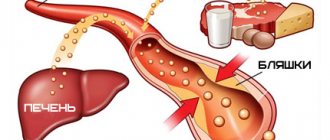
Systemic blood pressure (BP) depends on cardiac output and peripheral vascular resistance. The value of systolic blood pressure is predominantly influenced by the stroke volume of the left ventricle, the maximum rate of blood expulsion from it and the elasticity of the aorta. Diastolic blood pressure is determined by total peripheral vascular resistance and heart rate. Pulse pressure, calculated as the difference between systolic and diastolic blood pressure, reflects the elasticity of the great arteries. It increases with atherosclerotic damage to the arteries. The classification of blood pressure levels is presented in Table 1.
In the group of hypertensive patients with low and average risk of cardiovascular diseases, treatment begins with lifestyle changes: – quitting smoking; – refusal to abuse alcohol; – low-salt diet; – regular exercise in the fresh air; – for obesity – reduction in body weight by at least 5 kg.
The observation period for low risk is 1–6 months, for medium risk – 3–6 months. In the group of patients at high and very high risk, drug treatment is started immediately. In young people, blood pressure should be reduced to 130/85 mm Hg, in older people - to 140/90 mm Hg. Some categories of patients require an even more pronounced reduction in blood pressure. For example, in case of diabetes mellitus it should be reduced to 130/80 mm Hg, in case of kidney disease with proteinuria - to 125/75 mm Hg.
Drug therapy is considered optimal if the hypotensive effect continues throughout the day and the physiological circadian rhythm of blood pressure is maintained. An important criterion for effectiveness is the normalization of morning blood pressure, since stroke and myocardial infarction occur more often in the morning. At 8 a.m., the hypotensive effect of the drug taken in the evening should be at least 50% of the maximum effect. An even more informative indicator of the risk of cardiovascular complications is increased pulse pressure. It is necessary to reduce both diastolic and systolic blood pressure. Elevated systolic blood pressure, 2–4 times higher than diastolic blood pressure, increases the risk of stroke, coronary heart disease and heart failure.
Currently, the drugs of choice for the treatment of hypertension are diuretics, β-blockers, angiotensin-converting enzyme inhibitors (ACEIs), angiotensin II receptor blockers (ARBs), calcium antagonists, α1-blockers, imidazoline receptor agonists.
Basic principles of antihypertensive therapy
Antihypertensive drugs are prescribed in small doses, then titrated over weeks to an effective dose. Such tactics provide the opportunity to approach the treatment of hypertension individually in each specific case, taking into account the characteristics of pathogenesis and concomitant pathology.
– If monotherapy is insufficiently effective, it is advisable to use optimal combinations of drugs with different mechanisms of action. – After normalization of blood pressure, antihypertensive drugs are taken in maintenance doses.
Classification
Drugs that reduce the stimulating effect of adrenergic innervation on the cardiovascular system: 1. Agonists of imidazoline I1 receptors 2. Central a2-adrenergic agonists 3. Receptor blockers: • a-adrenergic blockers; • b-adrenergic blockers; • a, b-adrenergic blockers.
Vasodilators: 1. Calcium channel blockers 2. Potassium channel activators 3. Arteriolar vasodilators 4. Arteriolar and venous vasodilators.
Diuretics (diuretics).
Drugs that affect the functions of angiotensin II: 1. Angiotensin-converting enzyme inhibitors (ACEIs) 2. Angiotensin II AT1 receptor blockers 3. Vasopeptidase inhibitors.
The vasomotor center of the medulla oblongata regulates blood pressure with the participation of presynaptic receptors - imidazoline I1 receptors and a2 adrenergic receptors. These receptors, stabilizing the presynaptic membrane, prevent the release of norepinephrine to pressor neurons, which is accompanied by a decrease in central sympathetic tone and an increase in vagal tone. I1 receptors localized in the ventrolateral nuclei of the medulla oblongata are of primary importance in maintaining normal blood pressure; a2 receptors in the nucleus of the solitary tract play a lesser role. A probable endogenous ligand of imidazoline receptors is the decarboxylated metabolite of arginine, agmantine.
Imidazoline I1 receptor agonists
Drugs belonging to this group - moxonidine, rilmenidine - stimulate imidazoline receptors, which leads to inhibition of the activity of the vasomotor center and the sympatho-adrenal system. Peripheral vascular resistance, cardiac output and, as a result, blood pressure decrease.
It should be remembered that imidazoline receptor agonists can increase bradycardia and inhibition of atrioventricular conduction when used together with beta-blockers.
Despite the fact that imidazoline receptor agonists effectively control blood pressure, the therapeutic potential of this group of drugs (impact on the prognosis of patients with hypertension, rational combination with other drugs) requires further study.
a1-blockers
The mechanism of antihypertensive action of α1-adrenergic blockers (prazosin, doxazosin) is the competitive blockade of α1-adrenergic receptors of vascular smooth muscle cells, which prevents excessive stimulation of these receptors by catecholamines. There is a decrease in peripheral vascular resistance and a drop in blood pressure.
An important property of a1-blockers is their beneficial effect on the lipid profile (increasing the content of anti-atherogenic high-density lipoproteins, reducing atherogenic low-density lipoproteins and triglycerides). When using a1-adrenergic blockers, there is a high risk of developing hypotension of the first dose, compensatory tachycardia, and increased urination (a1-adrenergic receptors of subtype A are located in the prostatic part of the urethra).
β-blockers (BB)
BBs neutralize the influence of the sympathoadrenal system on the heart, which leads to a decrease in heart rate and contractility, and the parameters of cardiac activity are maintained at a level sufficient to ensure full hemodynamics (subject to correct dose selection).
In addition, BBs reduce the activity of the RAAS, blocking the hyperactivity of the juxtaglomerular apparatus of the kidneys, equipped with b1-adrenergic receptors. As a result, the synthesis of renin, which is the initial link of the RAAS, is reduced. Thus, BBs contribute to the reduction of the total amount of angiotensin II, a powerful vasopressor. Abrupt withdrawal of BB can lead to a compensatory increase in renin synthesis, which underlies the development of withdrawal syndrome, therefore, when treating BB, it is necessary to ensure that patients take these drugs strictly.
It is believed that under the influence of BB, the normal function of the baroreceptor apparatus of the sinocarotid zone is restored, which contributes to the antihypertensive effect. Cardioselective b1-adrenergic blockers (bisoprolol, metoprolol, atenolol, etc.) do not reduce peripheral blood flow, since they have virtually no effect on b2-receptors, which is important in the treatment of people with occlusive arterial diseases. Currently, in most cases they are preferred due to poorer tolerability of non-selective BBs (high risk of bronchospasm, decreased muscle blood flow). Cardioselective BBs have little effect on carbohydrate and lipid metabolism, which allows their use in diabetes mellitus and widespread atherosclerosis.
The presence of so-called internal sympathomimetic activity (acetobutol, oxyprenolol, pindolol, etc.) indicates a lesser effect on heart rate at rest and the ability of these drugs to expand the lumen of peripheral arterioles, helping to increase the volume of the vascular bed and reduce blood pressure.
Adrenergic blockers with additional vasodilating properties have found widespread use. Thus, carvedilol blocks not only b1- and b2-receptors, but also a1-receptors of the peripheral vascular bed, which adds to the hypotensive effect. The vasodilating effect of nebivolol is due to the ability of this drug to increase the amount of nitric oxide (NO), a powerful local vasodilator. These drugs are used in patients with peripheral atherosclerosis.
BB in the treatment of hypertension can be preferred in case of concomitant sinus tachycardia, ischemic heart disease, CHF, and supraventricular tachyarrhythmias. It is rational to use the properties of BB if the patient has hyperthyroidism (relief of tachycardia), glaucoma (decreased production of intraocular fluid), migraine (prevention of attacks), hypertrophic cardiomyopathy. In case of liver dysfunction, it is rational to prescribe hydrophilic beta-blockers (atenolol, acebutolol, bisoprolol), which are excreted from the body by the kidneys. If the patient is a smoker, then the doses of fat-soluble BBs should be increased, since in this category of patients the activity of liver enzyme systems increases. In patients with concomitant chronic renal failure, it is advisable to use lipophilic BBs (pindolol, labetolol, timolol, metoprolol, betaxolol, talinolol, etc.).
VASOACTIVE DRUGS
Central antihypertensive drugs act on the adrenergic receptors of interneuron synapses of the vasomotor centers of the trunk, which reduce central sympathetic activation and thereby prevent an increase in blood pressure (BP). These include: agonists of a2-presynaptic receptors - clonidine, methyldopa, guanfacine; the central sympatholytic drug reserpine and other rauwolfia drugs (these VPs act in the same way as peripheral sympatholytics). The main indication for the use of these drugs is hypertension. Clonidine is also used for the interictal treatment of migraine, relief of hot flashes during menopause and the cardiovascular component of withdrawal syndrome. In addition, clonidine reduces hyperkinesis in generalized tics. Centrally acting drugs can cause a sedative effect (lethargy, physical inactivity, drowsiness, especially at the beginning of treatment); with prolonged use, a decrease in memory, libido and impaired ejaculation are possible. Nasal congestion and dry mouth are observed more often only at the beginning of treatment. Methyldopa, as a false precursor of dopamine, reduces its synthesis, and reserpine, as a sympatholytic, reduces its reserves, which can increase the manifestations of parkinsonism with long-term treatment. If clonidine is suddenly discontinued, especially when combined with b-blockers, a hypertensive crisis may develop. Ganglioblockers
lower blood pressure, reduce cardiac stroke volume and peripheral vascular resistance.
Cerebral blood flow remains unchanged or increases slightly because cerebrovascular resistance decreases more than total peripheral resistance. In a neurological clinic, ganglion blockers are used to control arterial hypertension in patients with cerebral hemorrhage, acute hypertensive encephalopathy, and in the event of a crisis in chronic dyscirculatory encephalopathy. For different types of vegetative-vascular dystonia (VSD), the use of “mild” (for example, ganglerone) ganglion blockers leads to equalization of the disturbed balance between sympatho- and parasympathicotonia. Some ganglion blockers (hygronium, pentamine, benzohexonium) are effective for pulmonary edema. Side effect: orthostatic hypotension is observed, therefore, when using these drugs parenterally, patients should remain in bed for 2-3 hours. Possible slowdown of intestinal motility (rarely paralytic ileus), urinary retention, mydriasis, impaired accommodation, dysarthria, dysphagia. These phenomena are reduced with the administration of proserin and carbacholine. Peripheral sympatholytics
(guanethidine, etc.) deplete narepinephrine reserves in the terminals of the neurosmooth muscle junction, moderately block sympathetic ganglia and stimulate b2 receptors of arterial smooth muscle.
Guanethidine reduces cerebral vascular tone. Due to the high risk of orthostatic complications, it is rarely used. Guanethidine is contraindicated in acute cerebrovascular accident (ACVA), myocardial infarction, pheochromocytoma. A relative contraindication to its use is chronic dyscirculatory encephalopathy (DEP). The a-receptor agonist ergotamine has a pronounced vasotonic effect on both arteries and veins with initially low tone, reducing their blood supply by 45%. Improving microcirculation is facilitated by blockade of pathological arteriovenous shunting. Ergotamine is most often used to treat patients suffering from migraine attacks. With an overdose of ergotamine (more than 8-10 mg/day), acute ergotism develops: vomiting, diarrhea, paresthesia, convulsions. With long-term use of the drug at an average therapeutic dose, chronic ergotism develops with peripheral circulatory disorders due to vasospasm. Ischemic necrosis of the soft tissues of the toes is described as a rare complication. Ergotamine is contraindicated in hypertension, atherosclerosis, angina pectoris, peripheral arterial sclerosis, liver and kidney diseases. Dihydroergotamine also has the properties of an α-adrenergic agonist, but it acts more mildly than ergotamine. a -Adrenergic blockers
inhibit the transmission of sympathetic innervation through a-adrenergic systems at different levels and block a-receptors of arterial smooth muscles.
As a result, the tone of the smooth muscles of the arteries decreases, which leads to a decrease in blood pressure, especially in the case of initial arterial hypertension. Indications: arterial hypertension and regional hypertension in the bed of organ arteries with VSD, DEP and stroke. They have a regulatory effect on energy metabolism in the brain. Side effects: dizziness, orthostatic hypotension, headache, general weakness, drowsiness, pain in the heart, frequent urination. Usually observed with an individual overdose, especially at the beginning of treatment (“first dose effect”), they disappear when the dose is reduced and the drug is discontinued. b -Adrenergic blockers
inhibit b-receptors in the central nervous system, nerve endings, smooth muscles of blood vessels and bronchi.
Non-selective b-blockers interact with b1- and b2-receptors, and cardioselective - with b1-receptors of the heart. Drugs of this group with high lipophilicity (alprenolol, metoprolol, oxprenolol, propranolol) penetrate well through the blood-brain barrier, reduce anxiety, agitation, fear, relieve cardiovascular and autonomic-somatic disorders caused by stress, reduce blood pressure, and normalize EEG parameters. b-Blockers slow the heart rate, reduce the force of myocardial contractions, reduce oxygen consumption by the myocardium, inhibit the automaticity of the atrioventricular node and ectopic foci of myocardial excitation, and increase exercise tolerance. They improve the tone and reactivity of brain vessels. These drugs provide a pronounced hypotensive effect in hyperkinetic type arterial hypertension. Indications for the use of b-adrenergic blockers in a neurological clinic are VSD, including sympathoadrenal crises, idiopathic orthostatic hypotension, migraine, DEP with arterial hypertension. When treated with drugs of this group, the mortality rate of patients with spontaneous subarachnoid hemorrhage and ischemic stroke, the frequency of relapses of ischemic stroke and mycardial infarction complicating it, are reduced. They have a sedative effect, stop hemodynamic changes that accompany psycho-emotional stress, and reduce trembling hyperkinesis caused by excitement. Effective in treating patients with withdrawal symptoms. Side effects: bradycardia, angina pectoris, impaired atrioventricular conduction to complete heart block, left ventricular failure and pulmonary edema, cardiogenic or anaphylactic shock. Non-selective drugs cause and intensify bronchospasm. When using blockers with sympathomimetic activity (oxprenolol, pindolol, alprenolol), such complications are less common. Disorders of the nervous system (insomnia, disturbing dreams, hallucinations or depression, muscle pain or fatigue) are observed in 3-15% of cases. Signs of myotonia may appear and signs of myasthenia may increase. More rare complications are fibrosis of the lungs and pleura, blepharitis, conjunctivitis, anorexia, gastralgia. Due to frequent complications, the use of practolol was discontinued. Contraindications for use: severe heart failure, bradycardia, sinus rhythm disturbances, bronchial asthma, intermittent claudication; relative contraindications: moderate heart failure, obstructive pulmonary diseases, depression, hypothyroidism, liver and kidney diseases, diabetes (non-selective b-blockers prolong the effect of insulin). If you suddenly stop taking these drugs, withdrawal syndrome is possible: deterioration of coronary blood supply, pain in the heart, arrhythmia, increased blood pressure. the properties of a- and b- blockers
.
Indications for its use are determined by a combination of these properties. The effectiveness of the drug in the treatment of hypertension-tachycardia syndrome that develops after multiple combined injuries has been established. Side effects are associated with blockade of both a-receptors - orthostatic episodes, dizziness, tinnitus, impaired urination and ejaculation (without decreased libido, erection, with preservation of orgasm), and b-receptors - broncho-obstruction, intermittent claudication, Raynaud's disease, depression with sleep disorders. Serotonin receptor antagonists
(ketanserin, ritanserin), acting primarily on the central nervous system, are used to treat diseases with vasospasm of the peripheral arteries - Raynaud's disease and intermittent claudication.
Cyproheptadine, pizotifen, and inprasochrome are prescribed for the interictal treatment of migraine. Angiotensin-converting enzyme inhibitors (ACEIs) inhibit the formation of the pressor peptide, angiotensin II. ACE inhibitors are used for all types of arterial hypertension, especially of renovascular origin, congestive cardiovascular failure, angiospastic form of Raynaud's disease, DEP with arterial hypertension or congestive heart failure. In these forms, ACE inhibitors are more effective than sympatholytics acting on sympathetic endings, a-blockers and calcium antagonists. The combination of ACE inhibitors with other antihypertensive drugs, including calcium antagonists, b-blockers and diuretics increases their pharmacotherapeutic effectiveness. Nonsteroidal anti-inflammatory drugs (NSAIDs), especially indomethacin, reduce the antihypertensive effect of ACEIs. However, it should be noted that taking acetylsalicylic acid as an antiplatelet agent (at a dose of 100-300 mg) does not affect the effectiveness of ACE inhibitors. Drugs that act primarily on
vascular smooth muscle depending on the effect on enzyme systems: adenylate cyclase (AC), phosphodiesterase (PDE) belong to different pharmacological classes.
In practice, it is these drugs with predominantly myotropic action that are often called vasoactive drugs (“antispasmodics”, “vascular” myolytics). Isoquinoline derivatives
- papaverine and drotaverine - exert their action by activating AC and inhibiting PDE in vascular smooth muscle cells and have a moderate ganglioplegic effect.
Papaverine dilates arteries and veins. The latter can worsen venous outflow from the cranial cavity, which, given the initially reduced venous tone, can be considered an unfavorable effect. Drotaverine does not reduce the tone of the veins. Derivatives of vicamine
(an alkaloid from plants of the periwinkle family) - vinpocentine.
This drug activates AC to a greater extent and moderately inhibits PDE. Vinpocetine selectively improves cerebral hemodynamics and can be considered as an “optimizer” of vascular tone, providing a selective antispastic effect on cerebral vessels, or restores vascular tone in case of its initial decrease. The drug normalizes the rheological properties of blood and improves microcirculation. Vinpocetine has a direct neurometabolic cerebroprotective effect and has an antihypoxic and antiparoxysmal effect. An important feature of vinpocetine is the lack of influence on systemic hemodynamics and the absence of “steal” syndrome. Vinpocetine is used for both initial and severe forms of cerebrovascular insufficiency, transient cerebral ischemia, all forms and stages of stroke (except for the acute phase of hemorrhagic stroke), vasovegetative disorders (including endocrine origin). as well as for the prevention of convulsive syndrome in children who have suffered a traumatic brain injury. Xanthine derivatives
- caffeine, theobramine, theophylline, aminophylline, pentoxifylline - can also be considered as “optimizers” of vascular tone, but they have a more clearly visible venotonic effect (improving the outflow of venous blood from the cranial cavity).
In addition, these drugs activate breathing, increase heart rate (HR), and diuresis. These VPs improve the rheological properties of blood. Calcium antagonists
(Ca2+ channel blockers) have the ability to have antispasmodic, antihypertensive, and coronary effects.
The group of phenylalkylamines is represented by verapamil, phendiline, and difril. The group of dihydropyridines includes nifedipine, foridone, nicardipine, nitrendipine, isradipine, nimodipine. Among these VPs, one can distinguish adalat in capsules (liquid dosage form) and tablets, which can provide a rapid antihypertensive effect, and nimodipine, which acts on the cerebral arteries to a greater extent than other Ca2+ antagonists. Peripheral vasodilators
- hydralazine, sodium nitroprusside, milsidomine, minoxidil - have a pronounced peripheral antispasmodic effect, reduce venous tone, and reduce venous return to the heart.
They are rarely used in neurological practice. The use of vasoactive drugs in the treatment of VSD
VP is used against the background of adherence to healthy lifestyle standards, treatment with tranquilizers or antidepressants. Patients with VSD with persistent manifestations of systemic hypertension and arterial hypertension are prescribed drugs that inhibit central sympathetic activation (clonidine, methyldopa, reserpine), ganglion blockers, a- and b-adrenergic blockers. To regulate regional vascular tone, myotropic antispasmodics, vinca preparations, dibazol, a-blockers, and calcium antagonists are used. For systemic hypotension and regional hypotension, ergotamine and drugs containing it, other sympathomimetics - ephedrine, fethanol, phenylephrine (Mezaton), as well as anabolic and steroid hormones are prescribed. In the case of predominant venous hypotension, drugs of the xanthine series, periwinkle, and a-stimulants are indicated. For mixed forms of VSD, combination drugs are effective - bellataminal, belloid, bellaspon. In all cases, drugs that improve metabolic processes in the central nervous system are useful: aminalon, pyriditol, piracetam, vitamin therapy (B1, B6, C, PP). Non-drug treatments include acupuncture and various methods of physiotherapy.
The use of vasoactive drugs in the treatment of DEP
DEP is a slowly progressive insufficiency of blood supply to the brain, accompanied by small focal changes in brain tissue. The main etiological factors of DEP are hypertension and atherosclerosis, systemic vascular diseases, especially those affecting the aortic arch and the great vessels of the head extending from it. In the vast majority of cases, progression of DEP occurs during episodes of cerebral circulatory decompensation. During intensive treatment of a crisis associated with increased blood pressure, the choice of antihypertensive drugs should be adequate to the severity of the crisis (preference is given to fast-acting drugs); Blood pressure should not be reduced below the patient’s usual level; it is necessary to choose a method of drug administration that provides a rapid but smooth and controlled decrease in blood pressure (usually intravenous drip infusion) and take into account the possible side effects of fast-acting drugs; minimize the risk of complications. VP is selected depending on the type of cerebral angiodystonia. For hypertonicity of the arteries, drugs with a predominant antispasmodic effect are prescribed; for symptoms of dystonia and hypotension of the cerebral arteries and veins, preference is given to vinpocetine, aminophylline, and trental. Ischemic cerebral crisis
In patients with DEP against the background of atherosclerosis, it develops as a type of cerebral circulatory failure. It may result from a decrease in the pumping function of the heart and a decrease in blood pressure, increased blood viscosity, and increased activity of the coagulation system. In these cases, adding small doses of cardiac glycosides (corglycon) to the therapy is effective. In case of crisis against the background of hypercoagulation, heparin administration is indicated. Of the indirect-acting anticoagulants, preference is given to those that exhibit a lesser tendency to accumulate: syncumar, pelentan, phenylin. With long-term (many months) prescription of VP for the treatment of DEP outside of exacerbation, effective medications are selected individually. Unfortunately, in practice this means an empirical approach (trial and error). If conditions exist, we can recommend choosing an individually optimal VP using an acute pharmacological test. It consists of sequential administration of therapeutic doses of each of the tested vasoactive agents once a day (screening). In this case, after intravenous administration of the drug, the patient’s condition is monitored and synchronous recording of blood pressure, pulse, REG, and EEG is carried out within 1 hour. Each of the other tested EPs is administered the next day. For therapy, a drug is selected that, during an acute test, caused the most favorable changes in the recorded parameters. Such studies can be carried out in a functional diagnostics room. Pharmacotherapy with means of individual choice increases the effectiveness of treatment and reduces its duration.
The use of vasoactive drugs in the treatment of stroke
It is beyond the scope of this article to describe in detail the intensive care of hemorrhagic and ischemic stroke. The use of VP as part of the combined treatment of stroke is certainly not of decisive importance. Monotherapy of VP in the acute stage of stroke cannot be considered adequate; VP must be combined with other means of pathogenetic treatment; in the acute phase of stroke, parenteral administration of VP should be considered effective; in a daily intensive care program, their repeated administration should be carried out depending on the duration of action of a single dose (for most VP, 3 times a day). In the first days after the abolition of acute stroke due to the loss or decrease in vascular reactivity, the administration of VP may not be accompanied by a change in the clinical condition or electrophysiological parameters. The absence of these signs does not indicate the ineffectiveness of the VP. The assessment of VP activity is facilitated by their administration in the intervals between the introduction of other agents of pathogenetic therapy and the dynamic observation of their effect on the patient’s condition and synchronously recorded blood pressure, heart rate, ECG, REG and EEG. When choosing the optimal drug in the first days after stroke, screening for CAP is justified; in the most acute stage, to obtain a faster effect, it is justified to administer VP into a vein in a single drip system with cardiotonic, decongestant (dehydrating), hemorheological drugs, hemodilution agents, antifibrinolytics and anticoagulants. When carrying out complex intensive therapy, simultaneous administration of drugs with opposite pharmacodynamic properties, administration of drugs with a similar pharmacodynamic effect (due to the unpredictability of the potentiating effect) or incompatible drugs (for example, heparin + Cavinton) should be avoided. Identification of the “penumbra zone” on a computer or magnetic resonance imaging scan (a perifocal area with brain perfusion at a pre-functional level) serves as the basis for continuing intensive therapy for VP and other means of combined pathogenetic treatment. Thus, the use of VP in complex stroke therapy should be considered not only justified, but also necessary. At the same time, the assessment of their action should not be limited to identifying only the vasomotor effect. Each of the drugs of this pharmacological class usually improves blood circulation and functional activity of the brain, since, although to varying degrees, EPs provide an indirect (through improved blood circulation, protection against ischemia) and direct nootropic effect due to the normalization of the metabolism of the affected brain.
Literature:
1. Mashkovsky M.D. Medicines, in 2 parts, part I - M.: Medicine, 1993 297-340, 369-370, 502-560. 2. Shtok V.N. Medicines in angioneurology. - M.: Medicine, 1984; 303 pp. 3. Shtok V.N. Pharmacotherapy in neurology. M.: Medicine, 1995; 10-28, 81-107. 4. Vidal reference book. Medicines in Russia. M.: AstraPharmServis, 1997.
Calcium antagonists (slow calcium channel blockers)
According to the classification according to B. Nauler, all calcium antagonists (CA) are divided into 3 groups: derivatives of dihydropyridines (nifedipine, isradipine, amlodipine, etc.), benzothiazepines (diltiazem), phenylalkylamines (verapamil). AA limit the flow of Ca2+ ions into the cell, reducing the ability of the muscle fiber to develop contraction. Due to the limitation of the entry of Ca2+ ions into the cell, 3 main effects develop, to one degree or another characteristic of all AKs: a decrease in myocardial contractility (negative inotropic effect), a decrease in the tone of smooth muscles of the arteries (vasodilating effect), a change in the excitation threshold of cardiomyocytes of the conduction system (characteristic of pulse – reducing AKs – verapamil and diltiazem). It is known that dihydropyridine AKs can increase heart rate, especially at the initial stage of treatment.
AKs reduce the tone of arterioles, which is mainly responsible for their antihypertensive effect. Due to this, renal blood flow increases in parallel, which provides a small natriuretic effect, complemented by a decrease in aldosterone formation under the influence of AA. Blocking Ca2+ ions at the level of platelets leads to a decrease in their aggregation readiness.
Being highly active agents, AKs have a number of advantages, which are often called “metabolic neutrality”: the drugs of the group do not affect lipid, carbohydrate, mineral and purine metabolism. Since AKs improve coronary and cerebral blood flow, their use is justified in hypertension with concomitant coronary artery disease or cerebrovascular insufficiency.
Calcium antagonists[edit | edit code]
When the cell membrane of cardiomyocytes or smooth muscle cells is electrically excited, changes in ionic currents occur, including the influx of calcium. Calcium antagonists block its entry into the cell without affecting the influx of sodium or outflow of potassium. These drugs are also called calcium entry blockers or calcium channel blockers. Calcium antagonists are divided into two groups according to their therapeutic effect: those acting on the heart and those acting on the blood vessels.
I. Dihydropyridine derivatives
. Dihydropyridines, such as nifedipine, are uncharged hydrophobic substances. They relax the walls of the arteries and in a therapeutic dose have virtually no effect on the function of the heart (in an experiment on an isolated heart they have a clear effect). These drugs are called vasoselective calcium antagonists. Vasodilation and a decrease in peripheral vascular resistance lead to a decrease in blood pressure. Afterload on the heart and oxygen consumption are reduced. The development of coronary spasm is prevented.
Indication
The purpose of prescribing nifedipine is not only the prevention, but also the relief of angina attacks.
Side effects
: palpitations (reflex tachycardia due to decreased blood pressure), headaches, and swelling of the legs.
Other dihydropyridine derivatives have similar effects and differ in pharmacokinetic behavior (slower elimination) and more stable blood concentrations.
Nitrendipine, isradipine and felodipine are used to treat hypertension. Nicardipine and nisoldipine are prescribed for angina pectoris. Nimodipine prevents vasospasm after subarachnoid hemorrhage. Amlodipine contains protonated nitrogen in the side chain and therefore can be in cationic form (i.e., carry a positive charge), which explains its very long half-life (t1/2 = 40 hours).
II. Verapamil and other cationic amphiphilic calcium antagonists
. Verapamil at physiological pH has a positive charge on the nitrogen atom and is a cationic amphiphilic molecule. It has an effect not only on smooth muscles, but also on the heart. The influx of calcium causes depolarization in the sinus node of the heart (electrical excitation), in the atrioventricular node (conducting excitation from the atrium to the ventricles), as well as in cardiomyocytes (electromechanical work). Therefore, verapamil has negative chronotropic, dromotropic and inotropic effects.
Indications
. Verapamil is an antiarrhythmic agent and is indicated for supraventricular tachyarrhythmias. During atrial fibrillation and flutter, it inhibits the conduction of impulses through the AV node and weakens ventricular contractions. Verapamil is also used to prevent angina pectoris and treat hypertension.
Side effect.
Reflex tachycardia as a result of exposure to the sinus node is not observed; the heart rate does not change or there may be a tendency towards bradycardia. AV block or heart failure may develop. Patients often complain of constipation, as verapamil blocks intestinal activity.
Gallopamip (a methoxy derivative of verapamil) is very similar in structure and action to verapamil.
Diltiazem (a benzodiazepine derivative) is a cationic amphiphilic compound and has similar effects to verapamil.
Diuretics
The mechanism of the antihypertensive action of diuretics is associated with the ability of the drugs to reduce the volume of circulating fluid, primarily by reducing the reabsorption of sodium ions in the renal tubules. As a result of a decrease in fluid load, the total peripheral vascular resistance and blood pressure levels decrease. The hypotensive effect of diuretics is complemented by the ability of these drugs to reduce the sensitivity of the vascular wall to natural vasopressors (including adrenaline), in the maintenance of which sodium ions participate.
The most widely used antihypertensive drugs are thiazide and thiazide-like diuretics: hydrochlorothiazide, chlorthalidone, indapamide, etc. Loop diuretics are used only to relieve hypertensive crises. Long-term use of any diuretics is dangerous for the development of electrolyte imbalance, therefore, when prescribing them, it is recommended to monitor the content of electrolytes in the blood plasma. When using thiazide and thiazide-like diuretics daily in small doses, the risk of complications of therapy is minimized without a significant loss of the necessary hypotensive effect.
Thiazide and thiazide-like diuretics may be preferred for isolated systolic hypertension in older people, with concomitant CHF, and in women with hypertension in the perimenopausal period. Diuretics are convenient to supplement already prescribed treatment regimens to achieve the target blood pressure level.
Newspaper "News of Medicine and Pharmacy" 3-4 (269-270) 2009
Multicenter studies in recent years have shown that β-blockers and diuretics (individually or in combination) in the treatment of patients with arterial hypertension (AH) lead to an increased risk of new cases of diabetes mellitus.
This position is reflected in the new European (2007) and national (2008) Guidelines for the treatment of hypertension, which indicate that β-blockers should not be prescribed to patients with concomitant metabolic syndrome. The only exceptions are vasodilating drugs - nebivolol and carvedilol.
In addition, as is known, the majority of patients with hypertension have increased total peripheral vascular resistance. Therefore, other things being equal, for long-term therapy in most patients with and without metabolic syndrome, the use of drugs with vasodilating properties is preferable.
The mechanisms of vasodilatory action of β-blockers are different: 1) pronounced intrinsic sympathomimetic activity (ISA) (pindolol and celiprolol); 2) blockade of a 1 -adrenergic receptors (carvedilol and labetolol); 3) release of nitric oxide from endothelial cells, which has vasodilating properties (nebivolol); 4) stimulation of β3-adrenergic receptors (nebivolol).
The mechanism of the vasodilatory action of vasodilating β-blockers is of clinical importance. It is generally accepted that vasodilating β-blockers with pronounced ICA do not have a cardioprotective effect. Therefore, their use is less preferable or even dangerous in high-risk patients, for example, in patients with CHF or patients who have had a myocardial infarction.
Since the treatment of hypertension has traditionally favored cardioselective beta-blockers and non-BCA drugs, nebivolol should now be considered as the drug of choice in this class for the majority of patients with high blood pressure.
It has been established that the vasodilatory effect of the superselective β-blocker nebivolol is associated with the effect of the drug on the arginine-citrulline-nitric oxide (NO) system in the vascular endothelium. Studies in animals, healthy subjects and patients with hypertension have shown that nebivolol increases the expression of the gene responsible for the synthesis of NO synthetase, which leads to an increase in the formation and release of nitric oxide from endothelial cells. The cellular mechanisms of the vasodilatory effect of nebivolol are not yet fully understood. Several mechanisms have been suggested through which a clinically significant increase in NO synthesis is realized in the vascular endothelium: by stimulation of vascular β2- and β3-adrenergic receptors, by stimulation of serotonin receptors of the 5-HT1A type, or by interaction with mechanisms mediated by estrogen receptors. Nebivolol, as a substance with proven antioxidant properties, also appears to reduce the inactivation of nitric oxide by free radicals. As a result of reduced degradation of nitric oxide, its bioavailability increases.
A superselective β1-adrenergic blocker, such as nebivolol, has a minimal blocking effect on β2-adrenergic receptors and a moderate stimulating effect on β3-adrenergic receptors, mediating, in particular, endothelium-dependent vasodilation (including in the cavernous bodies of the penis), as well as lipolysis and thermogenesis in the brown tissue. adipose tissue. As a result, nebivolol (unlike atenolol, metoprolol, bisoprolol and carvedilol) does not cause erectile dysfunction in men with hypertension. The drug does not contribute to weight gain and has a positive effect on lipid and carbohydrate metabolism. Therefore, it can be considered a first-line β2-blocker for the long-term treatment of sexually active men, as well as patients with metabolic syndrome and type 2 diabetes, not to mention patients with chronic obstructive pulmonary disease or peripheral atherosclerosis.
Thus, nebivolol has five very important clinically important properties that distinguish it from the currently available β-blockers:
1) vasodilating effect;
2) maximum β1-selectivity in the class;
3) absence of ICA;
4) high lipophilicity;
5) a positive effect on lipid and carbohydrate metabolism.
In addition, nebivolol has an antioxidant effect and inhibits platelet aggregation. No other β-blocker has all of the above properties at the same time. These properties fundamentally distinguish nebivolol from all other cardioselective β-blockers, which since 2007 have been called traditional.
Obviously, all of the above-mentioned properties of the drug led to its successful entry into the US market in 2008 and the appearance of a number of enthusiastic publications in the American scientific press.
In conclusion, there are 16 potential stereochemical isomers of nebivolol. The molecule of the original drug Nebilet consists of 10 (5 dextro- and levorotatory) isomers in strictly defined proportions. It is from this point of view that it becomes clear why only the original drug has all the above-described properties and why in everyday clinical practice it is often not possible to achieve clinical efficacy comparable to Nebilet with other nebivolol drugs.
Medicines affecting the renin–angiotensin system
The renin–angiotensin–aldosterone system (RAAS) plays an important role in the regulation of blood pressure, cardiac activity, and water and electrolyte balance. Its activity increases with arterial hypertension, chronic heart failure and diabetic nephropathy. In acute myocardial infarction, the activity of the RAAS increases already on the first day; in complicated myocardial infarction, excessive activation of the RAAS persists long after the patient is discharged from the hospital. High renin activity and increased levels of angiotensin II in the blood are indicators of an unfavorable prognosis in patients with cardiovascular diseases.
For pharmacological blockade of the RAAS, ACE inhibitors and non-peptide angiotensin II receptor antagonists are used.
Vasodilators (vasodilators)[edit | edit code]
Source:
Visual Pharmacology
.
Author
: X. Lulman.
Per. with him. Ed.
: M.: Mir, 2008
The lumen of blood vessels regulates the distribution of blood in the vascular bed. The lumen of the veins determines the blood flow to the heart, and, consequently, cardiac output and cardiac output (CMV). Peripheral vascular resistance is determined by the lumen of the arteries. The cardiac output and peripheral vascular resistance determine the value of blood pressure.
In Fig. And the most important vasodilators are presented, the order of their arrangement approximately corresponds to the frequency of use. The listed drugs have different effects on the arterial and venous beds.
Application
. Arterial vasodilators: lowering blood pressure in hypertension, reducing the strength and frequency of heart contractions in angina, reducing afterload on the heart in heart failure. Venous vasodilators: reducing preload on the heart in angina or heart failure. The practical use of individual drugs is discussed further by group of drugs.
Counterregulation when blood pressure falls due to vasodilation (B). As a result of activation of the sympathicus, the heart rate increases (reflex tachycardia) and, accordingly, the cardiac output (MV) increases. The patient complains of rapid heartbeat. Activation of the renin-angiotensin-aldosterone (RAA) system leads to an increase in blood volume (fluid retention is observed, edema is possible) and MOS.
Counter-regulatory pathways are blocked by various drugs (beta blockers, ACE inhibitors, angiotensin II antagonists, diuretics).
Mechanism of action[edit | edit code]
There are many ways to influence smooth muscle tone.
Protection against vasoconstriction
: ACE inhibitors, angiotensin receptor antagonists, α-adrenergic agonists protect against the effects of humoral mediators such as angiotensin and (nor)adrenaline. Bosentan (see below) is an endothelin receptor antagonist that can be released from the endothelium as a potent vasoconstrictor.
Replacement of vasodilators
: analogues of prostacyclin (iloprost) or prostaglandin E1 (alprostadil) act on the corresponding receptors, organic nitrates replace NO in the endothelium.
Direct effect on vascular smooth muscle cells
: Calcium antagonists, acting at the channel level, potassium channel activators (diazoxide, minok, satil) cause membrane depolarization and relaxation of smooth muscles. Phosphodiesterase inhibitors (PDE) inhibit the destruction of intracellular cAMP, which reduces vascular tone. There are many PDE isoenzymes with different localization and functions.
Erectile dysfunction.
Zildenafil, vardenafil, tardapafil are inhibitors of phosphodiesterase PDE-5 and promote erection. During sexual arousal, NO is released from the nerve endings in the corpus cavernosum of the penis, which causes the formation of cGMP in the smooth muscles of the blood vessels. cGMP is destroyed in the corpus cavernosum by the enzyme PDE-5, which is important for this tissue, which prevents erection. PDE-5 blockers “preserve” cGMP.
Pulmonary hypertension
. We are talking about a narrowing of the pulmonary vascular bed of unknown origin. The disease often progresses, overloads the right ventricle of the heart and is almost impossible to treat with vasodilators. An example of a new therapeutic agent is the endothelin antagonist bosentan. Inhalation administration of N0 is also being tested.
Angiotensin-converting enzyme inhibitors
ACE inhibitors are a group of drugs that affect numerous pathological links, leading to functional and structural changes that underlie various diseases of the cardiovascular system. The mechanism of action of ACE inhibitors is the binding of zinc ions in the active center of the angiotensin-converting enzyme, the key enzyme of the RAAS, and blocking the reaction of the transition of angiotensin I to angiotensin II, which reduces the activity of the RAAS both in the systemic circulation and at the tissue level (kidneys, myocardium, brain). In parallel, due to ACE inhibition, the degradation of bradykinin is inhibited, which also promotes vasodilation. As a result, systemic arterio- and venodilation occurs, pre- and afterload on the heart decreases, and in the presence of left ventricular myocardial hypertrophy, the process of its reverse development begins (cardioprotection). A similar process is observed in the muscle layer of arterial vessels (angioprotection). ACE inhibitors inhibit the proliferation of mesangial cells in the kidneys, which is used in nephrology (nephroprotection). Decreased aldosterone production leads to decreased reabsorption of sodium and water in the proximal and distal tubules of the nephrons.
ACE inhibitors are preferred if a patient with hypertension has concomitant CHF, post-infarction cardiosclerosis, diabetes mellitus and its complications (including nephropathy). The ability of ACE inhibitors to restore endothelial function is used when the patient has dyslipidemia and diffuse atherosclerotic lesions. When prescribing ACE inhibitors, the dose of other antihypertensive drugs (especially thiazide diuretics) should be adjusted if the patient is taking them. The decrease in blood pressure due to ACE inhibitors in most cases occurs smoothly over several weeks. Despite the fact that ACE inhibitors are quite well tolerated in most cases, you should always remember the possibility of developing the following adverse reactions characteristic of this group of drugs: dry cough, hyperkalemia and impaired renal function, angioedema (at any time of treatment).
ACE inhibitors are contraindicated during pregnancy due to the risk of teratogenies, unilateral or bilateral renal artery stenosis, aortic stenosis, mitral stenosis, and obstructive hypertrophic cardiomyopathy.
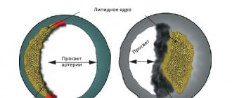
Obliterating atherosclerosis of the arteries of the lower extremities currently ranks third in the frequency of organ localization after damage to the coronary arteries and arteries of the brain. The increase in the number of patients puts this pathology among the most pressing problems of modern medicine, acquiring social significance [9].
In addition to widespread damage to the arteries of the lower extremities, the effectiveness of restoring blood circulation in the limb after reconstructive operations is influenced by the degree of damage to the microvasculature in ischemic tissues. Incompetence of capillary circulation is one of the leading factors in the development of severe trophic disorders in patients with this nosology [10].
Since in obliterating diseases of the arteries of the lower extremities, critical ischemia of soft tissues and, as a consequence, loss of a limb can be caused by both disruption of blood flow through the great vessels and irreversible changes in the microvasculature, the study of microcirculation of peripheral tissues is currently the most relevant. In this case, the study of microcirculation in severe forms of chronic arterial insufficiency of the lower extremities is of particular importance [11].
The purpose of the work is to assess the reserve capabilities of the peripheral microcirculatory blood flow (MCB) in patients with multifocal atherosclerosis, depending on the stage of ischemia of the lower extremities.
Material and methods
The study included 118 men (average age 60.8±0.9 years) with multifocal atherosclerosis, obliterating atherosclerosis of the arteries of the lower extremities, chronic ischemia of the lower extremities (CLI) stages IIB-IV according to the Pokrovsky-Fontaine classification. Exclusion criteria were the presence of chronic venous insufficiency and diabetes mellitus.
All patients were divided into two groups: group 1 included 68 patients with stage IIB CLI, and group 2 included 50 patients with stage III-IV CLI. The average age of patients in group 1 was 60.8±0.8 years, in group 2 61.4±1.1 years. The groups were comparable in age and anthropometric indicators.
In all patients, according to angiography, surgically significant damage to the arteries of the lower extremities was established. All patients were admitted to the Federal State Budgetary Institution NNIIPK named after. acad. E.N. Meshalkin of the Ministry of Health and Social Development of Russia for planned surgical treatment in the scope of direct and indirect revascularization. Before surgical treatment, the patients underwent laser Doppler flowmetry (LDF) of the lower extremities using functional tests, the purpose of which was to determine the functional state of the MCB depending on the stage of ischemia of the lower extremities.
Before surgical treatment, patients also underwent a general clinical blood test, a biochemical blood test with determination of lipid composition, and a rheological blood test (prothrombin index - PTI, blood clotting time).
Registration of the peripheral MCB of the lower extremities was carried out before surgery using a laser Doppler flowmeter (USA) with a set of surface sensors of the R type (rite angle) in combination with a personal computer. The microcirculation study was performed with the patient lying down at an ambient temperature of 24 °C and physical rest with preliminary rest for 15 minutes on the plantar surface of the nail phalanx of the first toe. When studying LDF indicators, the arithmetic mean value of the microcirculation indicator and its standard deviation (RMS) were determined. Since the LDF-gram is a superposition of a large number of different oscillatory processes performed by erythrocytes in the tissues under study, the value of the standard deviation is another important characteristic of the state of tissue blood flow, which determines the variability of microcirculation. The standard deviation index was calculated in each individual case and then averaged.
To assess the functional state, as well as determine the reactivity and reserves of the microcirculatory bed, functional tests were used, representing an artificially induced load on the vessels: occlusive and respiratory. These tests reflect pathophysiological disorders in vascular diseases of the lower extremities.
The occlusion test makes it possible to assess the functional reserves of the capillary bed and the reactivity of the smooth muscle cells of the precapillary unit [2, 6]. To conduct an occlusion test, a cuff from a blood pressure measuring device was placed on the lower leg area, the initial level of blood flow was recorded for 1 minute, then occlusion was created by inflating the pressure in the cuff to a level of 200 mm Hg. within 3 min. After cessation of occlusion, LDF recording continued until microcirculation was completely restored to the initial level. Using an occlusion test, we assessed the capillary blood flow reserve (CBF), which as a percentage of the initial level shows the increase in blood flow in the phase of post-occlusion hyperemia: (MCBmax - MCCbackground) ×100%/MCBbackground, where MCCbackground is the initial value of microcirculatory blood flow; MCBmax is the maximum value of microcirculatory blood flow recorded during the period of post-occlusion hyperemia. The MCKmax parameter, recorded after a three-minute occlusion, characterizes the degree of preservation of the NO synthase vasodilation mechanism [15] and is normally, according to various authors [2, 4], about 300%.
The time to reach MCCmax (s) from the moment the occlusion was removed was also calculated. This parameter characterizes the reactivity of precapillary microvessels [2]. According to the literature [7], MCCmax values in healthy individuals are achieved in the first 25–40 s after the cessation of occlusion.
The breathing test serves to assess the function of the reflex activity of sympathetic fibers, since it is known that deep inspiration is accompanied by activation of vasoconstrictor efferent sympathetic fibers and a decrease in perfusion [8]. When carrying out a breathing test, the background value of the volumetric blood flow velocity (MVV) was recorded for 1 minute, then, without interrupting the recording, the reaction of the MVV during a 15-second breath-hold at the height of a deep inspiration, after which the subject exhaled and gradually restored calm breathing within 1 min. The results were assessed by two parameters - the initial background MCB at rest and the level of decrease in MCB during a respiratory test in relation to the initial background, expressed as a percentage - ΔMBV. The reactivity of blood flow during a respiratory test was assessed using the formula: ΔMBV = MCBmin / MCBfon × 100%, where MCBmin is the minimum value of MCB recorded during the period of deep inspiration, and MCBfon is the initial value of MCB. According to the literature [2], in healthy people, the level of MCC in the skin of the pad of the first finger of the lower limb at the height of inspiration in relation to the initial background should be about 40%.
Statistical data processing was carried out using the Statistica 6.0 software package. The results are presented as means and error of the mean. Student's t-test was used to determine differences between groups, and Pearson's coefficient was used to conduct correlation analysis. Differences were considered statistically significant at p<0.05.
results
When analyzing the state of the peripheral MCB, it was revealed that on the affected limb in patients of group 2, the MCB level was statistically significantly lower than in group 1 (p <0.05), and amounted to less than 5 ml/100 g/min. It should be noted that the average SD, which characterizes blood flow variability, was also statistically significantly lower in the same group of patients (see table).
Carrying out a functional test allows you to assess the state of the reserve capabilities of the peripheral MCC. After the cessation of occlusion, a hyperemic reaction should normally be observed within 1 minute, which is due to the ability of the microvasculature to dilate in response to an ischemic stimulus. In our study, a hyperemic reaction in the first minute after vascular occlusion was observed more often in group 1 (41%; p<0.05) than in group 2 (24%). The maximum MCB was statistically significantly higher in patients of the same group. Moreover, in patients of both groups, MCBmax was recorded only at the 2nd minute of blood flow restoration (94.0±6.0 and 91.8±10.8 s, respectively; p>0.05). RCC indices in the 1st group of patients were on average higher than in the 2nd group (p>0.05).
Thus, the lowest reserve of microvascular vasodilation and the indicator of blood flow variability were observed in patients of the 2nd group with stage III-IV CLI; in the same group, the frequency of detection of post-occlusion hyperemic reaction in the 1st minute was lower than in the 1st group of patients with HINK stage 2B.
When analyzing a respiratory test reflecting a reflex increase in the neurogenic component of basal vascular tone, it was revealed that in patients of group 1 at the height of inspiration, the MCB on the affected limb was 51.1 ± 3.9% of the initial one, and this characterizes a moderate decrease in microvascular reactivity to sympathetic stimulus. Moreover, in this group, 2 (3%) patients showed a paradoxical reaction, expressed in an increase in the level of MCB at the height of inspiration.
In group 2, a paradoxical reaction was observed in 11 (22%) patients, as a result of which the average MCB values at inspiratory height amounted to 158.0±43.3% of the initial value and were statistically significantly higher than in group 1 ( see . drawing).
Figure 1. Indicators of microcirculatory blood flow (MCB) during a breath-hold test. The original MCC background is taken as 100% (dashed line); *— p<0.05 for intergroup comparison.
In the 1st minute of recovery, the MCB indices as a percentage of the initial background in the 1st group approached the initial level, while in the 2nd group the MCB remained higher than the initial level and was statistically significantly different from that in the 1st group (p< 0.05).
A comparative analysis of biochemical blood parameters with determination of lipid composition did not reveal statistically significant differences between the groups. At the same time, the lipid status of patients in both groups was disturbed: in the 1st and 2nd groups, the content of triglycerides was 1.88±0.11 and 1.92±0.23 mmol/l, respectively, and low-density lipoproteins - 3.75± 0.23 and 3.51±0.17 mmol/l, atherogenic coefficient - 4.95±0.24 and 4.72±0.29 rel. units
When comparing the indicators of a general clinical blood test, it was revealed that in the 2nd group of patients with stage III-IV CLI, the average platelet content was statistically significantly higher than in the 1st group (285.9±15.3 and 241.8±10. 7% respectively; p<0.05). No statistically significant differences were found in other indicators of the general clinical blood test. When comparing the rheological status of the blood, no statistically significant differences were revealed between the groups. The average PTI value in groups 1 and 2 was 89.8±2.2 and 90.1±1.5%, respectively, blood clotting time was 5.0±0.1 and 4.8±0.1 min respectively.
Thus, in patients of group 1 with stage IIB CLI, a moderate decrease in the reactivity of microvessels to a sympathetic stimulus was noted. In patients of the 2nd group with stage III-IV CLI, a disturbance in the reactivity of the microcirculatory unit to a respiratory test was revealed in the form of a paradoxical reaction of venules and arterioles.
When conducting a correlation analysis in the general group of patients, it was revealed that PTI has a statistically significant inverse correlation with RCC (r = –0.44), i.e. the higher the PTI, the lower the MCC reserve. In addition, a statistically significant direct correlation of the standard deviation with the MCMmax was revealed (r = 0.67), i.e. the higher the variability of microcirculation, the higher the ability of microvessels to dilate in response to an ischemic stimulus.
Discussion
Using the photoplethysmography method, the pathogenesis of microcirculation disorders in patients with CLI was studied. These works revealed that as the disease progresses, the pressure in the arterial and venous sections of the capillary equalizes and capillary stasis occurs, which is the central pathogenetic link of CLI, while a significant decrease in the partial pressure of oxygen is recorded [14]. In our study, the above disturbances in the microcirculatory system are expressed in the lowest volumetric velocity of peripheral blood flow in patients with stage III-IV CLI.
However, microcirculation disorders are caused not only by damage to the capillary bed, but also by pronounced disturbances in the hydrodynamics of the blood. The deforming ability of red blood cells decreases. Their rigidity, along with a slowdown in the speed of blood flow, leads to dynamic aggregation, which further aggravates tissue ischemia [3]. In our work, there were no statistically significant differences in clinical blood test parameters between patients with stage IIB and stage III-IV CLIN, except for platelet content. It should be noted that platelets are the first to respond to the rupture of an atherosclerotic plaque and form the basis for the formation of an arterial thrombus [5]. This should be taken into account to prevent complications of atherosclerosis.
In chronic obliterating diseases of the arteries of the lower extremities, morphofunctional changes in blood microcirculation occur, including structural changes in microvessels. As ischemia progresses, narrowing of the lumen of microvessels, deformation of venules, changes in the shape of capillary loops, increased tortuosity, and signs of unevenness of the lumen of capillaries intensify [1]. At the same time, it is undeniable that endothelial dysfunction is the first link in the development of atherosclerosis. Dysfunction of the vascular endothelium is accompanied by changes in the content of endothelins, von Willebrand factor, tissue plasminogen activator and nitric oxide (NO). Along with a decrease in the concentration of vasodilators, there is an increase in the level of vasoconstrictors and procoagulants [12].
As is known, reactive post-occlusive hyperemia serves as a protective adaptive reaction, in which blood flow is quickly restored after a period of its cessation. Impaired endothelial function together with structural changes in the microvasculature, as well as changes in blood hydrodynamics, will inevitably lead to a decrease in the reserve capacity of the MCC. In our study, the low vasodilator reserve was reflected in a mild hyperemic reaction after the cessation of occlusion according to MCC data in both groups of patients. At the same time, in patients with stage III-IV CLI, a hyperemic reaction in the 1st minute after vascular occlusion was observed less frequently than in the group of patients with stage IIB CLI. The maximum MCB volumetric velocity was statistically significantly lower in patients of the same group, and the time to reach MCBmax in both groups was recorded only at the 2nd minute of blood flow restoration.
The SCO characterizes the preservation of the MCB regulation mechanisms. The greater the SD, the better the functioning of the mechanisms of modulation of tissue blood flow: myogenic, neurogenic, respiratory, pressure changes [13].
In our work, the MSD was lower in the group of patients with stage III-IV CLIN with initially the lowest MCB indices.
It is known that the magnitude of the decrease in blood flow during a vasoconstrictor breathing test depends on the response of the vessel to the activation of adrenergic fibers, which is caused by both the influences of sympathetic innervation and the reactivity of the vascular wall [2]. A respiratory test in the group of patients with stage IIB CLI with an initial average MCV of 11 ml/100 g/min revealed a moderate decrease in microvascular reactivity to a sympathetic stimulus. In the group of patients with stage III-IV CINC with the lowest initial average MCB, below 5 ml/100 g/min, an increase in MCB to a sympathetic stimulus (paradoxical reaction) was recorded in 22% of cases, as a result of which the average value of ΔMBV exceeded the background MCB.
In this case, we can talk either about a violation of the autonomic regulation of vascular tone against the background of spasm and structural changes in microvessels, or about a kind of adaptive-protective mechanism of the microcirculation system in the form of activation of shunt blood flow. In both cases, the pathogenetic influence of atherosclerosis on the reserve capabilities of the microcirculatory unit is shown.
Thus, based on the data obtained, it has been established that as lower extremity ischemia progresses (from stage IIB to stage III-IV), the following occurs: 1) a decrease in the initial parameters of peripheral microcirculation; 2) a decrease in the reserve capacity of the peripheral microvasculature, which is reflected in the lowest post-occlusion hyperemic reaction in patients with stage III-IV CINC; 3) decreased variability of microcirculation and disruption of autonomic regulation of vascular tone.
Angiotensin II receptor blockers (ARBs)
At the end of the 1980s. It was found that in the heart, kidneys and lungs only 15–25% of angiotensin II is formed under the influence of ACE. The production of the main amount of this vasoactive peptide is catalyzed by other enzymes - serine proteases, tissue plasminogen activator, chymostatin-sensitive angiotensin I-generating enzyme (CAGE), cathepsin G and elastase. In the heart, the function of serine protease is performed by chymase. The presence of an alternative pathway for the formation of angiotensin II using tissue chymase, endopeptidases and other enzymes that can be activated when using ACE inhibitors explains why the use of these drugs cannot completely block the formation of angiotensin II and why in some patients with arterial hypertension and heart failure ACE inhibitors show insufficient therapeutic benefits. efficiency. Moreover, when using ACE, activation of alternative pathways for the formation of angiotensin II is possible. This was the basis for the creation of a group of compounds that block type 1 angiotensin receptors, through which the negative effects of angiotensin II are realized - vasoconstriction, increased secretion of aldosterone, vasopressin and adrenaline.
AT1 receptor blockers weaken the hemodynamic effects of angiotensin II, regardless of how it is formed, and do not activate the kinin system and the production of nitric oxide and prostaglandins. Under their influence, the aldosterone content decreases less than under the influence of ACE inhibitors; the activity of renin, the amount of bradykinin, prostaglandin E2 (PGE2), prostacyclin and potassium ions do not change (Table 2). In addition, AT1 receptor blockers reduce the production of tumor necrosis factor-?, interleukin-6, adhesion molecules ICAM-1 and VCAM-1. Penetrating the blood-brain barrier, they inhibit the function of the vasomotor center as antagonists of presynaptic AT1 receptors that regulate the release of norepinephrine. Angiotensin II receptor blockers reduce systolic and diastolic blood pressure by 50–70% over 24 hours (the day after taking the drugs, the degree of blood pressure reduction is 60–75% of the maximum effect). A persistent hypotensive effect develops after 3–4 weeks. course therapy. These drugs do not change normal blood pressure (there is no hypotensive effect of bradykinin), reduce pressure in the pulmonary artery and heart rate, cause regression of hypertrophy and fibrosis of the left ventricle, inhibit hyperplasia and hypertrophy of vascular smooth muscle, improve renal blood flow, and have a natriuretic and nephroprotective effect.
ARBs are used for the same indications as ACEIs; both groups are interchangeable.
The generally accepted indications for the use of angiotensin II receptor blockers are: – essential arterial hypertension, renovascular hypertension and hypertension resulting from the use of cyclosporine after kidney transplantation; – chronic heart failure caused by systolic dysfunction of the left ventricle (only in cases where patients do not tolerate ACE inhibitors well); – diabetic nephropathy (treatment and secondary prevention). For these diseases, drugs improve the quality of life of patients and long-term prognosis, prevent the development of cardiovascular complications, and reduce mortality. In heart failure in patients with normal or low blood pressure, angiotensin II receptor blockers less than ACE inhibitors cause arterial hypotension. It is assumed that ARBs have potential for use in acute myocardial infarction and for the prevention of arterial hypertension in people with elevated normal blood pressure (Table 1), as well as cerebral stroke and restenosis after balloon angioplasty.
ARBs were more often used in patients with intolerance to ACE inhibitors, but ARBs have now been proven to improve the prognosis (reduce morbidity and mortality) of patients with hypertension, CHF and diabetic nephropathy, so these drugs can be used as first-line agents.
The first and most well-known non-peptide angiotensin II receptor blocker is losartan, an imidazole derivative. One of the losartan drugs presented on the Russian market is Vazotenz (from Actavis). Losartan blocks AT1 receptors in 3-10 thousand. times stronger than AT2 receptors, it blocks the thromboxane A2 receptors of platelets and smooth muscles more than other drugs in this group, and has the unique ability to increase renal excretion of uric acid. The bioavailability of losartan (Vasotenza) when taken orally is only 33%. In the intestinal mucosa and liver, with the participation of cytochrome P-450 isoenzymes 3A4 and 2C9, it is converted into the active metabolite EXP-3174. The selective effect of the active metabolite on AT1 receptors is 30 thousand. times the effect on AT2 receptors, its hypotensive effect is 20 times stronger than that of losartan. Losartan is a first-line drug for hypertension in patients with diabetes mellitus. Combination drugs containing losartan and hydrochlorothiazide are also available. The losartan derivative irbesartan is oxidized by the cytochrome P-450 isoenzyme into an inactive metabolite, which is excreted in bile as a glucuronide. The non-heterocyclic drug valsartan binds 24,000 times more strongly to AT1 receptors than to AT2 receptors. It is excreted unchanged, which reduces the risk of unwanted interactions with other drugs.
Angiotensin II receptor blockers are well tolerated. Sometimes headache, dizziness, general weakness, and anemia occur during treatment. Dry cough occurs in only 3% of patients. Due to the long-term action of the drugs and their active metabolites, rebound syndrome does not occur after cessation of therapy. Contraindications for use: severe renal and liver failure, hyperkalemia, biliary obstruction, nephrogenic anemia, second and third trimesters of pregnancy, breastfeeding.
Peripheral vasodilators
Home Medical encyclopedia Medicines Cardiovascular medicines
See also diazoxide, dibazole, diprofen, minoxidil, neprosol (no-spa, papaverine hydrochloride, sections “Nitrates and nitrites”; “Calcium ion antagonists”.
APRESSIN (Apressinum)
Synonyms: Hydralazine, Hydralazine hydrochloride, Anaspamine, Aprelazine, Apresolin, Apresin, Dezelazin, Dralzin, Eralazin, Hypophthalene, Homoton, Hydrapress, Gipatol, Hyperazine, Idralazine, Ipolina, Lopress, Propectin, Pressfol, Radinol, Rolazin, Solezorin, etc.
Pharmachologic effect. Apressin belongs to the group of peripheral vasodilators (dilators that dilate the lumen of blood vessels). It reduces the resistance of resistant vessels (arterioles) and causes a decrease in blood pressure, stress on the myocardium (heart muscle), and increases cardiac output.
The peculiarities of the action of apressin include its ability, by reflexively activating the sympathetic nervous system, to increase cardiac output and cause tachycardia (increased heart rate), which can lead to increased angina in patients suffering from coronary insufficiency (inconsistency of blood flow through the cardiac arteries with the heart's need for oxygen). . Therefore, in recent years, apressin has been combined with beta-blockers (see Propranolol), which reduce circulatory hyperkinesis (increased tone of the cardiovascular system) and tachycardia.
Indications for use. Complex therapy of angina pectoris, heart failure. It is most indicated for patients with a hypokinetic or resistive type of blood circulation (a tendency to reduce the pumping function of the heart and stagnation of blood in the organs). It is also effective in the treatment of eclampsia (a severe form of late toxicosis of pregnancy). The drug increases renal and cerebral blood flow.
Method of administration and dose. Take apressin orally after meals, starting with a dose of 0.01-0.025 g (10-25 mg) 2-4 times a day, gradually increasing to 0.1-0.2 g (100-200 mg) per day (at 4 reception).
Higher doses for adults orally: single dose - 0.1 g, daily dose - 0.3 g.
The duration of treatment depends on the characteristics of the disease; Usually 1 course lasts 2-4 weeks. At the end of the course, treatment should not be interrupted immediately, but gradually, reducing the dose.
Side effect. Tachycardia, pain in the heart area, dizziness, headache, orthostatic collapse (a sharp drop in blood pressure when moving from a horizontal to a vertical position), nausea, vomiting, rash, swelling of various locations, sweating, lacrimation, fever. With long-term use, a syndrome resembling systemic lupus erythematosus sometimes develops.
Contraindications. Severe atherosclerosis.
Release form. Tablets of 0.01 and 0.025 g, film-coated, in a package of 20 pieces.
Storage conditions. List B. In a well-closed container.
MOLSIDOMINA (Molsidominum)
Synonyms: Corvaton, Sydnopharm, Morial, Motazomin.
Pharmachologic effect. It is an active peripheral vasodilator (vasodilator). It reduces the tone of peripheral capacitance vessels (venules) and reduces venous flow to the heart. Under the influence of molsidomine, the pressure in the pulmonary artery decreases, the filling of the left ventricle of the heart and the tension of the myocardial walls (the muscular wall of the left ventricle of the heart), as well as stroke volume, decrease. The drug improves collateral blood flow (bypassing the affected artery), reduces platelet aggregation (sticking together).
The vasodilatory effect of molsidomine is probably to a certain extent due to the presence of the NO group in its molecule, which indicates the presence of common mechanisms of action with nitrates and sodium nitroprusside.
The action is similar mainly to the action of nitrates. Compared to nitrosorbide, the effect occurs somewhat earlier, but the overall duration of action is somewhat shorter.
Indications for use. Molsidomine is used primarily as an antianginal (anti-ischemic) agent for the prevention of angina attacks. In these cases, it is taken orally. In addition, molsidomine is used in the complex therapy of pulmonary heart failure. To relieve angina attacks, molsidomine is sometimes prescribed sublingually (under the tongue).
Method of administration and dose. Take molsidomine 1-2 mg (1/2-1 tablet) orally 2-3-4 times a day after meals.
Sublingually, 2 tablets are prescribed.
When administered sublingually, the effect occurs within 5-10 minutes, when taken orally - after 20 minutes.
Side effect. When taking the drug, headaches and a slight decrease in blood pressure are possible, which disappear when the dose is reduced.
Contraindications. Cardiogenic shock and severe hypotension (low blood pressure). In case of acute myocardial infarction, the drug can be prescribed only under the strict supervision of a physician. The drug should not be used in the first 3 months. pregnancy.
Release form. Tablets of 0.002 g (2 mg) in a package of 40 pieces.
Storage conditions. List B. In a place protected from light.
SODIUM NITROPRUSSIDE (Natriinitroprussidum)
Synonyms: Sodium nitroprusside, Naniprus, Niprid, Nipruton, Hypotene, Niprus.
Pharmachologic effect. It is a highly effective peripheral vasodilator (vasodilator). Dilates arterioles and partially veins. When administered intravenously, it has a rapid, strong and relatively short-lived hypotensive (lowering blood pressure) effect; reduces the load on the heart and the need of the myocardium (heart muscle) for oxygen.
Indications for use. Sodium nitroprusside is used in complex therapy for acute heart failure, especially in cases resistant to conventional therapeutic measures. Administration of the drug quickly relieves (relieves) the signs of cardiac asthma and threatening pulmonary edema and improves cardiac hemodynamics. It is also prescribed for chronic heart failure, hypertensive crises (a rapid and sharp rise in blood pressure), acute myocardial infarction, hypertensive encephalopathy (a disease of the brain associated with impaired blood circulation).
Method of administration and dose. The drug is administered intravenously; when taken orally, it has no hypotensive effect.
A solution of sodium nitroprusside is prepared immediately before use. First, dissolve the contents of one ampoule (25 or 50 mg) in 5 ml of a 5% glucose solution, and then dilute an additional 1000; 500 or 250 ml of 5% glucose solution. When diluted in 50 mg of the drug in 500 ml of solution, 1 ml contains 100 mcg (when diluted in 250 or 1000 ml, 200 or 50 mcg, respectively).
The use of undiluted solution is not allowed.
For infusions lasting up to 3 hours, the following doses are recommended per 1 kg of body weight per minute: initial 0.3-1 mcg/kg per minute, average 3 mcg/kg per minute and maximum in adults 8 mcg/kg per minute . For controlled hypotension (controlled decrease in blood pressure) during surgery under anesthesia or while taking antihypertensive (blood pressure-lowering) drugs, it is usually sufficient to administer the drug in a total dose of 1 mg/kg over a 3-hour infusion.
When administered at a rate of 3 mcg/kg per minute, blood pressure usually decreases to 60-70% of the initial level, i.e. by 30-40%. For long-term infusion (days, weeks), the average rate of administration should not exceed 2.5 mcg/kg per minute, which corresponds to 3.6 mg/kg per day. In this case, it is necessary to constantly monitor the cyanide content in the blood or plasma, the concentration of which should not exceed 100 mcg per 100 ml in the blood, and 8 mcg per 100 ml in the plasma. If infusions continue for more than 3 days, the content of thiocyanate should also be monitored, the concentration of which should not exceed 6 mg per 100 ml of blood serum.
In case of tachyphylaxis (rapid decrease in the therapeutic effect upon repeated use of the drug) to sodium nitroprusside, when the hypotensive effect of the drug weakens due to the compensatory reaction of the body (more often this occurs in young people), the maximum doses indicated above cannot be exceeded.
The infusion rate, i.e., the dose of the drug entering the blood per unit of time, is determined individually with constant monitoring of blood pressure levels.
Freshly prepared solutions should be used. Immediately after preparing the solution and filling the system for drip administration, measures are taken to protect the drug from light by wrapping the container with the solution and the transparent parts of the system with opaque black paper, plastic film or metal foil attached to the package.
Sodium nitroprusside is a highly effective peripheral vasodilator, but it must be used with great caution.
The solution must be administered under careful monitoring of blood pressure; systolic pressure (“upper” pressure - blood pressure during the phase of ejection of blood by the heart) should decrease to no more than 100-110 mm Hg. With high concentrations and rapid administration, a rapid decrease in blood pressure, tachycardia (rapid heartbeat), vomiting, dizziness, and unconsciousness are possible. Then the dose should be reduced (slow down the rate of administration) or completely stop administering the drug.
Side effect. Headache, dizziness, nausea, loss of consciousness, severe hypotension (low blood pressure), tachycardia.
Contraindications. Cerebral hemorrhage, impaired cyanide metabolism, kidney disease, hypothyroidism
(thyroid disease), pregnancy, childhood and old age. With caution - with increased intracranial pressure.
Release form. Sodium nitroprusside lyophilized (dried by freezing under vacuum) in ampoules of 0.05 g.
Storage conditions. List B. In a cool place, protected from light, in sealed brown glass ampoules.
| print version | This information is not a guide to self-treatment. A doctor's consultation is required. |
Vasopeptidase inhibitors
Omapatrilat has the properties of an ACE inhibitor and a vasopeptidase inhibitor (neutral endothelial endopeptidase2). Blockade of vasopeptidase prevents the proteolysis of natriuretic peptides, bradykinin and adrenomedullin. This provides a pronounced hypotensive effect, improves renal blood flow, increases the excretion of sodium and water ions, and also inhibits the production of collagen by fibroblasts of the heart and blood vessels. The clinical effectiveness of omapatrilat in angina pectoris and CHF was revealed (decreased mortality, increased exercise tolerance, improved functional class).