Patient M., 55 years old. The result of treatment of varicose veins. Photos before and after the endovasal laser obliteration (EVLO) procedure on the left lower limb...
Patient M., 55 years old. The result of treatment of varicose veins. Photos before and after the endovasal laser obliteration (EVLO) procedure on the left lower limb for acute ascending thrombophlebitis in the v.saphena magna dextra pool.
Disease history:
Patient M., 55 years old, has been suffering from varicose veins for about 25 years, when he first noticed varicose veins on the right lower limb. Previously, he was examined for this disease, but was not treated. Pain and redness appeared about 3 days ago, after an air flight, I treated myself with ointments and creams, but without any noticeable effect. I contacted the Innovative Phlebology Center
“ ViTerra”
under the leadership of Dr. Semenov A.Yu. for examination and treatment.
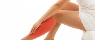
Symptoms of varicose veins
The disease can develop very slowly: years or even decades. The symptoms of the initial stage are varied, but for convenience they are combined into a certain complex under the general feeling of heaviness in the legs:
- fatigue in the legs without serious exercise,
- sore veins,
- night cramps in the calves,
- pulling sensation in legs.
If you do not track the symptoms of varicose veins at the initial stage, the vein will become more and more deformed. In the evenings, swelling in the feet and ankles will begin to appear, disappearing by morning; Spider veins will become visible, and then swollen veins and nodules on them will be felt. If you do not consult a specialist at this stage, then in addition to the obvious decrease in quality of life and obvious pain, the person risks getting skin pigmentation and trophic ulcers. The further course of varicose veins brings complications such as bleeding from the venous nodes and the risk of developing such a serious disease as thrombosis, which can be fatal without contacting a specialist.
Ultrasound scanning of the veins of the lower extremities:
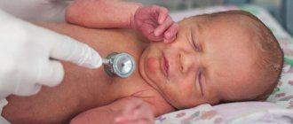
The deep veins are passable, the blood flow is phasic, synchronized with the act of breathing. The trunk of the great saphenous vein on the right has a straight course, is varicose along its entire length, thrombotic masses are located in the lumen in the middle and upper third of the leg with blood flow blocked up to 100%, flotation of blood clots was not detected at the time of the study. The valves of the saphenofemoral anastomosis and the trunk of the GSV on the right are incompetent. The diameter of the GSV trunk on the right in the SPS region is 13 mm, in the middle third of the thigh - 9-10 mm, in the lower third of the thigh - 7 mm. There are varicose perforators on the lower leg with incompetent valves up to 5-6 mm in diameter. The valves of the sapheno-popliteal anastomosis are wealthy. The small saphenous veins on both sides have a straight course, are not dilated, and blood clots are not located in them. The diameter of the SVC trunks in the area of the anastomosis is 3-4 mm.
Diagnosis: acute ascending varicothrombophlebitis in the pool v.saphena magna dextra. Varicose veins. Varicose veins of the right lower limb. Subcompensation stage. Trunk type on the right. Chronic venous insufficiency stage II.
CVD on the right:
C 2.3, Er, A1, Pr,o 2.3 LII.
The influence of pregnancy on the formation of varicose veins
During pregnancy, it is important to stop the progression of varicose veins for the health of the mother and the unborn child. Consultation with a phlebologist is required. Pregnancy creates the prerequisites for the appearance of initial varicose veins and its further development (6, 7 English):
- Hormonal imbalance between estrogens and gestagens. Already from the first days, the level of progesterone in the blood progressively increases, which prevents the onset of the next menstruation. But this hormone affects not only the condition of the endometrium and the entire uterus. Under the influence of progesterone, the tone of the smooth muscle layer in the walls of internal organs and blood vessels, including the veins of the lower extremities, decreases - this contributes to the deterioration of blood evacuation from the peripheral parts of the extremities with the development of venous hypertension.
- The growing uterus causes compression of nearby organs and vessels of the small pelvis. Already from the second trimester of gestation, most women experience difficulty in the outflow of blood through the inferior vena cava system. The resulting increase in pressure in the veins of the legs contributes to the decompensation of existing venous pathology and active remodeling of the vascular walls. There is often concomitant lymphostasis, which aggravates the situation.
- Physical inactivity at the end of the second and third trimesters of pregnancy. With insufficient physical activity, the efficiency of the muscle pump of the legs decreases, and there is a risk of early, initial varicose veins.
During pregnancy, symptoms of both cosmetic and classic varicose veins can develop. Women often do not attach much importance to them, considering them a natural manifestation of pregnancy. Therefore, a consultation with a phlebologist is included in the list of mandatory examinations.
Treatment:
On an outpatient basis, under tumescent (local) anesthesia, endovasal laser obliteration (EVLK) was performed using the German technology Biolitec v.saphena magna dextra . The duration of the procedure was about 30 minutes. 15 minutes later, after a follow-up examination by the operating surgeon-phlebologist, the patient left the clinic on his own.
Operating surgeon:
Rumyantsev A.Yu.
Assistant:
Voloshkin A.N.
The next day, an examination with ultrasound duplex scanning and dressing was performed.
C2 and C3 stages of varicose veins
C2 is the stage of appearance of varicose nodes or bumps on the legs. This is a serious stage of varicose veins, when a phlebologist diagnoses not only the presence of large dilated tributaries, but also, using ultrasound data, identifies a non-functioning main vein (one or more), which carries used blood not to the body for cleaning, but back to the leg (foot) . Treatment of this stage of varicose veins usually includes one or another surgical stage, which we will discuss below. The “gold standard” for the treatment of such veins today is endovenous laser obliteration (coagulation) in combination with procedures such as miniphlebectomy, as well as echopenic sclerotherapy of varicose tributaries.
C2 stage of varicose veins
C3 is the stage when varicose veins in the legs are accompanied by swelling. In such a situation, treatment of veins should be carried out comprehensively and include medications, compression therapy, surgical treatment (removal) of non-functioning veins, as well as long-term observation by a phlebologist.
C3 stage of varicose veins. Enlarged nodes and swelling of the lower leg
The main causes of the disease
Experienced doctors identify several main reasons that provoke the appearance of venous disease. These include:
- prolonged sitting and standing;
- physical exercise;
- genetic predisposition;
- sedentary lifestyle;
- presence of bad habits;
- hormonal and endocrine disruption in the body;
- constant stress and severe nervousness;
- incorrectly selected shoes;
- cancerous tumor in the pelvic area;
- pregnancy. This is a fairly common occurrence during this period.
Varicose veins during pregnancy quickly progress to stage 2. This pathology occurs due to heavy load on the venous system of the legs.
To determine the exact cause of venous disease, it is recommended to seek help from a highly qualified specialist. Passing a certain type of medical tests will reveal the entire clinical picture of the course of varicose veins.
Treatment of varicose veins using endovasal laser obliteration (EULO)
Varicose veins of the lower extremities
- a disease characterized by primary varicose transformation of superficial veins. Risk factors for the development of varicose veins of the lower extremities (LVL) are traditionally recognized as age, female gender, obesity, and heredity. Specific risk factors include pregnancy, taking hormonal drugs (estrogens, gestagens), and menopause.
The pathogenesis of varicose veins is complex and continues to be studied. All elements of the venous wall are involved in the pathological process with VVVN. Varicose veins are characterized by destruction of the valve apparatus of the veins of the lower extremities, as a result of which the outflow of blood through the veins becomes insufficient due to incomplete closure of the valves, and the veins begin to gradually expand. Or, on the contrary, there may be a primary weakness of the venous wall and its expansion, as a result of which the valves also cannot touch normally and, as a result, venous insufficiency develops. As a result of reflux (reverse movement of blood) along the main trunk of the saphenous vein, varicose transformation of the tributaries (branches) of the main trunk is formed.
At the beginning of the development of the disease, the patient may be bothered by symptoms and a visual defect in the form of subcutaneous dilated nodular veins, but as the disease progresses, such dangerous consequences as thrombophlebitis and thromboembolism, bleeding from varicose veins, as well as the formation of trophic long-term non-healing ulcers may appear.
The main treatment method for varicose veins remains surgery. The purpose of the operation in patients with varicose veins is to reduce the risk of developing thrombotic complications, prevent the progression of varicose transformation of the saphenous veins, bleeding from varicose veins, eliminate the symptoms of the disease, eliminate the cosmetic defect caused by the disease, prevent the progression and/or regression of edema, trophic disorders, reducing the frequency of recurrence of trophic ulcers and increasing the patient’s quality of life.
Objectives of surgical treatment:
- elimination of pathological vertical and/or horizontal reflux;
- elimination of varicose saphenous veins
Endovenous laser obliteration (EVLO) of the veins of the lower extremities
EVLO
is an effective and minimally invasive method of eliminating reflux (pathological reverse blood flow) through the main and perforating veins of the lower extremities in patients with varicose veins.
EVLO is a thermal method of vein obliteration, which is based on endovasal thermal damage to the venous wall, leading to the transformation of the vein into a connective tissue cord, which subsequently resolves. The intervention is characterized by low trauma, rapid restoration of working capacity and good cosmetic results. E VLO is an outpatient procedure and the patient returns home on the day of surgery.
The duration of the operation takes on average 40-60 minutes. At the Smitra clinic, for EVLO, the LAKHTA-MILON
with a diode laser with a wavelength of 1470 nm. The use of a more modern light guide with a radial type of radiation, rather than an end one, increases the absorption coefficient of laser radiation by the venous wall, which leads to an improvement in the outcome of the operation. The operation is performed using ultrasound guidance.
Visible varicose veins of the lower extremities, which are tributaries of the main saphenous veins, are also subject to removal after performing the EVLO procedure of the main trunk of the saphenous veins. Laser endovenous obliteration of varicose veins is not performed due to the high risk of thermal damage to the skin. Varicose saphenous veins are eliminated using sclerotherapy immediately after the EVLO procedure. During the procedure, a phlebosclerosant drug is used in the form of fine foam, which is injected into the vein and causes “gluing” of the vein walls from the inside. During the manipulation, ultrasound monitoring of vein puncture, administration and distribution of the drug is performed.
Preparing for EVLT
It is necessary to conduct an examination before starting treatment, undergo standard tests, and also undergo an ultrasound examination of the vessels of the lower extremities without fail.
List of required procedures:
- General blood and urine analysis.
- Biochemical analysis.
- Study of the blood coagulation system.
- Blood for HIV, syphilis, hepatitis B and C
- Fluorography.
- Examination by a therapist.
Postoperative period
After the operation, which lasts approximately 40 minutes, the patient must walk for 1-1.5 hours.
As soon as the required walking limit is met, you can go home. Do I need to make an appointment with a phlebologist after surgery?
Yes need. After the operation, it is necessary to undergo 4 follow-up examinations (on the 1st or 2nd day, after 2 weeks, after 2 months and 6 months from the date of surgery) by a vascular surgeon and, accordingly, 4 procedures for ultrasound examination of the veins of the operated lower limb.
Can the procedure on the second leg be done immediately?
No, you need to wait at least two weeks before having surgery on the veins of the second lower limb.
Should I wear compression stockings and for how long?
Yes, compression stockings must be worn continuously for the next two days of the procedure, and only during the day for the next 4 weeks.
Are there any recommendations for physical activity?
It is necessary to walk at a brisk pace for at least 1 hour every day, and you should also avoid prolonged static loads on your legs. In addition, you will have to stop visiting the bathhouse, sauna and taking a hot bath for at least two weeks.
Are there any visual consequences of the operation?
As a result of the procedure, there are no scars or bruises, but pigmentation and slight bruising may still occur at the puncture sites.
What complications are there after the procedure?
For two to three days, you may experience slight pain in the legs, discomfort when flexing and extending, a feeling of tension along the vein, and a short-term rise in temperature to subfebrile levels.
But all the consequences disappear within a few days.
You can learn more about the complications of varicose veins of the lower extremities, indications and contraindications for surgical treatment, preparation for surgery, the course of the operation, postoperative observation and treatment at an appointment with a vascular surgeon.
But we must understand that the elimination of large varicose veins does not mean getting rid of spider veins and reticular veins.
Percutaneous laser coagulation (PLC)
Telangiectasia (spider veins) are dilated intradermal veins with a diameter of less than 1 mm.
To combat this pathology, the method of percutaneous laser coagulation (PLC) is used, and it is also performed using the
LAKHTA-MILON device.
Laser technology allows for non-contact coagulation of blood vessels without damaging the skin. Hemoglobin, which is contained in the blood, absorbs thermal energy, which leads to heating of the vessel wall and its gluing. Then gradual destruction of the small vessel occurs, and it disappears within 2-3 weeks. The diode laser with a wavelength of 970 nm, which is used in our clinic, has low scattering in the tissues surrounding the vessel and is associated with a low likelihood of thermal damage to the skin. There is no need for anesthesia, as pain is minimal to moderate. The duration of the procedure is 10-30 minutes depending on the area of skin being treated. It is not advisable to treat large surfaces of the skin in one session, as severe swelling of the limb may occur.
Therefore, if the patient has multiple spider veins, then treatment is carried out in 3-4 sessions at 3-4 week intervals. Most vessels disappear after the first session, but some may require 2 or more sessions to remove.
Contraindications for PLK
Contraindications include tanned and dark skin, taking photosensitizing drugs, including some diuretics, antibiotics, vitamin A and its derivatives.
Laser treatment can be carried out two to three weeks after stopping medication, but it happens that you have to wait up to six months.
Postoperative period
Is there a recovery period needed?
There is no recovery period required after a PC procedure.
Do I need to wear compression garments?
Yes, it is recommended to wear compression stockings for several days after the procedure.
Are there any restrictions after the manipulation?
Yes, you should not expose your skin to sunlight, and you should also postpone visiting the sauna and bathhouse. Active sports should be replaced with walks for the first time after surgery.
Are there any complications after PCI?
Complications are very rare and do not pose a health hazard. There is a possibility of pigmentation, it depends on the characteristics of the skin.
There are also consequences that, with well-chosen measures, are reversible:
- burning in the area of manipulation
- skin redness and mild swelling
- the appearance of pinpoint crusts, but they disappear within 1 week.
You can learn more about the procedure, indications and contraindications, and preparation for the session at an appointment with a vascular surgeon.
If you need treatment for varicose veins in Novosibirsk, removal of spider veins, then you can make an appointment with a vascular surgeon at the Smitra clinic.
Prevention
In order not to feel what varicose veins are, we recommend following simple rules:
- alternate work and rest;
- limit physical activity and standing work;
- exclude visiting saunas and baths;
- switch to a healthy lifestyle;
- promptly contact a phlebologist when you notice the first symptoms of varicose veins;
- exercise therapy.
Read more in the article How to strengthen the walls of blood vessels. Prevention of varicose veins.
RISK FACTORS FOR VARICOSE DISEASE DEVELOPMENT
Heredity and its role in the development of varicose veins have not been unambiguously confirmed. One of the main counterarguments to the presence of a genetic predisposition is the different incidence of varicose veins in ethnic Africans and their fellow tribesmen who emigrated to the United States and Western European countries. At the same time, in comparison with settled fellow tribesmen, the incidence of varicose veins, which does not exceed 0.5%, among emigrants there is a significant increase in the incidence of 10-20%. In this regard, the obvious conclusion is that environmental factors, lifestyle characteristics and nutrition prevail in the pathogenesis of varicose veins.
Pregnancy is traditionally considered one of the main risk factors for the development of varicose veins, which explains, in particular, the more frequent (3-4 times) incidence in women. It is generally accepted that the main provoking factors are an increase in BCC (circulating blood volume) and compression of the pregnant uterus by the retroperitoneal veins. Meanwhile, the results of epidemiological studies indicate that only the second and subsequent pregnancies lead to a significant increase in the incidence of varicose veins by 20-30%. Moreover, the first signs of the disease appear already in the 1st trimester of pregnancy, when a sharp increase in the volume of blood volume or an enlargement of the pregnant uterus has not yet occurred.
Obesity is a proven risk factor for varicose veins. At the same time, an increase in body mass index to 27 kg/m2 and above leads to an increase in the incidence of the disease by 33%.
The high incidence of varicose veins in industrialized countries is to some extent due to dietary habits. A high degree of food processing and a decrease in raw vegetables and fruits in the diet has led to a constant deficiency of plant fibers necessary for the remodeling of the venous wall and chronic constipation, which causes a permanent increase in intra-abdominal pressure.
Lifestyle has a significant impact on the development and course of varicose veins. In particular, the adverse effects of prolonged static loads with heavy lifting or immobile standing and sitting have been proven. In Asian countries, the more frequent use of chairs and armchairs among the Europeanized part of the population led to a significant (3-4 times) increase in the incidence of varicose veins compared to people traditionally sitting on mats.
Dyshormonal conditions are involved in the pathogenesis of varicose veins. Their role has been progressively increasing in recent years, which is due to the widespread use of hormonal contraception, the popularization of hormone replacement therapy during the premenopausal period and in the treatment of osteoporosis. It has been proven that estrogens and progesterone, as well as their derivatives, reduce the tone of the venous wall due to the gradual destruction of collagen and elastic fibers.
Compression garments for varicose veins
In the early stages and after treatment, men are recommended to wear compression garments, which are selected individually. Anti-varicose underwear is similar in appearance to classic underwear, but there are no seams. The fabric consists of nylon, microfiber and lycra. The fabric is breathable and pleasant to the touch.
Compression jersey for men is divided into:
- stockings for varicose veins of the thighs,
- tights if both legs are affected,
- knee socks if the disease is on the calves or just above the knee.
You need to purchase underwear at the first sign of the disease.
Tips for wearing compression stockings:
- Put on your underwear carefully so as not to tear the product.
- File your fingernails and toenails to avoid tearing your underwear.
- Put it on in the morning, without waiting for swelling.
- Remove compression garments before going to bed.
- If it is difficult for an overweight patient to put on compression garments on their own, it is recommended to purchase a device for putting them on.
- Wash clothes in warm water with baby soap. To prevent water from getting on the silicone, tighten the upper part with thread, and clean the elastic band with a cotton pad in alcohol.
Device for putting on compression garments.
Phlebeurysm
Varicose veins are overstretched, irregularly shaped, tortuous venous vessels that have lost their elasticity.
They are increased in length and width and look like twisted blue strands that are visible under the skin. Veins become this way when the venous valves are missing or for some reason cannot perform their functions. If the valves do not work properly, blood flows backward through the veins, pooling in the lower extremities. As a result, the veins lose their natural shape, and a chain of various complications begins.
Diagnosis of varicose veins in men
The optimal solution for men with symptoms of varicose veins in Tver is to contact a competent phlebologist. Ultrasound examination of blood vessels at the time of treatment is a mandatory component of a consultation with a phlebologist! It is advisable that the ultrasound of the veins be performed not by a third-party doctor (no matter how highly qualified he may be), but personally by the phlebologist you contacted. The doctor who undertakes to advise you is responsible for both the correct diagnosis and the development of a treatment strategy.
Ultrasound in men is the main thing in making a diagnosis
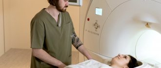
Ultrasound angioscanning in duplex and triplex mode (ultrasound of veins) is the basis for the diagnosis and treatment of venous diseases. Therefore, this test should be carried out by the doctor who will advise, treat or operate on you. Ultrasound of the veins should be performed in a standing position. For patients whose health condition does not allow ultrasound to be performed in an upright position, the examination can be performed in a lying position with straining, but its diagnostic value will be lower. Especially when it comes to diagnosing deep vein thrombosis. After interviewing the patient, examination, palpation and ultrasound of the veins, the phlebologist must write his report, which includes a brief history of the disease, diagnosis and recommendations. It is mandatory to write a protocol for ultrasound angioscanning indicating the date of the study. If necessary (complex cases, congenital anomalies, consequences of massive deep vein thrombosis), the doctor recommends additional clarifying studies (magnetic resonance venography, X-ray angiography or others).
Only this approach meets modern European standards. The expert examination will take no more than half an hour, and you will receive sufficient information about the venous system of your lower extremities.
Consultation with a phlebologist is absolutely safe and does not involve any traumatic examinations or procedures. No special preparation required. Timely diagnosis is extremely important for choosing modern and effective treatment, which will avoid complications and progression of the disease.