Ligation of the mouth of the great saphenous vein - crossectomy is an element of a surgical operation for varicose veins (phlebectomy), or an independent operation for ascending thrombophlebitis. Previously, this operation was used as an independent method of treating varicose trophic ulcers.
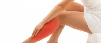
Acute thrombophlebitis of the saphenous veins most often should be treated conservatively. Indications for surgical interventions arise when there is a threat of complications. The most common reason for surgery is progressive thrombophlebitis of the great saphenous vein, with the threat of spreading to the deep veins. It is with this complication that ligation of the great saphenous vein is indicated. Bandaging allows you to block the thrombus from accessing the deep venous system.
Indications and contraindications
Previously, varicose veins were considered a disease that exclusively affected older people. Unfortunately, today the situation has changed significantly. Varicose veins are getting younger every day, and are now found even in teenagers.
Crossectomy is indicated in the following cases:
- The presence of thrombophlebitis of the lower extremities;
- Too frequent relapses of inflammation of the vein walls;
- Lack of positive effect from drug treatment;
- Thrombophlebitis in the area of the knee joint and above.
We can conclude that surgery is required for serious complications of varicose veins. Alas, even surgery does not guarantee a complete recovery. Stable remission can only be achieved with further comprehensive treatment.
Surgical combined phlebectomy, why the technique is not ideal
Combined phlebectomy at one time made a splash, a kind of revolution both in the treatment and understanding of the pathogenesis of varicose veins. More than a century has passed since then, today milder versions of anesthesia and other surgical instruments are used, but no fundamental changes have occurred in this surgical technique. Even with the advent of innovations in the form of: PIN stripping, intussusception stripping and cryostripping, the technique essentially remained the same.
Cryostripping - removal of varicose veins with cold
Classic combined phlebectomy using a Babcock probe still remains the most widespread for the treatment of varicose veins in the public sector of medicine due to the unobvious advantages of the above innovations.
Contraindications
Crossectomy has certain limitations. The operation cannot be performed in the following cases:
- The presence of malignant neoplasms in the body;
- Diabetes mellitus in advanced form;
- Obesity of various degrees;
- History of atherosclerosis;
- Cachexia;
- Narrowing of the lumen, blockage of deep-lying vessels;
- Severe renal or liver failure;
- Infectious diseases in the acute stage;
- Pregnancy period.
It is also worth noting that the procedure is not recommended in old age due to the high risk of complications.
What do we offer today instead of classical surgical treatment of varicose veins?
Everything changed about 20 years ago with the advent of the first successful attempts at endovasal thermoobliteration. This was a significant technological breakthrough that radically changed phlebology. The initial clinical experience with the use of thermoobliteration was on the verge of a medical experiment. Any innovative technology goes through this stage. During that period of the first steps and development of the thermoobliteration technique, one could talk about its competition with classical surgery.
Laser radial light guide
After the appearance of radial light guides for laser coagulation in 2008, there could no longer be any talk of a significant opposition between combined phlebectomy (in any form) and endovasal thermoobliteration. What are the benefits of endovascular varicose vein surgery:
- Fine manipulation control via ultrasound imaging
- Minimally invasive, only skin punctures.
- Mild anesthesia in the form of local tumescent anesthesia.
- Full outpatient service for all procedures.
- Good patient tolerance of both the manipulation and the postoperative period.
Preparation rules
To exclude contraindications to this procedure, the patient will need to undergo tests and, possibly, undergo some diagnostic procedures.
Mandatory examinations include:
- General blood analysis;
- Blood clotting test;
- Elimination of HIV, syphilis and hepatitis;
- Fluorography, ECG;
- Ultrasound examination of the veins of the lower extremities.
You will also need a certificate from a physician stating that the patient has no contraindications to crossectomy. If a person has chronic diseases, additional consultation with specialists may be required.
If you have to undergo this manipulation, check out the list of recommendations:
- Regardless of the type of anesthesia, the intervention is performed on an empty stomach - you cannot eat for at least 7-8 hours;
- Be sure to tell your doctor if you are taking any medications on an ongoing basis.
- If a woman is undergoing crossectomy, it is not recommended to carry out the procedure during or immediately before menstruation.
- If you are concerned about feelings of fear or anxiety, you can discuss taking mild sedatives with your doctor.
- You should stop drinking alcohol at least a week before surgery.
- You must stop smoking at least 3-4 hours before surgery.
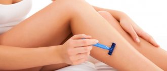
- If necessary, excess vegetation is removed at the site of the surgical incision.
Recommendations for conducting
Planned crossectomy surgery is possible only in a hospital by experienced vascular surgeons. Sometimes intervention is prescribed urgently, when minutes are counting and the patient needs to be saved. Indications for such urgent measures are:
Doctors who perform vein crossectomy must have sufficient experience and skills in performing complex interventions. You cannot trust your own health to the first specialists you come across. The Dobromed clinic has created the necessary conditions for ligation and removal of veins on the legs. Famous vascular surgeons and phlebologists work here and can help their patients.
How is crossectomy performed?
Crossectomy can be performed under local anesthesia or general anesthesia.
The procedure is carried out in several stages:
- The operated area is carefully treated with antiseptic agents.
- The doctor makes a small incision in the skin (usually no more than 5 cm in length) to gain access to the venous vascular bundle.
- The doctor ties the place where the great saphenous vein flows into the femoral vein.
- The great saphenous vein is isolated, carefully divided, and ligated.
- At least five tributary veins are also ligated, which reduces the likelihood of relapse and eliminates the return of blood into the veins of the limb.
- The incision area is sutured, the doctor installs drainage.
- The operated area is treated with antiseptic agents, after which a sterile bandage is applied.
The duration of the surgical intervention is about half an hour. In some cases, simultaneously with crossectomy, dilated veins and venous nodes can be removed or coagulated with electric current/cold, and a sclerosing solution can be introduced to stop blood circulation in the superficial veins.
Surgical treatment (combined phlebectomy) for the treatment of varicose veins - complications
Combined phlebectomy, like any extensive and fairly traumatic surgical procedure, is often complicated by the following unpleasant consequences:
- Bleeding from postoperative wounds.
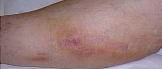
- Formation of hematomas, seromas.
- Infection of postoperative wounds.
- Formation of lymphorrhea and lymphocele.
- Neovasculogenesis (appearance of a network of varicose veins in the area of the saphenofemoral junction).
- Paresthesia and local permanent anesthesia for damage to cutaneous nerves.
- Thromboembolic complications up to the development of pulmonary embolism.
The last point should be discussed in detail, since despite the whole range of measures taken to prevent these complications, it is very difficult to avoid them completely. Despite the introduction of the latest anticoagulants into the practice of a modern surgeon, the presence of significant postoperative trauma and skin incisions does not allow the use of the main tool in the fight against venous thrombosis. Namely, early activation. The fear of bleeding and hematomas in operated patients is a weighty argument, but the longer the patient returns to normal activity, the higher the risk of thrombosis.
Rehabilitation period
For three days after surgery, it is recommended to remain in a hospital setting under constant medical supervision. If there are no complications, the sutures are removed, the drainage is removed, and the patient is transferred to outpatient treatment. During the recovery period, it is important to follow the following rules:
- It is mandatory to purchase compression stockings. The degree of compression is determined individually for each patient.
- For the fastest possible recovery, it is recommended to walk. The very next day after the operation, walking along the corridor is necessary, and on the following days, walking.
- At the same time, heavy physical activity should be abandoned - activity should be moderate.
- During the rehabilitation period, it is prohibited to take hot baths, visit saunas and steam baths. The timing is determined individually for each patient.
- It is recommended to eat well so that the body receives the necessary vitamins and minerals.
In addition, after the operation, the doctor prescribes medication:
- Antibacterial and anti-inflammatory drugs to reduce the risk of complications.
- Venotonic drugs to improve the tone of the venous wall.
- Painkillers to relieve discomfort after surgery.
- To prevent thrombosis, it is recommended to take anticoagulants.
- Vitamin complexes will support the immune system and speed up recovery.
More detailed information about crossectomy can be obtained during an in-person consultation with a doctor. Be healthy!
Our clinics in St. Petersburg
Structural subdivision of Polikarpov Alley Polikarpov 6k2 Primorsky district
- Pionerskaya
- Specific
- Commandant's
Structural subdivision of Zhukov Marshal Zhukov Ave. 28k2 Kirovsky district
- Avtovo
- Avenue of Veterans
- Leninsky Prospekt
Structural subdivision Devyatkino Okhtinskaya alley 18 Vsevolozhsk district
- Devyatkino
- Civil Prospect
- Academic
For detailed information and to make an appointment, you can call +7 (812) 640-55-25
Make an appointment
A complex approach
The algorithm described above is suitable for crossectomy as monotherapy for emergency situations when it is necessary to block possible complications of thrombosis immediately. But usually the operation becomes only part of an extensive phlebectomy, acting as the first step towards restoring the health of the legs. Here it is impossible to do without excision of the affected vessel.
The first step of combined radical intervention involves an inguinal incision in the area of the junction of the deep and superficial veins. The second vessel is cut off taking into account the degree of damage, and then ligated.
The second step involves making another cut at the top of the shin or near the ankle. Having identified the saphenous vein, a special probe made of metal material is launched there, which, as it moves, should reach the area of the first cut.
After the probe reaches the designated location, the veins are fixed. To do this, use a special thread, having previously placed it on the probe tip. The third part of the intervention is called the Babcock operation. It provides for long stripping on the left or right, which was made possible thanks to the use of a flexible probe tip. It is pulled through the incision, and the sharp edge of the instrumentation cuts off the vessel from the nearest intact tissue.
The strategy of miniphlebectomy, which is also called the Narata method, is discussed separately. This operative aspect involves removal of pre-marked venous nodules and tributaries followed by ligation of the perforating veins.
It is especially difficult for the surgeon if the vessels have a tortuous shape, which prompts the integrity of the integument to be broken in several places in order to remove the affected areas in parts. The nodes are removed using a special surgical device called a Müller hook.
To make the result look aesthetically pleasing, the punctures are made very small, up to 2 mm. Such wounds heal on their own without the need for stitches, and after a couple of months not even a trace remains of them.
Crossectomy: disadvantages of the method
Despite its high efficiency, the crossectomy method has several serious disadvantages:
- long rehabilitation period;
- the need for mandatory hospitalization of the patient in a 24-hour hospital.
Crossectomy (ostial ligation of the great or small saphenous vein) is considered an effective way to prevent pulmonary embolism in acute ascending thrombophlebitis, since it prevents the spread of the thrombotic process from the superficial to the deep veins [1, 2]. The operation is short, low-traumatic, has been used for decades and has become routine in clinical practice. The vast majority of surgeons believe it is so reliable that if they use anticoagulants, it is only in prophylactic doses, and more often they are limited to the use of antiplatelet and venotonic drugs.
How justified is such an unconditional belief that crossectomy for ascending thrombophlebitis solves the problem of preventing pulmonary embolism? Our experience indicates that surgeons' expectations are too high, as evidenced by two clinical cases that we would like to present to the attention of our colleagues.
Patient S.
64 years old, was admitted to the surgical department on January 12, 2011 with complaints of a painful lump on the inner surface of the right thigh, redness of the skin in this area, pain in the right lower limb when walking.
From the anamnesis it is known that she was sick for 2 days and noted the occurrence of the above complaints. She did not report shortness of breath, hemoptysis, chest pain, or episodes of loss of consciousness during the course of the disease.
Upon examination, the condition is satisfactory. Breathing in the lungs is carried out in all parts, vesicular, there is no wheezing. Pulse 84 per minute, rhythmic, blood pressure 130/90 mm Hg. The abdomen is soft, painless in all parts. Peristalsis is audible, but not increased. Rectal and vaginal examinations revealed no pathology.
Local status: the right lower limb is not swollen, the skin is of normal color and warm. The pulsation in the arteries is clear throughout. Along the inner surface of the upper third of the leg, lower, middle and upper third of the thigh there is a dense painful cord with hyperemia of the skin above it. There are no varicose veins. Homans and Moses symptoms are negative.
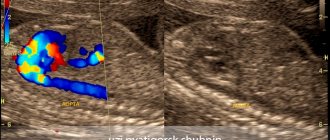
Ultrasound angioscanning of the veins of the lower extremities (USA): the right great saphenous vein is thrombosed along its entire length to the saphenofemoral junction (Fig. 1, a).
Figure 1. Angioscanograms of patient S. a - thrombus in the estuarine section of the great saphenous vein (arrow). The deep veins of both lower extremities are passable, their valves are intact. The perforating veins are not thrombosed.
In order to prevent deep vein thrombosis and pulmonary embolism, it was decided to perform a crossectomy. The surgical intervention was carried out without technical difficulties; no pronounced phenomena of periphlebitis were detected in the operation area.
In the postoperative period, the patient received compression (elastic bandaging), venotonic (troxerutin 300 mg 2 times a day) therapy. Taking into account the nature of the lesion, the age of the patient, the extent of the operation, and also taking into account the fact that there were no varicose veins, the risk of postoperative thromboembolic complications was assessed as high, and therefore unfractionated heparin 7500 units was prescribed 3 times under the skin of the abdomen until discharge from the hospital (7 days).
The patient in the hospital was additionally examined, an ultrasound examination of the abdominal organs, retroperitoneal space, esophagogastroduodenoscopy, colonoscopy was performed - no organic pathology was detected, examined by a gynecologist - no diseases of the internal genital organs were found.
During the control ultrasound examination of the veins of the lower extremities on the 5th day after surgery: the deep veins of both lower extremities are patent, no signs of thrombosis were detected.
The sutures were removed on the 8th day of the postoperative period, the patient was discharged from the hospital in satisfactory condition, recommendations: elastic compression of the lower extremities, taking antiplatelet and phlebotropic drugs.
At a follow-up examination 1 month after surgery, the patient did not have any complaints. Swelling and pain in the right lower extremity did not bother me, shortness of breath, hemoptysis, chest pain, or episodes of loss of consciousness were not noted.
Ultrasound examination revealed non-occlusive thrombosis of the right superficial femoral vein, which required the patient to be re-admitted to the hospital (Fig. 1, b).
Figure 1. Angioscanograms of patient S. b - non-occlusive thrombus of the right superficial femoral vein (arrows). Anticoagulant therapy was administered (unfractionated heparin 12,500 units 3 times under the skin of the abdomen for 8 days, followed by warfarin at a dose of 5 mg per day).
To exclude/confirm the asymptomatic form of pulmonary embolism, the patient underwent lung scintigraphy, which revealed disturbances in lung perfusion of the subsegmental type, which indicated the presence of thromboembolism of small branches of the pulmonary artery (Fig. 2, a).
Figure 2. Scintigrams of the lungs of patient S. a - impaired perfusion of the left lung according to the subsegmental type (arrows).
USAS on the 10th day of re-hospitalization: initial recanalization of the right superficial femoral vein. The patient was discharged from the hospital on the 14th day in satisfactory condition (INR at discharge 2.2). Elastic compression and warfarin for 6 months are recommended.
A follow-up examination after 2 months showed the absence of chronic venous insufficiency of the lower limb and cardiopulmonary failure. With USAS: good recanalization of the deep veins of the right lower limb. Lung scintigraphy indicated the absence of perfusion-ventilation disorders (Fig. 2, b).
Figure 2. Scintigrams of the lungs of patient S. b - after 2 months. Absence of perfusion-ventilation disorders.
An analysis of the medical history of this patient shows that the actions and prescriptions of the attending physicians in the hospital complied with all existing standards for the treatment of ascending thrombophlebitis of the main saphenous veins. However, this did not protect the patient from developing deep vein thrombosis and pulmonary embolism, which developed after discharge. The next incident ended more tragically.
Patient K., 48 years old, was admitted to the surgical department on March 28, 2011 with complaints of a painful lump on the inner surface of the right leg and thigh, redness of the skin in this area.
From the anamnesis: he was ill for 7 days, when a hyperemic, dense, sharply painful cord appeared along the inner surface of the right shin to the level of the knee joint.
He was treated on an outpatient basis under the supervision of a surgeon, using heparin ointment locally 2 times a day and taking troxerutin 300 mg 2 times a day. Despite treatment, over time the cord spread to the middle third of the right thigh. No shortness of breath, hemoptysis, chest pain, or episodes of loss of consciousness were noted during the course of the disease.
On examination the condition is of moderate severity. Breathing in the lungs is carried out in all parts, vesicular, there is no wheezing. Respiratory rate 17 per minute. Pulse 80 per minute, rhythmic, blood pressure 140/80 mm Hg. The abdomen is soft, painless in all parts. Peristalsis is audible, but not increased. A rectal examination revealed no pathology.
Local status: the right lower limb is not swollen, the skin is of normal color and warm. The pulsation in the arteries is clear throughout. Along the inner surface of the upper third of the leg, the lower and middle third of the thigh, there is a dense, painful cord with hyperemia of the skin above it. Homans and Moses symptoms are negative. Varicose dilation of the tributaries of the main veins on the legs. The left lower limb is not swollen, the skin is of normal color and warm. The pulsation in the arteries is clear throughout. Homans and Moses symptoms are negative.
With USAS: the right great saphenous vein is thrombosed along its entire length to the upper third of the thigh. The right small saphenous vein is thrombosed along its entire length to the middle third of the leg. The deep veins of both lower extremities are passable, their valves are intact. The perforating veins are not thrombosed.
In order to prevent deep vein thrombosis of the right lower limb and pulmonary embolism, it was decided to perform ligation of the small and great saphenous veins for the patient. The surgical intervention was carried out without technical difficulties, no pronounced phenomena of periphlebitis were detected in the operation area, and no thrombotic masses were detected upon palpation of the small and great saphenous veins within the surgical wound.
In the postoperative period, the patient underwent compression (elastic bandaging), venotonic (micronized purified flavonoid fraction 500 mg 2 times a day). Taking into account the patient's age and the extent of the surgical intervention, the risk of postoperative venous thromboembolic complications was assessed as moderate, and therefore unfractionated heparin was prescribed at a dose of 5000 units 3 times under the skin of the abdomen.
In the postoperative period, the patient complained of moderate pain in the area of postoperative wounds; there were no other complaints. Local status - no negative dynamics. On the 5th day after surgery, the patient suddenly developed severe shortness of breath at rest, a feeling of suffocation, and general weakness. On examination: the condition is serious, the skin is pale, cold sweat. Breathing is harsh, sharply weakened in the lower regions on both sides. Respiration rate 30 per minute. Heart sounds are muffled, pulse is 105 per minute, rhythmic. Blood pressure 90/60 mm Hg. The abdomen is soft, painless in all parts. Peristalsis is audible, but not increased. Local status: no negative dynamics, dressings are dry. The situation was assessed as massive thromboembolism of the pulmonary arteries.
Due to his serious condition and severe symptoms of respiratory and cardiovascular failure, the patient was transferred to the intensive care unit for further treatment. In order to clarify the extent of pulmonary lesions and determine further treatment tactics, the patient was decided to undergo computed tomography of the chest with intravenous bolus enhancement. However, despite the intensive therapy, the patient's condition quickly and progressively worsened, and the symptoms of cardiopulmonary failure increased. Against the background of severe bradycardia, resuscitation measures were started, but were unsuccessful. The patient was declared dead.
Autopsy revealed thrombosis of the left femoral vein and pulmonary embolism, which was recognized as the cause of death.
As in the previous case, it was hardly possible to blame the attending physicians for violating the protocol for managing patients with varicothrombophlebitis: an operation was performed to prevent the transition of thrombosis to the deep venous system, anticoagulant prophylaxis was carried out, as well as other conservative measures. Nevertheless, the patient died from a pulmonary embolism, the source of which was deep vein thrombosis of the contralateral limb that developed in the postoperative period.
These cases from everyday clinical practice clearly demonstrate that not always, not in every patient, even timely crossectomy is a guarantee against the development of such a formidable complication as pulmonary embolism. We are confident that similar situations can be reported by employees of any surgical clinic that has even a small flow of patients with ascending thrombophlebitis. Moreover, in our observations, anticoagulant prophylaxis was used, which also turned out to be ineffective. It is quite possible, although there is no consistent data on this in the literature, that in patients with ascending thrombophlebitis it is necessary to use other dosages of anticoagulants - therapeutic or intermediate. This should be the subject of close attention by the surgical community, as well as the duration of anticoagulant therapy for this pathology.
Conflict of interest
absent.
What operations are performed for varicose veins in the legs?
There are three main types of operations for varicose veins of the lower extremities: classic venectomy (phlebectomy), stripping and vein ligation.
Classical venectomy (phlebectomy). During this intervention, a longitudinal skin incision is made on the leg, corresponding to the course of the varicose vein. The vessel is isolated, ligated in the area of the ankle and knee (if we are talking about the lower leg) or the knee and groin (if a vein on the thigh is removed), large branches of the vein are ligated and it is cut off, after which the incision is sutured. Phlebectomy is an outdated technology that is practically not used these days, as it has been replaced by new, less traumatic and more effective methods of surgery.
Stripping. A more advanced and modern modification of phlebectomy and less traumatic than the classic operation. The essence of the intervention is that small punctures are made in the area of the ankle and knee (or knee and groin), the vein is isolated, ligated, a special flexible wire probe is inserted into its lumen and with its help the vein is “pulled out” from under the skin. Scarring after such an intervention is minimal.
Vein ligation. With this intervention, the varicose vein is not completely removed, it is only tied at the base of the varicose nodes, due to which blood circulation in the vessel stops and the nodes collapse. This type of intervention is considered ineffective for large veins, but for small-diameter vessels it is more appropriate than their complete removal. Also, vein ligation makes sense if the vessel is naturally very tortuous, has sharp bends or sharp narrowings, which makes it impossible to pass the probe along the entire length of the vein.
Reviews of doctors providing the service - crossectomy
I fell into the hands of the best phlebologist - Malakhov Yuri Stanislavovich.
Explains everything clearly, to the point, and is always open to questions. After the operation, we kept in touch via SMS. I was satisfied and, most importantly, healthy. Read full review Daria Alexandrovna B
08.12.2021
Many thanks to Dr. Malakhov Yuri Stanislavovich for the RFO operation performed on my mother in June 2021. Mom no longer complains of pain in her leg, cramps or leg fatigue. She works a lot in the garden, walks several kilometers without getting tired, although she is already over 70. Good health to you... Read full review
Svetlana Vladimirovna Ch
06.12.2021
Surgical treatment of varicose veins of the lower extremities, why did we completely abandon it?
As mentioned above, classical surgical treatment of varicose veins is actively used in European medical practice, including Moscow, the Moscow region and other Russian cities.
To the question: why is this, and why use horses when there are already good cars? By using a century-old operation, we lose the following advantages of innovation:
- No ultrasound imaging available. The intervention is controlled through the surgical wound; to improve visibility, it is necessary to enlarge the incision.
- Surgical trauma increases significantly. This not only increases the rehabilitation period, but also affects possible complications and side effects.
- The effectiveness of treatment decreases.
- In most cases, separation of the treating doctor from ultrasound diagnostics.
- Trauma, sometimes significant, at the sites of incisions and phleboextraction sites.
- Pseudo-radicalism. As practice shows, with phlebectomy, with very rare exceptions, significantly fewer varicose veins are removed than using modern techniques.
In the practice of the Moscow City Phlebological Center, we use only the best modern techniques and combined phlebectomy has long been out of place there.
Crossectomy: indications for performance
Crossectomy (Troyanov-Trendelenburg venectomy) may be recommended for patients at the Yusupov Hospital Therapy Clinic for the following diseases:
- in acute thrombophlebitis with a steadily ascending nature of thrombosis;
- with purulent thrombophlebitis, panphlebitis of various localizations (in the knee, femoral segment and above);
- with forms of thrombophlebitis resistant to antibacterial drugs (with immunodeficiency).
Thus, crossectomy is used for acute thrombophlebitis, i.e. complicated course of varicose veins.