When carrying out diagnosis and treatment, it is necessary to establish the location of the damage, the degree of stenosis and its causes. Therefore, patients are prescribed a wide diagnostic program, as well as treatment methods - today they are varied and effectively eliminate the problem.
Most often, the disease affects extracranial vessels, but pathology of intracranial arteries also occurs. This condition is responsible for half of the clinical cases of cerebral ischemia and about 30% of strokes. There are complete and incomplete obstruction (blockage) of the vascular lumen, which lead to disruption of the blood supply.
Normally, the common carotid arteries arise: the left one from the aortic arch, and the right one from the brachiocephalic trunk. They are directed vertically and are located on either side of the cervical spine. Next, it divides into the external and internal carotid arteries. The branches of the arteries supply blood to the intracranial parts, parts of the face, and organs of the head and neck.
Why does carotid artery stenosis develop?
The disease does not have a single cause. Most often, it is a combination of risk factors. With their prolonged action, sufficient intensity and propensity of the body, the disease develops. This complicates the situation because there is no single factor that needs to be eliminated to correct the situation. There are often situations when the body is affected by several reasons why pathology develops. Let's look at the possible reasons.
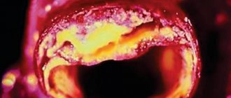
First of all, you should pay attention to atherosclerosis. This is the most common condition that leads to arterial stenosis and can cause a complete interruption of blood flow. Atherosclerosis is a disorder of fat metabolism, in which the amount of cholesterol and saturated fatty acids in the blood increases, which leads to the formation of a plaque on the vessel wall, gradually, it becomes larger and interferes with the normal rheological properties of the blood - its flow slows down. The larger the fat accumulation becomes, the more blood elements accumulate on it, increasing in size. A thrombus is formed from platelets and red blood cells that adhere to the site of wall damage, which can lead to embolism. Sometimes atherosclerotic formations can become covered with small ulcers, disrupt the integrity of the vessel, and sometimes block its lumen, stopping the movement of blood.
A blood clot attached to the wall leads to thrombosis. It is worth distinguishing this condition from thromboembolism, in which a blood clot displaces from its place, moves further along the vascular bed and, at the point of narrowing of the diameter, clogs it. The plaque itself can act as an embolus.
Causes of vasoconstriction
Vasoconstriction, also called stenosis, is familiar to a large number of people, especially those who spend a long time at the computer for work. But constant overstrain of the anatomical structures of the neck in a sitting position is far from the main cause of the development of pathology. There can be a lot of reasons, and they are divided, first of all, into:
- Congenital.
- Purchased.
We need to look at the reasons in more detail.
Congenital pathology
In the first case, narrowing of the vessels of the cervical spine is preceded by congenital hypoplasia of the veins and arteries of the cervical spine, as well as stenosis of the carotid arteries and disorders in the development of the spine in this area, vertebral arthrosis. Congenital causes may occur due to:
- distribution of toxins throughout the pregnant woman’s body;
- injuries to a pregnant woman;
- infection of the expectant mother's body.
In general, any stressful situation for a pregnant woman’s body can result in abnormal development of the fetus.
Purchased
In the case of acquired causes of the development of pathology, it is worth noting that the development of the disease is provoked by: atherosclerosis, a decrease or increase in blood pressure, an increase in blood sugar levels, injuries and hernias in the cervical spine, as well as various tumors. In addition, the disease can appear with intense mental or physical work, neglect of sports and even minimal physical activity, bad habits and stress.
What else causes stenosis?
More rare cases are associated with a specific pathology. Narrowing of the arterial vessel is observed with a disease of the connective tissue that forms the stroma of the artery. This is called fibromuscular dysplasia - the vessels cease to maintain tone and collapse, stopping blood flow. Stenosis can accompany some inflammatory diseases of the vascular wall - arteritis, systemic vasculitis, Takayasu's disease, Horton's disease. There are rare pathologies, for example moyamoya disease, in which there is a slow progressive narrowing of the lumen of the arteries.
Stenosis can occur after an injury - a strong blow, a concussion. After some traumatic brain injuries, a special hematoma is observed - subintimal, which has the appropriate localization. A blood clot that forms due to increased blood clotting and rare types of anemia can block the lumen. There is antiphospholipid syndrome, in which young patients experience heart attacks and strokes precisely because of increased coagulation. Blood clots form when heart rhythm disturbances occur while taking certain medications.
The narrowing may be due to congenital structural features of the vessels - they can be tortuous and hypoplastic. Vascular pathology accompanies systemic diseases, diabetes, obesity and metabolic disorders.
Conditions for the occurrence of narrowing of veins and arteries
The causes of vasoconstriction are very diverse. They depend on the type of vessel, external and internal factors, and the duration of their exposure.
External reasons
Arteries have a pronounced muscular layer, so they more often react with spasm to unfavorable factors. During spasm, small arteries narrow temporarily, but frequent repetition can lead to loss of the ability to relax and become stable.
Contributing factors are:
- smoking,
- stressful situations,
- alcohol consumption,
- hypothermia.
A similar external effect on the arteries is observed:
- with the development of vegetative-vascular dystonia;
- in the initial stages of hypertension and ischemic disease;
- with frostbite of the extremities;
- with Raynaud's syndrome.
Longer-term narrowing of arterial vessels from external factors is observed with mechanical compression:
- during severe injuries (long-term compartment syndrome);
- tumor growth near blood vessels;
- the pressing action of the bone tissue of the spinous processes of the spine;
- prolonged incorrect use of a tourniquet to stop bleeding (this is why first aid requires placing a note indicating the time of application).
Internal reasons
Internal causes of vasoconstriction include:
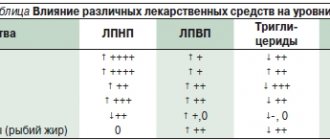
- atherosclerotic damage to the wall - between the middle and inner membranes of arteries of the muscular-elastic type, a low-density fraction of lipoproteins is deposited with the formation of plaques, over time they are supplemented with calcium salts, the lumen of the vessel loses its diameter;
- inflammatory changes (vasculitis, arteritis) - swelling of the walls reduces blood permeability;
- endarteritis - an unclear allergic reaction from the intima of the arteries of the legs and arms, leading to complete obliteration of the vessel;
- congenital pathology (aortic stenosis);
- thrombosis and embolism - play an important role in the development of pathology of the brain and heart;
- metabolic disorders in diabetes mellitus, thyrotoxicosis, obesity.
What manifestations are accompanied by the pathology?
The clinical manifestations of the disease depend on where the problem is located. Also, the nature of the onset of the disease, which can be sudden or gradual, plays an important role. It is worth remembering that in the first stages of the disease, compensatory mechanisms always turn on. For example, if the blood flow is partially disrupted, a system of collaterals develops, which will provide alternative routes for blood supply to the brain. The lack of oxygen supply is also compensated - the pulse and breathing become more frequent, intracellular processes change. For short-term conditions this is enough, but for serious pathologies it is not. In such cases, symptoms of the condition develop. Naturally, with an abrupt onset and significant stenosis, compensatory forces do not work and the clinical picture has a pronounced character, which indicates a stroke.
One of the first alarm signals is transient cerebral ischemia. These are short-term episodes of circulatory disturbance that can be compensated by collateral blood flow. The condition can be recognized by decreased sensitivity and motor activity, as well as visual disturbances. Patients usually complain of a feeling of numbness in the upper limb, half of the face, and loss of visual fields. Decreased muscle strength in the arm or forearm. The patient feels the movement of dark spots in front of the eyes, as well as a decrease in visual acuity. In some cases, the section of the artery that supplies the retina is affected. In some localizations, corresponding disorders occur - difficulty speaking, hearing, swallowing, facial asymmetry, severe pain. More common symptoms are dizziness, feeling unwell, weakness, and convulsions.
If stenosis develops gradually, the clinical picture also develops slowly. In some cases, it is so nonspecific that it is difficult to recognize the disease. For a long time, both the patient and the doctor may mistakenly believe that the cause of poor health is fatigue or increased stress. Also, the symptoms of occlusion overlap with the symptoms of a tumor process and dementia. This is indicated by increased irritability, depressive disorders, sudden changes in mood, increased drowsiness, decreased memory and mental abilities.
Nonspecific symptoms require a high level of responsibility from the patient and the doctor, since high-quality diagnosis is necessary. The diagnosis determines how correct the treatment of the pathology and the result will be.
Get ahead of the disease and help yourself
The prognosis for a disease such as cerebral vasoconstriction depends mainly on how quickly it was diagnosed and how quickly treatment was started. The technique should only be determined by the attending physician. Because in each specific case, tactics must be individual. However, the following can serve as general recommendations:
- It is necessary to change the food system, where the daily diet must certainly consist of fresh vegetables and fruits containing useful substances and always greens! Be sure to include fish in your daily menu, but it is better to limit sweets, smoked foods, sausage, carbonated water, and salt or eliminate them altogether.
- You should reconsider your lifestyle and engage in more sports, go for walks in nature more often, get fresh air, start an active, eventful life, prevent excess weight, and move a lot.
Diagnostic program for the disease
First of all, a timely start to the diagnostic search is necessary. It is better to rule out the disease and continue to live peacefully than to postpone a visit to the doctor “for later.” For symptoms that cause concern, professional medical attention is necessary. The doctor begins making a diagnosis by interviewing the patient and general clinical examinations. If there are neurological symptoms, reflexes, sensitivity, motor activity, muscle and analyzer function are assessed. The following instrumental methods are used:
- Ultrasound of the arteries of the head and neck with Doppler sonography - triplex scanning. A safe, fast and inexpensive method that allows you to exclude or confirm the diagnosis;
- Functional tests during ultrasound examination;
- Computed tomography angiography - visualization of blood flow through vessels using a contrast agent;
- Magnetic resonance imaging of the brain.
To diagnose atherosclerosis, a laboratory method is used - the patient donates blood for a lipid profile. Also, it is necessary to evaluate the blood coagulation system and its coagulation properties. It is mandatory to measure pulse and blood pressure. To identify systemic vasculitis, specific laboratory tests and immunological studies are used.
MRI and CT are the “gold standard” in the diagnosis of ischemic disorders, including stroke. Depending on the location and extent of the lesion, treatment is prescribed.
When do veins narrow?
The venous system is more prone to lose tone, but there is a pathology that leads to a persistent gradual loss of the diameter of the venous capillaries, and then the cessation of blood supply. We are talking about cirrhosis of the liver. All blood flowing from the tissues necessarily passes through this organ. With chronic inflammation of the lobules of liver cells (hepatocytes) and replacement of the interlobular space with scar tissue, the venules narrow. Then the blood flow through them completely stops. Problems arise in the area of the portal vein. Due to its sharp narrowing, hypertension and stagnation occur in the underlying sections, and overload of “excess” into the veins of the esophagus.
On the right is the liver of a patient with cirrhosis, there are no vessels between the compacted lobules
Thrombophlebitis (inflammation + thrombosis) cannot be excluded as a cause. In diseases that cause a decrease in blood flow velocity (stagnation), the process of parietal thrombus formation is activated. The spread of infection from chronic foci increases the narrowing of the affected area of the vein.
Symptoms of circulatory disorders due to altered vascular lumen depend on the specific location of the affected area. Let's look at the manifestations of the most significant diseases.
Is it possible to be treated with traditional methods?
You cannot straighten a narrowed vessel with folk remedies. You should not collect and test on yourself numerous “tips” for cleaning and getting rid of atherosclerosis.
Using a combination of garlic, lemon and honey is more of a dietary recommendation. It has a good effect on the immune system, so it is always useful. The product will help in the recovery period after stress, injury or infection. But it is impossible to “dissolve plaques” at the current level.
Medicines will help delay further narrowing. People's advice can be tried against the background of their application.
Symptoms of insufficient blood supply to the legs
Signs of narrowing of the arteries of the lower extremities are detected in obliterating endarteritis, vascular atherosclerosis, and Leriche syndrome.
Concerned:
- pain in the legs, first only when walking, then at rest;
- symptom of “intermittent claudication”, after stopping the pain disappears;
- numbness and chilliness of the feet;
- increased sweating;
- cramps in the calf muscles;
- in severe stages, trophic disorders - ulcers, non-healing cracks in the feet, gangrene.
Clinical manifestations of impaired blood supply to the brain
Signs of cerebral insufficiency are caused by narrowing of the carotid and vertebral arteries, which carry blood to the brain.
For adults, the main importance is given to:
- atherosclerosis;
- cervical osteochondrosis;
- congenital pathology of vertebral vessels;
- hypertension;
- thromboembolic complications.
The disease can take a long-term chronic course or occur suddenly in the form of a stroke.
For the child, the most important are:
- mother's condition during pregnancy;
- birth injuries;
- previous vasculitis due to childhood infections;
- congenital pathology of the heart and blood vessels.
If you faint, a stroke cannot be ruled out.
In acute cases, concern:
- severe headaches;
- dizziness to the point of loss of consciousness;
- noise in ears;
- impaired vision;
- decrease or disappearance of sensitivity and movements in the limbs;
- impaired speech.
Chronic deficiency occurs gradually, all symptoms begin with irritability, insomnia, and impaired attention. Then memory loss, fatigue, headaches, and insomnia increase. Further narrowing of blood vessels leads to a change in personality, the inability to contact others, and movement disorders.
Clinical features for narrowing of arteries in the neck
Atherosclerosis of the carotid artery is considered the first manifestation of the disease. It is detected during Doppler sonography in individuals who do not yet have symptoms. Osteochondrosis affects the processes of the vertebrae in the cervicothoracic region, which compress the vertebral artery. The patient is concerned about:
- headaches in the temples, back of the head, forehead;
- feeling of "pressure";
- connection of well-being with turning and tilting the head to the side;
- dizziness, darkening of the eyes;
- possible loss of consciousness;
- nausea, vomiting.
Less often they complain of numbness of the tongue and hands.