According to statistics, vascular diseases of the brain account for about 42% of all abnormalities of the nervous system. In the field of neurology, cerebrovascular accidents are recognized as one of the most common ailments. Affecting a significant number of elderly people and patients of older working age, these diseases pose a serious threat not only to normal life activities, but also to the patient’s life itself. Vascular pathologies require great attention from specialists and timely and effective treatment.
When it comes to vascular pathologies, they most often mean discirculatory encephalopathy. This is a slowly progressive vascular lesion of the brain. The course of the pathology is divided into three stages in accordance with the severity of the clinical picture. At the initial stage, the symptoms are not clearly expressed, so problems arise with making a diagnosis. Some symptoms indicate mental disorders, so errors in the anamnesis often occur. As a rule, an accurate diagnosis is made after a prolonged presence of the main symptoms (dizziness, impaired coordination and cognitive functions).
Classification of vascular diseases of the brain
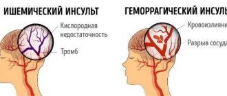
There are two main groups of vascular pathologies: acute and chronic cerebrovascular accidents. The first group is divided into three subgroups: transient cerebrovascular accidents, cerebral strokes, mixed strokes. Transient disorders of cerebral circulation include transient ischemic attacks and hypertensive crises. Brain strokes are ischemic and hemorrhagic. Hemorrhagic strokes are divided into subarachnoid hemorrhage, parenchymal-subarachnoid hemorrhage, and ventricular-parenchymal-subarachnoid hemorrhage. Ischemic strokes, in turn, are divided into lacunar, atherothrombotic, cardioembolic and hemodynamic stroke.
The second group includes: chronic subdural hematoma, initial manifestations of cerebrovascular insufficiency, subcortical vascular dementia, discirculatory encephalopathy. The initial manifestations of cerebrovascular insufficiency are divided into two stages. The first stage is characterized by the absence of neurological manifestations. At the second stage, neurological manifestations are clearly expressed. Discirculatory encephalopathy is the most common among chronic cerebrovascular accidents. It is divided into three main stages:
- The first stage is moderately severe, characterized by diffuse symptoms. The patient may complain of short-term headaches, periodically suffer from sleep disorders and increased irritability.
- The second stage is more pronounced; clinical manifestations form in it. The person often experiences surges in blood pressure, loss of space, and cognitive impairment. As a rule, at this stage the disease is most often diagnosed.
- The third stage is pronounced, during which irreversible changes occur in the body. Serious disturbances in memory and cerebral circulation in general appear. In rare clinical situations, complications in the form of strokes are observed (in the absence of timely consultation with a doctor).
Difficulties in diagnosing thrombosis of the cerebral veins (CV) and venous sinuses (SV) are associated with the variety of clinical manifestations, the localization of thrombosis, the rate of its development, and the cause of the disease [1–3]. The MV and dural sinuses contain about 70% of the blood flowing to the brain, however, thrombosis of the MV and VS is much less common than arterial thrombosis. The course of thrombosis of MV and VS is extremely variable - from progressive and recurrent to benign and curable [4].
This disease has been known for more than 200 years - the first description of thrombosis of MV and VS was made by J. Morgagni in 1761 [5]. In 1825, the French physician M. Ribes [6] described a patient with headache, epileptic seizure and delirium, who was found to have thrombosis of the MV and VS.
Large epidemiological and clinical studies are limited by the small number of patients with CF and VS thrombosis. Most publications about this disease are casuistic reports. Results from multicenter studies have only been published within the last 15 years, including the International Study on Cerebral Vein and Dural Sinus Thrombosis (ISCVDST) [7], based on 624 observations. The Italian registry includes data from 706 cases of MV and VS thrombosis [8]. One of the latest retrospective studies [9], including 152 patients with MV and VS thrombosis, was conducted in 2021.
Epidemiology
To date, there are no epidemiological studies on MV and VS thrombosis, so the exact incidence rate is unknown. The first data on the incidence of MV and VS thrombosis were obtained from autopsy results. H. Ehlers and C. Courville [10] in 1936, summarizing the results of 12,500 autopsies, identified 16 cases of thrombosis of MV and VS, but data from modern studies [4] indicate that the incidence is 10 times higher.
The prevalence of MV and VS thrombosis is heterogeneous. In most patients, the disease develops between the ages of 20 and 50 years. Children and elderly people also get sick. In this regard, the epidemiology of the disease is separately described for different age groups. According to ISCVDST, the incidence of thrombosis of MV and VS in adults is 3-4 cases per 1 million, and in children and newborns - 7 cases per 1 million child population. Until the mid-60s, the incidence of thrombosis of MV and VS in men and women was considered equal, however, according to recent reports [7], thrombosis of MV and VS occurs more often in women, especially in the age group from 20 to 35 years (male: female ratio equals 3:1). The high prevalence of the disease in women of childbearing age is most likely associated with pregnancy, the postpartum period, and the use of oral contraceptives [11]. According to a study conducted in the USA in 1993-1994, about 12 births out of 100,000 are complicated by the development of MV and VS thrombosis [7]. An increase in the proportion of women among patients with MV and VS thrombosis over the past decade has been shown; currently it is about 70%. The predominance of female patients is explained by hormonal factors. About 1/3 of women of childbearing age in Western countries use oral contraceptives, which accounts for approximately ½ of patients with MV and VS thrombosis [12].
There are no reliable data regarding geographic or ethnic differences in incidence, but several studies have reported noteworthy results. In 2021, data from a study were published in the Adelaide region (Australia) with an adult population of about 1 million. In a retrospective analysis of all cases of MV and VS thrombosis for the period 2005-2011. the incidence was 15.7 cases per 1 million population per year; an almost equal distribution of patients by gender was found (52% women, 48% men) [13]. Prevalence of MV and VS thrombosis in the Netherlands in 2008–2010. amounted to 13.2 cases per 1 million population; to Hamadan (Iran) in 2009-2015. — 13.5 cases per 1 million population [12].
Localization
Thrombosis is more often localized in the dural sinuses than in the M.V. The most common thrombosis is the superior sagittal, sigmoid and transverse sinuses, and then - in descending order: thrombosis of the cortical veins, cavernous sinus, cerebellar veins. Thrombosis of the superior sagittal sinus occurs in 46% of cases, sigmoid or transverse sinuses - in 32%, several sinuses - in 20%, superior sagittal sinus and superficial cerebral veins - in 40% of cases [4]. In 2/3 of cases, thrombosis is not limited to one sinus, but spreads to adjacent sinuses and veins. Isolated thrombosis of the superior sagittal sinus is observed in 13-55% of cases, sigmoid and transverse sinuses - in 10%; in 40% of cases, sinus hypoplasia is observed, which is difficult to distinguish from thrombosis [14].
In a retrospective study by J. Liang et al. [15] the following data were obtained: thrombosis of the superior sagittal sinus occurred in 72.7% of cases, left transverse sinus - in 43.2%, left sigmoid sinus - in 43.2%, right transverse sinus - in 36.4%, right sigmoid - in 36.4%, straight sinus - in 9.1%. In 47.7% of cases, thrombosis of the MV and VS was accompanied by secondary changes in the brain (infarctions were detected in 43.2% of cases, hemorrhages in 27.3%). Most often, secondary changes were localized in the frontal (31.8%) and parietal (36.4%) lobes. Subarachnoid hemorrhages occurred in 13.6% of cases, subdural hematomas - in 4.5%.
L. Zhou et al. [16] revealed thrombosis of the transverse sinus in 65.0% of patients, sigmoid sinus in 55.6%, and superior sagittal sinus in 54.7%. Secondary brain changes were found in 56.4% of patients. Edema of limited areas of the brain was observed in 30.8% of patients, massive edema - in 4.3%, intracranial hematomas - in 15.4%, subarachnoid hemorrhages - in 4.3%.
Risk factors
The diseases that are most often associated with thrombosis of the MV and VS include infections of the orbital region, mastoiditis, inflammatory diseases of the middle ear and face, and meningitis. Inflammatory processes in the area of the mastoid process or face are a predisposing factor for the development of thrombosis of the transverse and sigmoid sinuses. Cavernous sinus thrombosis is caused by infections of the paranasal sinuses (ethmoid and sphenoid) [3, 4]. S. Imam et al. [17] described thrombosis of MV and VS in a 39-year-old patient with chickenpox.
Risk factors for the development of aseptic thrombosis of MV and VS are taking oral contraceptives, pregnancy, the postpartum period, traumatic brain injury (including mild), implantation of a pacemaker or prolonged standing of a subclavian venous catheter with its thrombosis, tumors, collagenosis (systemic lupus erythematosus, disease Behçet, Sjogren's syndrome), blood diseases (polycythemia, sickle cell anemia, thrombocytopenia), antiphospholipid syndrome, thrombophilia (most often caused by mutations in the factor V Leiden and prothrombin genes, deficiency of antithrombin III, protein C), nephrotic syndrome. A history of indications of episodes of increased blood clotting (deep vein thrombosis, thromboembolism of the arteries of the pulmonary trunk system, stroke, myocardial infarction, multiple miscarriages) suggests a state of primary hypercoagulation. Conditions accompanied by secondary hypercoagulation include late pregnancy, childbirth, and the presence of a brain tumor. Hemodynamic disturbances include dehydration, anemia, and congestive heart failure. Changes in the vascular wall can occur as a result of injury, brain tumor, or viral encephalitis [3, 4]. In some cases, a connection between MV and VS thrombosis and temporal arteritis, Wegener's granulomatosis [18] and Churg-Strauss syndrome [19] has been suggested.
In obstetric and gynecological practice, thrombosis of MV and VS mainly occurs in the postpartum period, especially during the first 3 weeks after birth. Risk factors for the development of the disease include dehydration, anemia, cesarean section, arterial hypertension, infectious diseases, maternal age (15-24 years), thrombophilia [4]. Thrombophilia accounts for 34.1% of all causes of thrombosis of MV and VS [4]. Most often we are talking about an abnormality of factor V, which gives it resistance to activated protein C; mutations of the antithrombin III genes and proteins C and S are less common. Thrombosis can also be caused by a deficiency of factors of the anticoagulation system of the blood and resistance of factor V to protein C. In addition, hyperhomocysteinemia predisposes to thrombosis. According to a study by Y. Kapessidou et al. [20], hereditary and acquired forms of thrombophilia were the cause of thrombosis of MV and VS in 64% (7 out of 11) of cases, including protein S deficiency detected in 4 patients, idiopathic thrombocytopenic purpura - in 2 patients (in one case associated with systemic lupus erythematosus, in another - with antiphospholipid syndrome).
Based on the results of several case-control studies [21–23] and one meta-analysis [8], there is a strong association between the use of oral contraceptives and the development of MV and VS thrombosis. The risk of thrombosis may vary depending on the composition of the drugs. Third-generation oral contraceptives are characterized by an increased risk of developing the disease [21], and the risk of its development when using contraceptive patches is similar to the risk when taking oral forms [24]. In approximately 1/3 of the cases cited in the literature, the etiology of MV and VS thrombosis remains unclear [4].
Clinical picture
The clinical picture of MV and VS thrombosis is variable. The average period from the onset of symptoms to diagnosis is 7 days [7]. The most common initial symptom of thrombosis of MV and VS is a sudden intense headache, which, as a rule, is diffuse and difficult to relieve with analgesics. Its course can be acute (less than 48 hours), subacute (from 48 hours to 30 days) and chronic (more than 30 days). Then, focal and general cerebral symptoms such as depression or confusion, epileptic seizures (usually partial with secondary generalization and post-attack paralysis), hemiparesis, aphasia, congestive optic discs, and mental disorders occur. Neurological symptoms in thrombosis of MV and VS most often develop subacutely, in a period from several days to 1 month (in 50-80%), although an acute onset can also be observed (in 20-30%) [25].
The International Headache Society has defined diagnostic criteria for headache in thrombosis of MV and VS:
A. New-onset headache that meets criteria C and D.
B. Thrombosis M.V. and VS diagnosed using neuroimaging methods.
C. Simultaneous development of headache, focal neurological symptoms and thrombosis of the MV and VS.
D. Headache that regresses within 1 month after the start of specific treatment.
With thrombosis of the MV and VS, venous congestion develops in the brain due to obstruction of venous outflow. Irritation of the receptors located in the wall of the MV and the interoceptors of the meninges with increased pressure in the venous system of the brain causes the appearance of headaches. Disruption of venous outflow leads to an increase in intracranial pressure and the development of hypoxia, resulting in necrotic changes in brain tissue. About 90% of patients describe persistent intense headache, more pronounced in the morning, as the main complaint with thrombosis of MV and VS. Typically, pain increases when the patient is in a horizontal position [25, 26].
Headache due to thrombosis of MV and VS has no specific features. Its clinical manifestations depend on the location and severity of thrombosis, the age of the patient, and the time from the onset of the disease. It is less common in elderly patients than in young patients, which may be due to a lower incidence of intracranial hypertension and decreased activity of the pain perception system in the elderly [26].
Headache due to thrombosis in 50% of cases has a subacute onset, intensifies over several days, but can be paroxysmal in nature. In 10% of cases, a thunderclap headache occurs. Gradually, the pain becomes persistent, refractory to painkillers and persists at night, intensifying with physical activity and the Valsalva maneuver.
Subjective characteristics of headache have no diagnostic value. Diffuse, predominantly bursting, headache is more common, and local pain is less common (42%). The intensity of pain is usually moderate or high. In most cases, headaches worsen at night and early morning, accompanied by nausea, vomiting and phonophobia. It can imitate migraine, tension headache, cervicalgia and is in no way related to the localization of the pathological process [27].
Headache may be a consequence of the chronic course of thrombosis of MV and V.S. Its peculiarity is that there are no indications of an acute episode of headache or the occurrence of neurological symptoms, however, MRI in the venosinusography mode reveals signs of thrombosis of one of the V.S. The predominant type is a bilateral headache of a pressing or bursting nature with a tendency to persist, aggravated by physical stress or emotional experience [2].
A prospective study examined headache characteristics in 123 patients with MV and VS thrombosis. Patients with intracranial hypertension and meningitis were excluded from the study. It was shown that in 14% of patients, headache was the only manifestation of the disease. In 88% of patients, thrombosis involved the transverse sinuses, and almost all (except one) cases of unilateral venous thrombosis had ipsilateral headache. Patients often characterized the headache as constant, one-sided, of high intensity and pulsating in nature. In most cases, patients noted a sudden onset of the attack; 3 patients described the “thunderous” nature of the headache. The pain syndrome regressed within several days or weeks after the start of treatment for MV and V.S. thrombosis. The prognosis was favorable in all cases. The authors suggested that the pathogenesis of headache in the absence of intracranial hypertension is associated with irritation of nerve endings in the walls of occluded MVs and VSs [28].
Swelling of the optic discs is observed in 45-86% of patients with thrombosis of the MV and V.S. The initial manifestations may be epileptic seizures, the frequency of which ranges from 10 to 60%. Focal neurological symptoms (paresis, dysarthria, visuospatial disturbances, homonymous hemianopsia) occur in 15% of patients [7, 27].
The results of a three-year clinical study of patients with MV and VS thrombosis showed that 60% (29 out of 48) of patients developed headaches: migraine (14), tension-type headache (13), other types of headache (2). Upon further evaluation (mean period 44 months), chronic migraine was diagnosed in 25% (6 of 24) of patients. Microhematomas were detected in most patients. In a study by R. Cumurciuc et al. [29] in 30% of patients with thrombosis of MV and VS, chronic headache was observed within 3 months. In a retrospective study by L. Zhou et al. [16], which included 117 patients with thrombosis of the MV and VS, it was shown that headache was observed in 87.2% of patients, epileptic seizures - in 31.6%, focal neurological symptoms - in 29.9%, visual disturbances - in 26.5%, disturbances of consciousness - in 15.4%.
Diagnostics
Due to the absence of pathognomonic clinical symptoms, instrumental research methods are of utmost importance in the diagnosis of MV and VS thrombosis. In recent years, the improvement of neuroimaging technologies has opened up new opportunities for diagnosing the disease (MRI, MRI and CT venosinusography). Under K.T. and MRI in standard modes can reveal cerebral edema, foci of necrotic changes in brain tissue, a filling defect in the area of confluence of sinuses (“delta sign”), as well as signs of venous thrombosis (increased signal intensity from an altered VS in T1 and T2 modes, and also T2-FLAIR) [2, 3, 30].
If after MRI or CT the diagnosis remains unclear, it is possible to perform digital subtraction angiography, which can detect not only VS thrombosis, but also the rare isolated MV thrombosis. Also, during digital subtraction angiography, it is possible to detect dilated and tortuous veins, which is an indirect sign of V.S. thrombosis. At the same time, a careful assessment of neuroimaging data is necessary to exclude diagnostic errors that may be caused, for example, by hypo- or aplasia of the sinuses [2, 3]. In a recent study by J. Kang et al. [31] it was shown that the ASL perfusion method with MRI allows diagnosing MV and VS thrombosis with high accuracy.
If hereditary thrombophilia is suspected, the level of homocysteine, the concentration and activity of antithrombin III, proteins C and S, and thrombophilic mutations are determined: mutations in the methylenetetrahydrofolate reductase gene - C 677 T
;
mutations in the blood coagulation factor V gene - G 1691 A
(factor V Leiden);
mutation in the prothrombin gene - G 20210 A
[2, 3, 25].
When taking oral contraceptives and liver disease, the concentration of all three factors of the blood anticoagulation system decreases simultaneously, and during pregnancy, primarily proteins C and S. With nephrotic syndrome, the concentration of antithrombin III decreases, and proteins C and S increase. Disorders of the anticoagulation system of the blood are far from the only cause of thrombosis of MV and VS: the disease can be caused by antiphospholipid syndrome and coagulopathy. The diagnosis is confirmed by the study of lupus anticoagulant, antibodies to cardiolipin and β2-glycoprotein, blood clotting factors, and determination of von Willebrand factor.
Treatment
The main goal of treatment for thrombosis of MV and VS is to restore their patency. According to various studies, the use of direct anticoagulants in the acute period of thrombosis of MV and VS improves the prognosis and reduces the risk of disability. The effectiveness of heparin in therapeutic dosage has been proven in double-blind, placebo-controlled studies. The hemorrhagic component in thrombosis of MV and VS is not a contraindication to the administration of heparin in a therapeutic dose. The initial dose of heparin is 5000 units and is administered intravenously as a bolus, after which they switch to intravenous drip administration at a rate of 1000 units/hour. The average daily dose of the drug is 20,000-40,000 units. Activated partial thromboplastin time should be monitored every 3 hours; it should be doubled compared to the norm. If the desired result cannot be achieved, the heparin dosage is gradually increased every 6-8 hours by 100-200 units. Upon completion of the acute phase of the disease, it is recommended to transfer the patient to oral warfarin. In this case, while the administration of direct anticoagulants continues, warfarin is prescribed under the control of the international normalized ratio (INR) (INR 2-3), then heparin is discontinued, and oral administration of warfarin is continued for 8-12 months [4, 32].
In the ISCVDST study [7], 80 patients out of 624 with MV and VS thrombosis received low molecular weight heparins. 79% of them recovered, 8% had mild symptoms, 5% had significant neurological impairment, and 8% died. These data indicate the effectiveness and safety of the use of low molecular weight heparins in the acute period of thrombosis of MV and VS.
J. Scott et al. [33] were the first to report the effectiveness of local thrombolytic therapy (TLT) for MV and VS thrombosis. Along with drug TLT [34], in recent years, for thrombosis of the MV and VS, instrumental methods of removing blood clots from the sinuses of the dura mater have been used - thrombectomy [35] and decompressive craniotomy [30].
Mortality
The mortality rate for thrombosis of MV and VS is 5-10% [5, 30]. The decrease in mortality is due to an increase in the quality of hospital care and improved diagnosis of the disease [36], as well as a decrease in the incidence of traumatic brain injuries and severe infectious diseases. The use of anticoagulant therapy and decompressive craniotomy had a positive effect on patient survival [12, 30].
Recurrent course
The incidence of recurrence of MV and VS thrombosis remains unknown. In a study by P. Palazzo et al. [37], which included 187 patients, the average follow-up period was 73 months, the average duration of anticoagulant treatment was 14 months. Repeated thrombosis of MV and VS was diagnosed in 6 patients, extracranial venous thrombosis - in 19. The rate of recurrence of venous thrombosis after 1 year was 3%, after 2 years - 8%, after 5 years - 12%, after 10 years - 18%. According to the results of the ISCVDST study [7], the incidence of recurrent thrombosis of MV and VS was 2.2%, extracranial venous thrombosis - 3%; the observation period was 16 months.
Forecast
When treatment is started in the early stages of MV and VS thrombosis, the prognosis is favorable in more than 90% of patients. In the absence of treatment for thrombosis of MV and VS, the prognosis is unfavorable in 15% of cases, mortality reaches 10%, but even with significantly pronounced neurological disorders, complete spontaneous recovery is possible. Risk factors for the development of an unfavorable prognosis of the disease are the rapid progression of thrombosis with depression of consciousness, generalized epileptic seizures at the onset of the disease, childhood and old age of patients, localization of thrombosis (cerebellar veins and deep MVs) [4, 7].
The authors declare no conflict of interest.
Causes of pathology
Vascular diseases of the brain can occur for a number of reasons. As a rule, several reasons lead to the appearance of pathology. The most common ones include:
● genetic predisposition;
● narrowing and hardening of intracerebral arteries (with hypertension);
● multiple focal and/or diffuse brain lesions;
● long-term somatic diseases of the expectant mother (and during pregnancy);
● atherosclerosis of cerebral vessels;
● rheumatic lesions;
● disturbances in the functioning of the respiratory system (for example, pulmonary edema);
● abuse of alcohol and tobacco products during pregnancy;
● disturbances in metabolic processes;
● increased blood viscosity;
● fractures of the skull bones;
● heart rhythm disturbance (with atrial fibrillation, blood hemodynamics are disrupted);
● serious abnormalities in the nervous system (increased anxiety, chronic stress);
● serious abnormalities of the endocrine system (diabetes mellitus, obesity);
● development of intrauterine infections;
● difficult long labor (with injuries);
● hypertension;
● hypercholesterolemia;
● various anomalies of the cardiovascular system (for example, hypoplasia of cerebral vessels, mitral valve prolapse).
MRI or CT scan of cerebral vessels?
MRI and CT have their advantages in different situations. People who have electronic devices (pacemaker) or objects made of magnetic metals in their body are allowed only CT scanning.
An MRI scan takes tens of minutes and requires you to lie still. A CT scan takes less time, but this procedure uses X-rays. CT scans should not be done too often. The procedure is contraindicated for pregnant women at any stage. MRI is performed as many times as necessary; it is contraindicated only in the 1st trimester of pregnancy.
MRI of cerebral vessels is a safe procedure that will provide a detailed picture of the disease. At the medical office, you can undergo an MRI scan by appointment on any convenient day and time.
Risk group
The risk group includes people with infectious pathologies during the neonatal period. The likelihood of developing vascular diseases of the brain increases significantly with hemolytic anemia, surgery and head injuries. The mother’s poor lifestyle (abuse of alcohol and tobacco products) can also negatively affect the condition of the child’s brain. A family history of cardiovascular pathologies (for example, myocardial infarction or arterial hypertension in close relatives) doubles the risk of developing cerebrovascular diseases.
2. Reasons
Considering the critical, priority importance of the brain, evolution took care of its maximum protection: powerful skull bones, upper location, as well as “reserve circuits” of cerebrovascularization (blood supply to the brain). The brain receives nutrition from several main vessels - the carotid and vertebral arteries; the latter, merging, form the unpaired basilar artery, or vertebrobasilar arterial system. If there are disturbances in any of the blood supply channels, other channels compensate for the deficiency in order to preserve the functioning of the brain at any cost. The compensatory capabilities of the brain in this sense are very great (and have not yet been sufficiently studied), however, unfortunately, they also have their limits. With intense sudden ischemia, the cerebrovascular system simply does not have time to react and prevent irreversible necrotic changes.
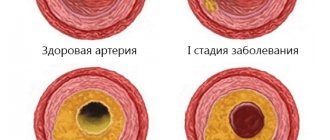
Spasm of the main cerebrovascular vessels can occur for various reasons: massive bleeding, sudden blow, rapid hypothermia, poisoning with certain substances, etc. However, all these factors occupy only a small proportion compared to the leading cause of ischemic stroke: embolism, i.e. blockage of the arteries by a detached atherosclerotic plaque, a clot of coagulated blood, etc. In general, arterial hypertension and atherosclerosis, lipid metabolism disorders with the accumulation of mushy deposits on the walls of blood vessels, are typical “diseases of civilization”, since they are largely caused by the very lifestyle and destructive habits of modern man: physical inactivity, extremely unhealthy diet and overeating, smoking etc. It is no coincidence that circulatory disorders in various systems of the body (primarily the coronary and cerebral arterial systems) consistently occupy first place in the lists of causes of non-violent mortality.
Visit our Neurology page
Diagnosis of pathology
If the symptoms described above appear, it is recommended to immediately consult a cardiologist or neurologist. During the initial examination, the specialist collects an anamnesis of the patient’s life, asks in detail about his complaints (how long ago and under what circumstances they appeared), about diseases of the cardiovascular system of his closest relatives. Based on these data, further examination is prescribed.
To diagnose vascular diseases of the brain, they resort to laboratory and instrumental diagnostics. First of all, the patient is prescribed a general and biochemical blood test to determine the level of cholesterol, glucose, triglycerides, lipoproteins and prothrombin index (PTI). These substances affect the functioning of the heart and blood vessels. Biochemical indicators are very important in making a diagnosis. An increased level of leukocytes indicates the presence of an inflammatory process in the body. Among the instrumental diagnostic methods, the following are in particular demand:
● electrocardiography (ECG). This is a simple technique that does not require additional preparation. The essence of an ECG is to record the electrical potentials of the heart, due to which it is possible to detect changes in rhythm, electrolyte deficiency and other initial signs of cardiovascular pathologies;
● Doppler ultrasound of the vessels of the neck and brain (USDG). This is a modern, highly informative diagnostic method. It is used to determine damage to the arteries and indicates insufficient blood supply to the brain. With its help, it is possible to assess blood flow in the vessels of the neck and head;
● duplex scanning of blood vessels. Thanks to color contrast of flows during duplex scanning, it is possible to more clearly distinguish between moving and stationary objects (blood and vessels, respectively);
● ultrasound examination of the heart (ultrasound). This technique makes it possible to visually view the contractions of the heart muscle, assess the condition of its valves, and diagnose heart failure;
● echocardiography (EchoCG). Sometimes specialists prescribe echocardiography in addition to an ECG. This is a highly accurate examination method, with its help it is possible to assess the condition and determine the size of the valves;
vascular magnetic resonance imaging (MRI). This is a completely safe technique for the body (there is no radiation), it shows the lumen and patency of large vessels, and makes it possible to detect abnormalities in vascular development.
How to do an MRI of cerebral vessels
After the patient enters the radiologist's office, the doctor asks several questions about bothersome symptoms and contraindications for the procedure. The patient is asked to remove all metal objects and lie down on the tomograph bed. Normal operation of the tomograph is accompanied by noise, knocking and other loud sounds; this should not be alarmed. In our medical center, each patient is given headphones that play pleasant, calm music during the examination.
Preparation for MRI of the head and blood vessels
The patient is given a special button in his hand, which can be pressed during the procedure to inform the doctor about a sudden deterioration in health.
Prevention of vascular diseases of the brain
Vascular diseases of the brain are easier to prevent than to cure. To do this, experts recommend leading a healthy lifestyle: engaging in moderate sports, drinking about 1-1.5 liters of water per day (to avoid dehydration), eating properly and following a diet. The diet involves limiting table salt and foods containing large amounts of animal fats (for example, sour cream and fatty meats). Fresh vegetables and fruits should prevail in the daily diet. A split diet is recommended (4-5 meals per day).
The intake of alcoholic beverages should be minimized (or completely abandoned). The emotional component is the key to good health, so you should not be exposed to stressful situations. Sleep should be complete (at least 7 hours of sleep per day are required). Moderate physical activity should be regular; it significantly improves the condition of the body. These include: swimming, yoga, Pilates, cycling.
Exercising and proper nutrition help prevent the appearance of excess weight, while they train the cardiovascular system. At least once a year it is required to undergo a study of the state of cerebral circulation (especially for those who are at risk). This will allow timely detection of pathology.
In order to prevent the progression of multi-infarction conditions, patients are prescribed combination therapy (antiplatelet and anticoagulant). The most suitable anticoagulants are selected depending on blood clotting parameters. If any signs of bleeding appear, it is important to contact a specialist promptly.
Vascular diseases of the brain are often accompanied by dizziness. To prevent them, doctors prescribe medications that affect the autonomic nervous system. To prevent cognitive impairment (memory deterioration, increased inattention), drugs that improve metabolism are prescribed. In the presence of movement disorders, therapeutic exercises, physiotherapy, massage and other methods of rehabilitation therapy are useful.
Contrast MRI of cerebral vessels
Magnetic resonance angiography (MRA) creates images based on signals from hydrogen atoms, which are abundant in blood and other fluids. The image on the monitor screen is very clear, so a contrast agent is rarely required. When performing computed tomography, unlike MRI, contrast is used much more often.
In MRI diagnostics, substances based on gadolinium are used. A contrast agent is injected into a vein, changing the magnetic properties of the tissue for a short time, and is excreted through the kidneys. It was possible to create gadolinium compounds that rarely cause unwanted and allergic reactions. There is no evidence that they cause neurological problems after contact with the human body. However, they are used with caution in severe kidney disease.
Before a contrast study, be sure to tell your doctor about:
- pregnancy (confirmed or probable);
- breastfeeding;
- kidney diseases;
- previous allergic reactions to any medications.
Contrast makes it easier to decipher images, as it shows the condition of the vessels in more detail.
Gadolinium-based contrast agent
Diagnostic methods
Neurologist
It is not enough to interview and examine the patient to identify narrowing of the blood vessels of the spine; in any case, additional examinations will be prescribed. These include:
- Duplex scanning
- despite the fact that this is a rather dangerous diagnostic method in this situation, it is informative and accessible, it allows you to determine the degree of narrowing and its nature. - Angiography - the process uses a contrast agent to examine the vessels.
- and MRI
- allow you to obtain layer-by-layer 3D images, with which you can make a diagnosis.
All this will allow the doctor to determine not only the patient’s health status, but also the reasons that led to the occurrence of stenosis.
Consequences of cerebral circulatory disorders
The brain is the organ most vulnerable to lack of oxygen. The circulating blood volume in the brain remains virtually unchanged and is about 750 ml. Since nervous tissue is not able to retain oxygen, its reserve lasts for 8–10 seconds. When brain cells are deprived of blood supply, that is, there is no access to vital nutrients and oxygen, they die - a stroke occurs.
The consequences of a stroke can vary widely depending on the area of the brain, the severity of the impairment, and the general health of the person.
Cerebral circulatory disorders include:
- cerebral hemorrhage,
- thrombosis,
- embolism (blockage),
- subarachnoid hemorrhage.
They are usually the result of pre-existing vascular disease, congenital pathology, or may be triggered by trauma.
Symptoms
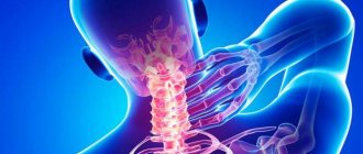
Symptoms of vasoconstriction in the cervical spine do not appear for a very long time, and a person with this disease does not notice any deterioration in well-being. However, later in the development of the disease it begins to manifest itself:
- constant dizziness;
- lack of strength even with constant rest;
- sudden loss of consciousness for no reason;
- flickering before the eyes;
- noise or ringing in the ears;
- pressing or aching pain in the head, particularly in the back of the head, may spread to the neck.
In general, two groups of symptoms can be distinguished. The first group includes manifestations that reduce a person’s level of performance, but do not lead to a desire to visit a doctor. The second group includes obvious pain manifestations and symptoms that indicate significant blockage of blood vessels and the need for urgent hospitalization of the person.
Causes of deterioration of blood circulation in the brain
In some pathologies, stenosis (narrowing) of the lumen of the vessel or its obstruction (blockage) occurs. As a result, the speed of blood flow slows down, ischemia (oxygen starvation of the tissue) occurs in certain areas of the brain, leading to necrosis (death) of the tissue.
Risk factors are:
- hypertension;
- cardiac ischemia;
- diabetes;
- hormonal imbalance, including the use of contraceptives;
- atherosclerosis;
- overweight;
- physical inactivity;
- stress;
- alcohol abuse, smoking;
- lipid metabolism disorder.


