Deep vein thrombosis of the extremities is a serious disease characterized by the formation of a blood clot (thrombus) in the lumen of the vein, which creates obstacles to normal blood flow. Clots can form in the veins of any organ, but the deep veins of the extremities are the “favorite” place, accounting for about 90% of all cases of the disease. Also, deep vein thrombosis of the lower extremities is one of the most dangerous pathologies; it is here that floating blood clots are most often formed, which can lead to pulmonary embolism, which can result in death.
According to statistics, for every 100 thousand people, about 160 cases of the disease are diagnosed. In Russia, this diagnosis is made to 240 thousand patients every year. But you need to know that the formation of a blood clot is a protective mechanism, without which a person would die from blood loss even with a minor injury. Blood coagulates and forms a clot that clogs the damaged vessel. After the wound heals, the blood clot dissolves on its own. But when the balance between the coagulation and anticoagulation systems in the body is disturbed, a problem appears. In addition, the cause of thrombosis may be:
- Impaired blood flow - blood stagnation can occur due to immobility, cardiovascular failure, vascular injuries, as well as malignant tumors, when a tumor compresses a vessel. The presence of spasm, scar, atherosclerotic plaques, congenital vascular anomalies, varicose veins, infections - all this can disrupt blood flow.
- Bleeding disorders, for example due to genetic defects or autoimmune diseases.
- Adhesion (agglutination) of platelets - occurs in any shock condition for the body: heart attack, extensive trauma, burn, severe intoxication, sepsis, etc.
In most cases, the disease at the initial stage occurs without any significant manifestations. It has been established that clinical symptoms in the case of thrombosis appear when more than 10% of the normal blood supply is disrupted; complete blockage of blood circulation can cause a dangerous complication.
Risk group
- Women get sick 5 times more often (affected by taking hormonal medications, pregnancy, etc.)
- Overweight people - with obesity after 40 years, the risk of developing the disease increases 5 times.
- Patients after operations
- People leading a sedentary lifestyle
- Heavy physical activity
- Patients with pathologies accompanied by vascular damage (diabetes mellitus, rheumatism, endarteritis, etc.)
Symptoms depend on the location of the thrombus and the degree of obstruction.
- In case of defeat:
- coronary or cerebral vessels - heart attack or stroke,
- retina - loss of vision,
- abdominal aorta - abdominal pain, vomiting, symptoms of peritonitis,
- renal arteries - lower back pain, blood in the urine, vomiting, stool retention,
- superficial and deep veins - swelling of the limb, pain, cyanosis, feeling of fullness, trophic ulcers on the legs.
Diagnostics
Postthrombotic disease of the lower extremities is detected based on an external examination by a doctor, using instrumental examination methods and anamnesis data.
In the latter case, the patient is interviewed and the history of the previous illness is studied - if the patient was treated for thrombosis, the likelihood of PTFS is very high. Using duplex scanning, the condition of the venous wall, the speed of blood flow, the evacuation of blood and its outflow from the extremities are revealed. Also, ultrasound, passing through hard and soft tissues, provides information about the presence or absence of blood clots.
As an addition to the diagnosis of PTFS, the patient may be prescribed an x-ray using a contrast agent. Once the disease is confirmed, appropriate treatment is prescribed.
Advantages of thrombosis treatment at the Swiss University Hospital
- In our Center for Cardiovascular Surgery, treatment of deep venous thrombosis is carried out not only by conservative methods. We have widely introduced our own minimally invasive surgical interventions into clinical practice, thanks to which we were able to reduce not only the trauma of surgical intervention, but also save the patient from complications, which leads to rapid healing and a reduction in the duration of the rehabilitation period, improving the quality of life.
- To prevent venous thromboembolic complications, our clinic widely uses complex methods of preoperative, intraoperative, postoperative prevention of venous thromboembolic complications in accordance with the degree of risk of their occurrence.
- Vascular surgeons (phlebologists) at our clinic have many years of experience in performing surgical interventions, each of our specialists is fluent in all the techniques used in our clinic. Each surgeon has performed more than 2,500 successful operations for varicose veins, including those complicated by varicothrombophlebitis and deep vein thrombosis, with analysis of long-term results.
- Ultrasound diagnosis of thrombosis of the main veins of the lower extremities, its nature and the exclusion of local contraindications before surgery is carried out by vascular surgeons (phlebologists) using expert-class equipment at the highest professional level. Doctors of the Center for Vascular Surgery are certified specialists of the highest qualification category with an academic degree.
- We have developed and put into practice a protocol for ultrasound duplex scanning of the veins of the lower extremities, which allows not only to map in detail varicose veins and their sizes, but also to accurately determine the variants of pathological reflux with the location of sources of reflux, routes of spread of reflux, channels of blood return to the deep vein system, variant of thrombotic lesions of various venous segments of the limb. This protocol allows you to adequately select the method and scope of minimally invasive surgical intervention. Thus, in our practice, the number of tactical errors associated with the choice of method and volume of surgical intervention for venous thrombosis has significantly decreased, which has a qualitative impact on the results.
- The Vascular Surgery Center is equipped with expert-class equipment from world-famous manufacturers (such as Karl Storz, Covidien, ACUSON-Siemens, Valleylab, etc.). Cooperation with leaders in equipment production makes it possible to be several years ahead of other clinics and be one of the first to apply innovative technologies, scientific and medical developments in practice. To perform surgical intervention for deep venous veins, we use, in addition to proprietary low-traumatic surgical instruments, certified disposable surgical instruments.
- We provide treatment in accordance with international standards, the quality of the services provided is assessed by international organizations that regularly visit our clinic to verify compliance with international standards.
- For each patient, we select a set of surgical intervention techniques, based on the individual characteristics of the course and severity of varicose veins, the presence and severity of concomitant pathology, physical and social activity, attitude towards one’s disease, and motivation for a good long-term result.
- We strictly and unswervingly adhere to the principles of continuity of treatment and family medicine, the principle of “open doors”, when the patient has the right to contact the operating surgeon if necessary at any time.
- In addition to vascular surgeons (phlebologists), patients in our clinic, if necessary, can count on the help of other specialists, which makes it possible to undergo a comprehensive examination and treatment.
- Considering the chronic nature of the course of chronic venous diseases, the tendency to relapse, and thrombotic complications, we have moved away from the impersonal “operate and forget” system to a lifelong monitoring system, including annual ultrasound examination.
- We will determine the frequency of further follow-up examinations and examinations for the prevention and timely detection of relapse of venous thrombosis, correction of conservative treatment.
- When determining whether a patient is suitable for surgical intervention for venous thrombosis, we do not follow his wishes, observing, first of all, the principle of “do no harm.”
- Together with you, we will try not only to save you from severe manifestations of chronic venous diseases and possible relapse of varicose veins, but also to significantly improve the quality of your life in the future.
Etiology and pathogenesis
Postthrombotic disease develops after thrombosis, since the veins can no longer fully recover and irreversible consequences occur, provoking the development of pathology. As a result, the vessel is deformed, the venous valves are damaged - their function is reduced or completely lost.
The main reasons for the development of PTPS cannot be described point by point, since the formation of post-thrombophlebitis syndrome leads to one persistent disorder - thrombosis of a venous vessel. This disease leads to blockage of the vein and disruption of blood flow. As the treatment continues, after a few days the blood clot begins to gradually dissolve, and the damaged vessel fills with blood again.
But at this stage there is one peculiarity - after restoration, the vein is no longer able to fully perform its functions - it is deformed, its walls are not so smooth, and the valve apparatus functions poorly. All this leads to stagnation and the development of insufficient pressure in the venous system of the extremities. Blood is not discharged through perforating veins from deep vessels to superficial ones - therefore postthrombophlebitis syndrome affects all vessels of the lower limb.
Over time, the subcutaneous and internal veins expand, a compression drop in pressure, a slowdown in blood flow and the appearance of new clots. As a result, the disease becomes chronic and constant signs and symptoms appear that bother the patient.
How blood clots block veins and damage valves
Blood clots form in a vein just as a dam forms in a river bed. Just like water floods fields during a flood. Swelling of the leg also occurs due to thrombosis, and just like fields, it can only be drained through streams outside the main riverbed. Over time, changes occur and these small venous channels enlarge and protrude through the skin, increasing blood flow. This explains why a person who has had deep vein thrombosis has dilated saphenous veins. In some cases, these veins are mistaken for varicose veins, and if the valves in these veins work normally and they are removed, the condition of the leg may worsen. That is why any person needs an ultrasound duplex scan before undergoing surgery for varicose veins after deep vein thrombosis. Since the small outflow channels are enlarged, leg swelling decreases. At the same time, several mechanisms occur in the thrombosed vein and the blood clots are broken down through a process called thrombolysis.
Thrombosis and thrombolysis
This process can be divided into several stages:
- To prevent blood loss, a blood clot forms on the damaged vascular wall, partially or completely blocking the lumen of the vessel (the size of the clot depends on the nature of the damage);
- partially or completely blocked blood flow reduces the load on the vessel and makes it possible to fully restore damaged tissues;
- after healing of the damaged area, the thrombolysis mechanism is launched, which is necessary to restore vascular patency;
- under the influence of blood-thinning enzymes, the blood clot dissolves and full blood flow is restored.
These mechanisms are typical for a healthy person when he or she receives an injury, but sometimes this is also possible during the initial stages of thrombosis. Natural thrombolysis during thrombosis can occur by changing the diet (including blood thinning products in the menu), but only if the formation has a loose blood structure.
But in most patients, the formations that narrow the vascular lumen consist not only of platelets; atherosclerotic deposits are additionally attached to them and fibrinous threads are deposited on them. The body can no longer destroy such a dense structure on its own, and medications are used for this.
Doctor's comment
Are you worried about pain along the vein, a dense cord appears on your leg, swelling has become noticeable, perhaps the sensitivity of the skin of the limb has changed or a feeling of “pins and needles” has appeared? The most reasonable thing in this situation is to contact a phlebologist. These manifestations may indicate such a serious disease as thrombosis. If treatment is not timely, there is a high risk of complications that pose a very serious danger to both health and life. Our Vascular Surgery Center is equipped to conduct a thorough assessment of venous blood flow and confirm or rule out deep vein thrombosis. In addition, our clinic uses the most modern treatment methods. A disease that was considered intractable just a few years ago is now completely treatable. Moreover, if your condition allows, conservative treatment is possible - medications, injections, compression therapy. The main thing is timely application. If the disease has reached an advanced stage, you can count on the help of experienced surgeons. Don't wait until your only choice is emergency surgery. It’s so easy to make an appointment and finally get rid of the debilitating symptoms of the disease.
Head of the Vascular Surgery Center Girsiashvili Aleko Givievich
Surgical recanalization
The following types of surgical recanalization are performed:
- Removal of a blood clot is carried out minimally invasively, using endovascular therapy methods. An incision is made under local anesthesia, a catheter is inserted into the damaged vessel and, under the supervision of the operating physician, it is brought to the location of the thrombus. Next, the clot is captured and removed from the vein.
- Bypass surgery is used when the clot cannot be removed. A bypass path of blood flow is formed. The material used is your own vessels - veins taken for plastic surgery, or synthetic analogues.
- Ligation involves applying a ligature above and below the site of the clot, blood flow is redistributed among small arteries and veins.
- Stenting is the insertion of a balloon that dilates the vessel. Blood circulation improves and platelet aggregation on the affected wall decreases, but such an operation is advisable only if thrombus formation is gradual.
Surgery is not always performed to restore blood flow. Recanalization of the umbilical vein is performed to provide access to the liver and gallbladder in case of their pathology. Infusion solutions are administered through a catheter; in case of purulent damage to organs, antibacterial agents are delivered to the site.
Recanalization of a thrombus is often a lengthy process that requires the attention of not only doctors, but also patients. To achieve a better result, as well as to prevent further thrombus formation, the patient must reconsider their lifestyle, follow a diet and promptly consult a doctor at the first symptoms of the disease. Advances in modern medicine can significantly increase the likelihood of a full recovery.
Vitamins and nutrition to strengthen the walls of blood vessels
Have you been struggling with HYPERTENSION for many years without success?
Head of the Institute: “You will be amazed at how easy it is to cure hypertension by taking it every day...
In addition, it is useful to eat porridge from various cereals - oatmeal, buckwheat, corn and rice. It would be better to replace pasta with porridge.
How can we strengthen the walls of blood vessels? For this, it is also useful to eat legumes such as peas, beans, lentils, and soybeans. Soy is an indispensable product for keeping the walls healthy, since it contains all the minerals and compounds required by the body that help remove cholesterol from the body.
There are also vitamins to strengthen the walls of blood vessels. Vitamin P is very important, which is extremely effectively absorbed by the body in combination with vitamin C. Vitamin Z reduces fragility and restores elasticity to the walls. It is for this reason that your daily diet must include foods that are rich in this vitamin compound.
Especially invaluable plant products for strengthening the walls of blood vessels are onions, garlic and eggplants. They rid blood vessels of excess fatty deposits and free the walls from fragility. The active substances contained in cucumbers are also effective.
As for fruits, grapefruit is the leader among citrus fruits; among berries it is worth noting red and black currants, as well as chokeberry. If it is impossible to eat fresh fruits and vegetables, it is better to include green tea, chokeberry and rose hip decoctions in your diet.
In order to strengthen it, it is recommended to harden it with a contrast dousing. The temperature difference and water pressure on the walls of the vessel trains the cardiovascular system well and develops a normal response to climatic, seasonal and weather changes. In addition, water procedures have a positive effect on the nervous system. There are also drugs that strengthen the walls of blood vessels.
See the continuation of this article here: strengthening blood vessels part 2
Recanalization of fallopian tubes and umbilical vein
Recanalization of the fallopian tubes is the restoration of their patency by removing connective tissue adhesions in their lumen.
This is a mechanical obstacle that often causes female infertility.
The process is asymptomatic and develops after previous infectious processes, abortions, or prolonged use of spirals.
This manipulation can be performed on an outpatient basis. A catheter is inserted and, under camera control, adhesions are dissected.
Through the restored umbilical vein it is possible to administer infusion solutions for a long time during operations on the liver and biliary tract.
For purulent diseases and abscesses, antibiotic solutions are administered.
The technique is contraindicated in case of local inflammatory processes, kidney tumors, compressing the portal vein.
Determination of vein recanalization and options for solving the problem
When they first contact a specialist and are offered to carry out recanalization, patients are overtaken by fear due to a lack of understanding of what will be done to them. You need to know that recanalization of veins after thrombosis is the restoration of vascular patency, which can be achieved in various ways. There are 3 main methods of restoring venous patency:
- Independent, or natural.
- Conservative or medicinal.
- Operative or surgical.
Under normal conditions, the formation of a blood clot is a protective reaction of the body, which is aimed at preventing the development of bleeding.
Under certain conditions: blood thickening, increased activity of the coagulation system, hereditary predisposition, impaired venous outflow, this process becomes pathological.
The resulting thrombus can partially or completely block the lumen of the vessel; when it breaks off, an embolism develops, which can cause death. If the blood clot is overgrown with connective tissue, it will not resolve on its own.
With a balanced functioning of the blood coagulation system, immediately after the formation of a blood clot, the fibrinolytic system is activated. Its activities are aimed at disorganizing and resolving the blood clot using special substances. This type of thrombus destruction is possible in the initial stages, while it has a loose structure.
Medical recanalization is carried out in the absence of a threat of blood clot rupture or partial blockage of the vessel, which cannot lead to loss of function of a vital organ. There are several groups of drugs used separately or in combinations
The list of groups, main representatives and the effect they have are presented in the table.
| Group of drugs | Name | Action provided |
| Anticoagulants of direct and indirect action | Unfractionated heparin, low molecular weight heparin, warfarin | They have little effect on the resorption of an existing blood clot, but prevent its growth and increase in diameter and length |
| Antiplatelet agents | Aspirin, Curantil | Inhibits the process of platelet aggregation, that is, gluing them together |
| Fibrinolytic agents | Streptokinase, Alteplase | Quickly dissolve a blood clot |
| Angioprotectors | Detralex, Venarus | They do not take part in the resorption of a blood clot, but inhibit thrombus formation |
In situations where there is no serious danger to life, direct-acting anticoagulants - heparins - are more often used.
The duration of therapy reaches a year or more. Additionally, antispasmodics, antioxidants, and drugs that improve blood rheology are prescribed.
What is this – surgical recanalization of veins? Surgical methods are used in cases where it is necessary to quickly eliminate the problem, with complete occlusion of the vessel, with a high risk of blood clot rupture and the development of embolism. Several methods are used:
- thrombectomy – removal of a blood clot from the lumen of a vessel. In 1946, the first successful operation of this type was performed, and today they are performed in all major surgical centers in the world;
- bypassing - creating bypass paths;
- stenting – installation of a special stent (a device that widens the lumen of a vessel), which restores normal blood flow.
The choice of surgical treatment method is carried out by a vascular surgeon based on the results of additional research methods. The location of the thrombus, its prevalence, and the condition of other vessels are taken into account.
Treatment
Treatment of thrombosis should begin as early as possible to prevent the development of life-threatening conditions that occur when a blood clot breaks off and carries it through the bloodstream into the pulmonary artery.
Conservative treatment
Conservative treatment is carried out in all cases, regardless of the nature of the thrombosis. The main components of conservative treatment are: active motor regimen, elastic compression, anti-inflammatory drugs, phlebotonics, anticoagulants. Treatment of deep thrombosis can take from several weeks to 3-6-9 months.
Surgical treatment
If drug therapy is ineffective, or if there is a threat to the health or, less often, the life of the patient, surgical treatment is recommended. Surgery is indicated for severe development of the disease and for a floating or rapidly growing thrombus, as well as for the presence of blood clots in the iliac and femoral veins. The operation involves removing the blood clot and ligating the deep vein. The decision on the advisability of a particular technique is made only individually after examination and exclusion of contraindications to surgery. If thrombosis is caused by varicose veins, recovery is possible only if the varicose veins are eliminated. In this case, varicose and thrombosed veins are removed, which relieves the patient of both the disease and the complication. The patient quickly returns to normal life.
At the Clinic’s Center for Cardiovascular Surgery, we widely use a combination of the following surgical interventions in the treatment of venous thrombotic complications:
Crossectomy
- high ligation of the great or small saphenous vein with mandatory ligation of all estuarine tributaries and excision of the trunk of the saphenous vein within the surgical wound. This is the minimum necessary intervention for acute varicothrombophlebitis. The operation is feasible in any category of patients. It is usually performed under local anesthesia.
Thrombectomy from the main veins.
We perform it when thrombosis spreads beyond the safenofemoral/safenopopliteal anastomosis. The operation can be performed under regional anesthesia or using intubation endotracheal anesthesia. The choice of access and thrombectomy method is determined by the level of location of the proximal part of the thrombus.
Thrombectomy from the perforating vein.
We perform it in case of thrombosis of the perforating vein.
Ligation of the superficial femoral vein.
The indication for intervention is simultaneous embolic thrombosis of the femoropopliteal segment.
Plication of the inferior vena cava.
The indication for intervention is embolic ileocaval thrombosis. Surgery is performed using laparoscopic techniques.
Implantation of vena cava filter.
The indication for intervention is embolic ileocaval thrombosis.
Vein recanalization. What is this Classification?
Recanalization is the process of restoring the patency of a vessel whose lumen is closed by a thrombus. Vein recanalization occurs in one of three ways:
- natural;
- medicinal;
- surgical
Natural recanalization is a physiological process. It occurs under the influence of aseptic fibrinolysis. The clot resolves on its own in almost half of the cases. In addition to the destruction of the blood clot, its revascularization is possible: it grows with microvessels and collagen structures. The patency of the vessel is restored and the destruction of the clot is accelerated. Independent removal of blockage is possible at the stage of a “loose” blood clot. When it is overgrown with connective tissue, fibrinolysis is difficult.
However, sometimes the body cannot cope with the dissolution of a blood clot: blood thickening, increased clotting activity, genetic predisposition, and impaired venous circulation complicate this process. The resulting clot blocks the lumen of the vessel and can cause embolism, a life-threatening condition. In these cases, medical recanalization is indicated.
Deep vein thrombosis of the lower extremities
The deep venous system of the lower extremities plays a major role in the outflow of venous blood from the lower extremities, normally providing the outflow of 80-90% of the blood.
When deep vein thrombosis occurs in the lower extremities, the outflow of most of the blood from the lower extremities is hampered. Deep vein thrombosis is a condition that occurs as a result of the formation of a blood clot in the lumen of the veins of the lower extremities.
This condition is life-threatening due to the possibility of a blood clot breaking off and migrating through the bloodstream to the pulmonary artery, which in this case leads to pulmonary embolism.
Deep vein thrombosis is one of the main causes of pulmonary embolism. In turn, approximately a third of all cases of sudden death are caused by pulmonary embolism.
Causes of deep vein thrombosis
Various reasons lead to the occurrence of deep vein thrombosis, including limb injuries, recent surgical interventions, cancer, chemotherapy treatment, prolonged immobilization and physical inactivity, hematological diseases, varicothrombophlebitis of the superficial veins, taking hormonal contraceptives, pregnancy and the postpartum period, obesity, old age and etc.
Symptoms of thrombosis
The clinical picture is directly proportional to the extent of the thrombotic process. In the initial stage of thrombosis, pain in the lower limb, swelling, and fever occur.
In cases of total thrombosis of the deep system with a transition to the iliac veins, blue phlegmasia may develop, which is characterized by a violation of the general condition, high temperature, a change in the color of the skin to a purple hue, an increase in the circumference of the limb several times and severe pain.
There are several stages of the thrombotic process: the stage of thrombus formation, the stage of organization and the stage of recanalization. Thrombosis is most dangerous in the initial stage, when thrombotic masses are not yet fixed to the vein wall. During this period, the likelihood of thromboembolism is highest.
Subsequently, the blood clot is organized and fixed to the wall of the vein. In certain cases, a floating thrombus occurs - a freely “dangling”, not fixed top of the thrombus, posing a threat of detachment.
After time, recanalization of the thrombus occurs - resorption of the thrombus with restoration of the lumen of the vein to one degree or another. But, unfortunately, the valves that regulate unidirectional blood flow permanently die, which leads to the development of postthrombophlebitis syndrome.
Treatment methods for deep vein thrombosis
Treatment methods depend on the stage of the disease, the state of the thrombotic masses and the extent of the thrombotic process. Treatment is predominantly conservative; in some cases, surgical treatment is indicated.
The earlier treatment is started, the more favorable the prognosis - the risk of thromboembolism is greatly reduced, further spread of the blood clot stops, recanalization (restoration) of the vessel lumen occurs to a greater extent, and therefore the manifestations of postthrombophlebitic syndrome are minimized for the rest of life.
https://www.youtube.com/watch?v=9R8tIqtxOPM
If there are complaints, clinically and with the help of instrumental research methods, it is possible to make a correct diagnosis and prescribe a course of necessary treatment, and in some cases, save the patient’s life.
At ACMD-Medox you will be consulted by a vascular surgeon; if necessary, you will undergo ultrasound diagnostics of blood vessels (duplex scanning of blood vessels) and other instrumental studies.
Remember! Early contact with a vascular surgeon contributes to more effective treatment and a better long-term prognosis.
What problems does scarring of veins cause?
First of all, this leads to leg pain and swelling. If the valves in the deep veins are damaged, blood flows in the opposite direction - to the foot. This causes an increase in pressure in the vessels at the ankle level, which leads to swelling. Many people with incompetent deep vein valves after venous thrombosis (the valves don't work) have pain when standing up. And this pain intensifies during the day and goes away if the person lies down again. Some people experience a feeling of blood overflowing in the lower leg due to incompetence of the deep veins.
Methods for diagnosing vascular damage
They resort to the use of laboratory and instrumental diagnostic methods. For laboratory analysis, venous blood is used, a coagulogram, or hemostasiogram, is examined. It reflects the activity of the coagulation and anticoagulation systems. The level of fibrinogen, thrombin, prothrombin, prothrombin index and activated partial thromboplastin time (aPTT) are determined. Each of these indicators is important and diagnostically valuable.
In routine practice, non-invasive, that is, not requiring damage to integrity, instrumental diagnostic methods are used. This is vascular ultrasound with Doppler sonography, vascular angiography with contrast agent, and, if necessary, MRI with contrast.
Drug recanalization
If there is no life-threatening condition (risk of blood clot rupture or blockage of vital vessels that has already occurred), then conservative treatment is first applied. Depending on the location of the thrombus formation, a medication is selected.
It can be:
Fibrinolytics. Drugs of this group (Alteplase, Streptokinase) are administered intravenously and promote rapid thrombolysis, but their use causes many adverse reactions. The need for rapid thrombolysis appears when vital arteries are blocked (heart attack or PE - pulmonary embolism).
- Angioprotectors. They hardly participate in the lysis process, but prevent further thrombus formation. These include drugs such as Detralex and Aescusan.
- Antiplatelet agents. Agents that reduce the ability of platelets to aggregate (stick together). The most famous drug is Aspirin, which is used in cardiology to prevent thrombotic complications. This group also includes Curantil, Thrombo-Ass, Tirofiban, etc. (the list of drugs is very long).
- Anticoagulants. Drugs that promote active blood thinning. The most famous representatives of this group are Warfarin and Heparin.
But if there is no life-threatening condition, the recanalization process occurs slowly and takes about six months (sometimes maybe more). Additionally, patients are prescribed vasodilators and medications that improve blood flow.
In most cases, if the disease was detected in a timely manner, the walls of the artery are completely cleared and restore their tone, and on the veins, a slightly expanded and slightly deformed area may indicate the presence of the disease after successful treatment.
FAQ
How is thrombosis diagnosed?
Our clinic has modern equipment for assessing blood flow in the deep veins, including ultrasound duplex (triplex) examination of blood vessels, functional tests, scintigraphy, as well as laboratory tests of hemostasis and a number of other studies. The clinic also offers the possibility of a comprehensive examination: in addition to vascular surgeons (phlebologists), patients, if necessary, are given consultations with other specialists, including urologists, cardiologists, etc.
Why is thrombosis dangerous?
As a result of thrombosis, in the absence or untimely initiation of treatment, chronic venous insufficiency may develop, which can lead to swelling of the extremities and impaired trophism: trophic ulcers, lipodermatosclerosis, eczema. In addition, with widespread thrombosis, gangrene of the limb cannot be excluded. But the most dangerous complication is pulmonary embolism: particles of a blood clot enter the pulmonary artery through the bloodstream, which leads to its blockage. As a result, acute respiratory and heart failure develops. In this case, the risk of mortality is high. If clot particles block a small branch of an artery, a pulmonary infarction develops.
Is it possible to prevent the development of thrombosis?
Prevention of thrombosis is extremely important for people at risk, especially those suffering from inactivity. Any physical activity helps improve blood circulation, which reduces the risk of developing pathological changes in blood vessels. In addition, you should follow the drinking regime. Elastic compression is also recommended to improve blood circulation in the deep veins. But some patients require prophylactic anticoagulants.
What types of blood clots are there?
Depending on how much the thrombus blocks the lumen of the vessel, thrombosis is divided into occlusive and non-occlusive - in this case, a parietal (attached to the wall) or floating (floating) thrombus, attached only at the base to the vascular wall, is also distinguished. The latter is the most dangerous due to the risk of developing pulmonary embolism. As for the occlusive thrombus, it completely fills the lumen of the vessel, so there is no blood flow in the vein.
What are the clinical symptoms of thrombosis?
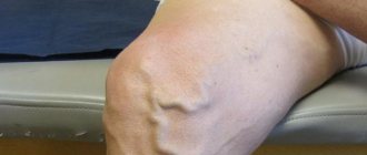
Most often, the disease begins in the veins of the leg; in the absence of adequate treatment, the process spreads higher. However, thrombosis, although less frequently, can develop in the veins of the pelvis, but there are no disturbances in venous outflow, as in the extremities, so the disease is asymptomatic, which complicates diagnosis. In general, the manifestations of the disease are varied: from the complete absence of any clinical signs to significantly pronounced ones, sometimes gangrene. Sometimes in the early stages of the disease, the only manifestations are suddenly increasing swelling and pain of varying localization and intensity. Blueness of the skin caused by overflow of the saphenous veins is also possible. The severity of symptoms is influenced by the degree of impairment of venous outflow and the location of thrombosis. With thrombosis of the saphenous veins, a swollen nodular vein is visible under the skin, the skin around it has a reddish tint, small veins are clearly visible and look like bluish winding lines.
How to properly perform limb compression during thrombosis?
In the acute period, with severe swelling, it is recommended to compress the leg with an elastic bandage for the first 7-10 days. The limb should be bandaged in the morning, before getting out of bed, starting from the toes. On the lower leg and thigh, the elastic bandage should be applied in a spiral, each new turn half covering the previous one. The bandaging boundary should be 10–20 cm above the level of thrombosis. During the day, the bandage can be removed for 20 minutes, always in a supine position, then bandage the limb again. Elastic bandaging is an art that the patient must be taught by the attending physician, and the quality of its formation must be monitored repeatedly. An incorrectly performed elastic bandage will only make things worse.
After 7-10 days, when the swelling begins to decrease, you can switch to medical knitwear, it is more aesthetic and comfortable. The compression class should be recommended by the attending physician.
Is physical activity allowed during thrombosis?
If there is a risk of pulmonary embolism, if surgical treatment cannot be resorted to, the patient is recommended to rest in bed, with the leg in an elevated position. In other cases, dosed walking or a set of special exercises are recommended, which prevents further spread of the process and allows the patient to be activated as soon as possible.
Are thermal procedures allowed for thrombosis?
Patients with thrombosis are contraindicated from taking hot baths, visiting a bathhouse or sauna. You should also avoid other thermal procedures (for example, ozokerite), compresses, massages, because they all increase blood flow, which, if outflow is impaired, aggravates the clinical manifestations of the disease. Patients are recommended to only take a shower.
Prognosis and complications
The prognosis for postthrombophlebitic vein damage is relatively favorable in cases where the patient adheres to the doctor’s basic recommendations - does not violate the treatment program and follows the basic rules for preventing relapses of the disease. With this approach, it is possible to achieve a maintaining optimal state for a long time.
If the rules of the health program are violated, the patient experiences complications in the form of poor circulation in the extremities, which can lead to gangrene, requiring amputation. The second serious complication is infarction of the brain or internal organs when a blood clot enters the general bloodstream.
Treatment and symptoms of thrombophlebitis of the deep veins of the lower extremities
The formation of blood clots in the lumen of the veins is quite common and is called acute deep vein thrombosis of the lower extremities. With this disease, 27–35% of patients experience thromboembolism of the arteries in the lungs.
Thrombophlebitis of the veins of the lower extremities occurs relatively rarely in healthy people, and the number of diseases increases every year
Therefore, the development and application of conservative treatment of the disease is an important task in vascular surgery.
Thrombophlebitis of deep veins
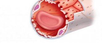
Vein thrombi occur for various reasons and develop with a normal epithelial layer on the vessel wall. Their formation begins in the veins of the leg - on their valves, where blood clotting factors accumulate due to the vortex of blood flow through the valve leaflets and in the area of the vein division.
Platelets provoke early thrombus formation by settling on the valves of the veins and at points where the integrity of the epithelial layer is compromised.
They attach to the endothelium, or the exposed collagen layer on the vein walls. The subsequent event is platelet aggregation, release of thromboplastin from the tissues and the appearance of a red thrombus.
The latter is retractable, capable of aseptic lysis and consists of fibrin, erythrocytes and platelets.
Subsequently, the behavior of the thrombus is influenced by the processes of fibrinolysis and coagulation.
The action of fibrinolysin leads to lysis within three to four days, most of the thrombus is destroyed, fragmented, dislodged and can move into the pulmonary arteries.
Subsequently, the formation may resolve without affecting the venous wall or be replaced by connective tissue if the thrombus is of significant size and the area of adhesion to the wall is quite extended.
With extensive varicose veins, the thrombus settles in the overlying veins or spreads to the perforating veins and deep veins of the affected surface of the lower limb. Deep venous thrombosis involves the spread of the thrombus to the femoral and popliteal veins, and the discharge of blood from them can stop thrombosis along the ascending line.
Clinic of the disease
Symptoms of thrombophlebitis of the deep veins of the lower extremities depend on the location of the thrombus and the degree of its spread and changes in venous patency (obstruction or stenosis of the lumen), the occurrence of collaterals. Clinical manifestations are varied - from an asymptomatic course of the disease to severe pain and extensive swelling, sometimes it ends in gangrene of the extremities.
Asymptomatic progression is typical for the case when venous outflow occurs without obstacles, while the situation is difficult to recognize and the indicators are characteristic of only one limb. Sometimes the first noticeable sign is only thromboembolism of the arteries in the lungs. Manifestations of the disease occur quickly - within two to three hours to two days from the appearance of a blood clot:
- the ankle, foot, and distal part of the lower leg swell;
- pain is felt when light pressure is applied to the lower leg muscles;
- pain appears in the calf when the foot is dorsiflexed and calms down at rest;
- at the site of the lesion, the lower leg becomes hot due to inflammation and increased blood flow;
- dilated superficial veins are observed;
- There is a difference in circumferential size between the affected and normal limbs.
What can happen if postthrombotic syndrome occurs?
Changes in the skin occur, the addition of induration, eczema, hyperpigmentation and, as a finale, trophic ulcers are sometimes very large, and the leg may look like an inverted bottle of champagne. What can help? The best thing is to prevent possible deep vein thrombosis. But it’s like the proverb: “It’s better to be rich and healthy than...”. If you have postthrombotic syndrome, only three treatment methods can help: compression
(compression stockings, tights, bandages, in combination with specially designed physical therapy),
drug therapy
(phlebotonics and disaggregants) and valve surgery (various options
for restoring the valve apparatus
with preventing the discharge of blood through incompetent perforating veins). No other methods have yet been invented. In order to help patients with post-thrombotic syndrome, knowledge, patience and modern treatment methods are needed - this is, first of all, a method of restoring the valve apparatus through a low-traumatic operation without the use of incisions. We are ready to help you.