The tricuspid valve is a movable septum between the right ventricle of the heart and the right atrium. The valve is involved in blood circulation, through it blood flows from one part of the heart to another. Violations in this part lead to overload of the right side of the heart, provoke a circulatory failure, worsen the patient’s quality of life, and even threaten death. An effective treatment method is valve surgery, during which its structure and mobility are restored.
Description of the procedure
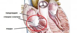
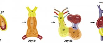
The heart is divided into chambers, and blood circulates between them through valves. There are four valves in total in the heart, and failure of even one causes severe damage to health. The tricuspid or tricuspid consists of three leaflets that open and close during the heartbeat. When the valves open, blood passes through them and enters the right atrium from the right ventricle. The flaps close tightly to prevent blood from flowing back.
Content:
- Description of the procedure
- When is plastic surgery indicated?
- Preparation for valve repair
- Carrying out the operation
- Postoperative period
- Plastic results
- Where is tricuspid valve repair performed?
If the valve does not open completely, it is called stenosis. In this case, the blood flow is disrupted and the ventricle is overloaded. If the leaflets do not close completely, this is called tricuspid insufficiency.
Tricuspid valve repair is intended to restore its function.
This is a rather complex surgical intervention, which is always performed under general anesthesia, most often using a cardiopulmonary bypass machine (ACB). Depending on the problem (stenosis or insufficiency), the appropriate tactics are chosen.
With stenosis, the valves may fuse or thicken; during surgery, they are manually separated and sutured in the right places. If insufficiency is diagnosed, sutures are placed around the circumference of the valve to restore the valve.
As a result of such actions, normal blood circulation is restored and the load on the heart is reduced.
Often, for effective treatment, special rings and half-rings are installed at the time of surgery. These structures can be artificial or biological; the latter are preferable, as they are more easily accepted by the body and do not require lifelong medication. The rings are sewn along the perimeter of the valve apparatus, they restore the required lumen and ensure normal closure of the valves.
Moreover, such reconstruction provides a long-lasting result, since it does not allow the fibrous ring to re-expand. Also, plastic surgery of the tricuspid valve may involve complete excision of its leaflets and replacement with an implant. Such measures are resorted to when it is impossible to rehabilitate the original valves.
Prosthetics of the tricuspid valve, along with plastic surgery, is one of the surgical treatment options, which is used when it is impossible to preserve the valve due to gross changes in the leaflets and subvalvular apparatus. In recent years, in the domestic literature [1, 2], there has been a small number of studies devoted to the study of long-term results of tricuspid valve replacement. In this regard, we decided to present our experience on this problem. According to various authors [4-7, 10], tricuspid valve replacement has a high level of complications, and mortality remains within 20%.
Material and methods
From 2000 to 2009, 218 tricuspid valve replacement surgeries were performed in our center. In this work, data from 186 patients were analyzed. The main characteristics of the patients are presented in Table. 1
.
Systolic pressure in the pulmonary artery, according to sounding of the right side of the heart, in half of the patients exceeded 40 mm Hg.
Most patients were admitted for treatment with severe heart failure, with a mean NYHA functional class of 3.5 ± 0.7. 51% of patients already had persistent atrial fibrillation. Almost all patients had cardiomegaly due to the right side of the heart. 94 (51%) patients had undergone valve correction in the past, of which 67 (36%) had previously undergone closed mitral commissurotomy or mitral valve replacement. 27 (15%) patients had previously undergone intervention on the tricuspid valve, of which 13 (7%) had a recurrence of tricuspid insufficiency after previously performed repair, and 14 (8%) required re-replacement of the tricuspid valve due to degeneration of the bioprosthesis. All operations were performed according to standard methods. Cardiopulmonary bypass was performed through bicaval cannulation and cannulation of the ascending aorta. Until 2005, operations were performed under conditions of moderate hypothermia (32 °C); subsequently, under normothermia conditions. Cold blood cardioplegia was used in the majority of patients; pharmacocold cardioplegia with Custodiol solution was used in 9 (4.8%). The duration of artificial circulation and the time of aortic clamping were 118.9±48.8 and 93.8±41.8 minutes, respectively.
Isolated tricuspid valve replacement was performed in 43.5% of patients, the rest underwent two-valve (32.8%) and three-valve (23.7%) corrections. Moreover, the majority underwent mitral-tricuspid (52 cases) and mitral-aortic-tricuspid replacement - 41 (50%). Tricuspid valve replacement with a mechanical prosthesis Meding-2 was performed in 19 patients. Until 2006, KemKor xenoprostheses were mainly used (82 cases), since 2006, as a rule, Pericor pericardial prostheses began to be implanted (68); 9 patients were implanted with Perimount prostheses (Carpentier-Edwards Inc.), 8 with Bio-Lab prostheses.
results
In 37.6% of cases, patients required prolonged (more than 24 hours) artificial ventilation, in 38.7% - a long (more than 48 hours) stay in the intensive care unit, in 72% - inotropic support of varying duration. In the early postoperative period, 24.4% of patients suffered various complications. The most common complications were third degree atrioventricular block (9.1%), pericardial effusion (4.8%), infectious complications (4.9%) and postoperative bleeding (3.8%). Neurological disorders were recorded in 1.6% of patients. Mortality rate was 13.4%. The main causes of death were progressive cardiovascular failure (7%) and multiple organ failure (2.8%). Other causes include gastrointestinal bleeding (1.2%), acute respiratory failure, acute cerebrovascular accident, postoperative bleeding and generalized peritonitis (0.6% each). Prognostic factors associated with mortality were rheumatic genesis of tricuspid disease ( p
=0.017) and initial NYHA functional class (
p
=0.03).
Long-term results were studied in 126 patients over a period of 13 to 130 months (median - 43 months). The dynamics of the main echocardiographic parameters are presented in table. 2
.
After surgery, the size of the right parts of the heart decreased in patients (from 71.7±17.2 to 60.0±10.6 mm, p
<0.001) and right ventricular size (from 38.7±7.0 to 34.7±6.2 mm,
p
<0.001).
The functional class of heart failure decreased significantly (from 3.5 to 2.4, p
<0.001).
The need for implantation of a permanent pacemaker arose in 32.7% of those observed. 41.8% continued to require diuretics, most of them were patients with multivalve repair. On mechanical prostheses, in contrast to biological ones, a more pronounced peak gradient was observed (11.1 ± 4.8 mm Hg versus 8.1 ± 3.8 mm Hg, p
= 0.006).
Dysfunction of bioprostheses was detected in 8.2% of patients in the period from 50 to 117 months after surgery (median - 71 months). Thrombosis of the mechanical prosthesis was detected in 10.5% of patients (median follow-up period: 16 months). The need for tricuspid valve replacement occurred in 13.5% of patients. Mortality in the long-term period was 12.7%. In the majority of patients (81.3%), the causes of death were related to cardiac pathology. The figure
shows the actuarial curve for 10-year survival, which was 87.3% (95% CI 81.5–93.1) at 10 years.
Figure 1. Actuarial 10-year survival curve after tricuspid valve replacement.
Discussion
Our study included a heterogeneous group of patients who required tricuspid valve replacement. The rheumatic genesis of the defect predominated. The choice to replace rather than repair the valve was, as a rule, dictated not so much by the etiological essence of the defect as by the severity of organic changes in the valve. These were patients with heart failure of functional class III-IV. Patients with rheumatism had the highest number of two- and three-valve corrections. The severity of the initial condition of the patients was also evidenced by the fact that more than ⅓ of the patients needed prolonged artificial ventilation and a long stay in the intensive care unit. Implantation of a permanent pacemaker was required in 32.7% of patients due to acute postoperative third-degree block or the development in the long-term period of high-grade atrioventricular block and sick sinus syndrome. Thus, in total, various complications were observed in 57.1% of patients. Some authors [8] cite a lower frequency of need for pacemaker implantation. Other authors [4, 6, 7] present approximately similar data. Moreover, F. Filsoufi et al. [5] when treating 81 patients, certain complications were observed in all cases.
Among the factors associated with mortality, our study noted rheumatic origin of tricuspid valve disease, which is likely explained by several factors. Firstly, this is a long course of the disease and, as a result, more severe heart failure. Secondly, the frequent combination of rheumatic tricuspid disease with mitral or aortic disease and, accordingly, the combined nature of the operation. Despite this, our observation showed an improvement in echocardiographic parameters and a decrease in the severity of heart failure, which in most patients corresponded to NYHA functional class II.
The initial severity of heart failure is also a predictor of poor surgical outcome. Similar data are presented by Y. Topilsky et al. [9].
R.M. Muratov et al. [3] noted an earlier occurrence of dysfunction of bioprostheses when the subvalvular apparatus of the tricuspid valve was preserved.
Our study did not reveal such a pattern. We also found no differences in the need for reprosthetics between different types of bioprostheses ( p
=0.37). There is no complete agreement among surgeons regarding the priority in choosing the type of prosthesis. A number of researchers [11-13] do not note the advantages of bioprostheses over mechanical ones in the tricuspid position.
In other studies [1, 4, 14], on the contrary, it is recommended to use bioprostheses in the tricuspid position.
Our study has a number of limitations and limitations. First, this is a retrospective study, and it is not possible to draw any conclusions regarding the superiority of biological or mechanical prostheses. Secondly, the presented population is heterogeneous in terms of the etiology of the defect, which also has a great impact on long-term results. Thirdly, long-term results could not be studied in all patients.
conclusions
1. Patients undergoing tricuspid valve replacement constitute a high-risk group.
2. Prosthetics of the tricuspid valve in case of its organic changes is the optimal method of surgical treatment, although it is accompanied by an increased level of mortality and complications.
3. The development of dysfunction (thrombosis) of mechanical prostheses in the tricuspid position occurs at an earlier time in comparison with biological prostheses, which requires further study of the long-term results of the use of mechanical prostheses.
When is plastic surgery indicated?
Such manipulations are carried out in two cases: when the doors open poorly and when they do not close completely. This can happen due to acquired diseases or congenital anomalies. Congenital pathologies of the tricuspid valve are very rare. Typically, valvular disorders are caused by acquired ailments:
- heart injuries;
- infectious diseases;
- systemic lupus erythematosus;
- rheumatism of the heart muscle;
- carcinoid syndrome.
Stenosis and insufficiency in the tricuspid septum are very rarely independent diseases. As a rule, these are consequences of other pathologies. Problems with the mitral valve, located on the left side of the heart, place stress on the right side.
As a result, the right atrium and ventricle cannot cope with the load, and the valve is deformed. Disturbances in the functioning of the valve apparatus may not manifest themselves at first, but over time the patient feels:
- strong heartbeat;
- shortness of breath;
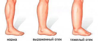
- swelling of the lower extremities;
- chest pain;
- bloating;
- weakness, frequent fatigue.
Also, disruptions in this area are accompanied by enlargement of the jugular veins. Even the patient himself can distinguish swollen veins in the neck. The skin becomes an earthy yellow color, as the liver often also suffers due to impaired blood circulation.
Nausea, belching, and heaviness in the abdomen may occur periodically. The need for plastic surgery is determined only after a thorough examination and clarification of the diagnosis.
Preparation for valve repair
The first step in patient preparation is diagnosis. To clarify the diagnosis, a chest ultrasound, ECG, blood and urine tests are prescribed. In some modern clinics, the heart is examined using a computed tomograph. Angiography of the heart vessels and or CT scans show the heart muscle and blood circulation in detail; during the studies, the radiologist can accurately determine tricuspid stenosis or insufficiency. After the tests, the patient undergoes a consultation with a cardiac surgeon, neurologist, and therapist.
If valve disorders are accompanied by additional heart problems, they try to get rid of them before surgery. For example, in case of arrhythmia, the heart rhythm is first normalized. If the clinical picture is complicated by heart failure, a sodium-excluding diet is prescribed. All patients are prescribed a diet a few days before the procedure; they need to avoid salt, too fatty foods, too spicy foods, and drink less water. You cannot eat 8 hours before plastic surgery. As part of the patient's preparation, the attending doctor explains to his patient how tricuspid valve repair is performed. It is important that the person being operated on does not worry and does not overload the heart and nervous system. If you have concerns or fears, you should talk to your doctor about it.
Diagnosis of tricuspid heart valve disease in Israel
In the presence of cardiovascular pathologies, it is very important to conduct the examination quickly and efficiently, since any delay may worsen the patient’s condition.
- Day 1
- Day 2
- Day 3
First day. From the airport to a doctor's appointment
Immediately upon arrival in the country, the patient, accompanied by a representative of the international department, goes to the clinic. A cardiac surgeon is already waiting for him there. The specialist conducts an examination, studies the data from the studies performed, and, if necessary, prescribes additional ones.
Second day. Comprehensive examination in one day
- Laboratory tests of urine and blood.
- CT, MRI.
- Angiography of blood vessels.
- Chest X-ray.
- Auscultation.
- Electrocardiographic study.
- Echocardiography traditional and transesophageal.
- Bicycle ergometry.
- Cardiac catheterization if necessary, etc.
The third day. Individual treatment plan
Assessing the results of tests and studies, the council of doctors decides which drugs and in what dosage should be chosen for drug therapy and whether surgical intervention is necessary (if so, to what extent). Taking into account the characteristics of the course of the disease, the condition and age of the patient when planning treatment tactics allows us to achieve high efficiency and avoid medical errors.
Carrying out the operation
Reconstruction of the valve apparatus occurs in a hospital setting, in the operating room. Before this, the patient is given premedication: sedatives and hypnotics are administered intravenously. Throughout the entire process, assistants will monitor the patient’s condition using connected equipment. When sleep comes, the surgical team begins manipulations.
- The surgeon makes an incision in the chest. If you get to the heart through the intercostal space, you won’t have to cut the sternum bones. In most cases, the doctor opens the chest, then gets to the valve layer by layer.
- The heart is forcibly stopped and a heart-lung machine is connected. During the process, this device will saturate the blood with oxygen and give it the desired temperature while the heart is “turned off.”
- Then the surgeon’s actions will depend on the pathology itself. To restore the lumen, the valves can be cut and sutured; most often, sutures are placed around the circumference of the valve. There are several suturing techniques; for moderate dilatation, de Vega sutures are usually used. If valve closure is corrected, rings, half rings or tape are most often sewn in. The implantation of such structures is called “annuloplasty”. If it is impossible to restore the function of the tricuspid valve, its leaflets are removed and a prosthesis is placed in their place.
- Before turning off the AVR, the doctor checks the operation of the operated or prosthetic valve. The shortcomings are corrected, the heart-lung machine is turned off.
- The incision site is sutured in layers, the external seams are treated with sterilizing agents. Then apply a tight bandage.
- The patient's condition is assessed again and he is transferred to the intensive care unit. When the anesthesia wears off, the patient is placed in an intensive care unit.
After the operation, the patient remains under the supervision of doctors for another week. This is necessary in order to monitor the results of plastic surgery and prevent complications. In the first days, bed rest, moderate drinking, and diet are indicated. After the final check, the patient is discharged home.
Postoperative period
During discharge, the doctor gives his patient instructions on nutrition and lifestyle, and sets a date for the next examination. In the postoperative period, you must follow a prescribed diet, which will exclude:
- fat;
- roast;
- too spicy and salty.
During recovery, stress has a very adverse effect, so it is important to ensure a calm and measured pace of life. If necessary, the attending physician can prescribe medications to support the nervous system if the patient is overly sensitive. Alcohol is completely avoided in the first month after valve reconstruction. It is also important to quit smoking. Visits to the doctor will be scheduled; at first, examinations are scheduled every month, if there are no complaints. It is worth visiting a doctor unscheduled in the following cases:
- bleeding from the wound, swelling, itching in this part;
- constant chest pain (may be at first);
- causeless fever;
- frequent dizziness and nausea.
During the appointment, the cardiologist will listen to your heartbeat, breathing rhythm, and measure your blood pressure. Diagnostics are also periodically indicated: ultrasound, ECG, tests. This is necessary in order to track the rehabilitation process.
Physical activity should be moderate, it is recommended to walk in the fresh air more often, and the step should be leisurely.
Your doctor will tell you when you can return to full activity. Drug treatment is also prescribed to maintain the functioning of the heart and the whole body. If a biological prosthesis was implanted, anticoagulants are indicated in the first three months. If an artificial prosthesis was sewn in, anticoagulants are indicated for life.
Lifestyle after surgery
The first four weeks after surgery are considered the early postoperative period. At this time, the patient should treat his body as carefully as possible and listen to his feelings. After discharge from the hospital, if you experience the slightest pain, shortness of breath, or wheezing in the chest, you should immediately consult a cardiologist or therapist.
Also at this time, the patient should monitor his diet more carefully, eliminating all harmful foods - fatty, fried, salty and spicy. It is advisable to limit fluid intake to one and a half liters per day so as not to overload the cardiovascular system with excess volume.
It is imperative to quit smoking and stop drinking alcohol.
One month after surgery, you can allow light physical activity, such as short walks in the park.
At the end of the first month after surgery, you should contact your doctor and perform a full examination - cardiac ultrasound, ECG, 24-hour blood pressure and ECG monitoring, and, if necessary, transesophageal ECHO-CG. You should see a doctor monthly in the first year, and then every six months if there are no complaints.
Plastic results
Symptoms of stenosis or tricuspid insufficiency disappear immediately after surgery. In the first 1-2 months after it, the patient’s condition will gradually improve if the manipulations went without complications. Negative consequences may include:
- bleeding;
- heart attack;
- formation of blood clots and blockage of blood vessels;
- disturbances of vision, appetite, sleep (this is normal, goes away within a month);
- infection;
- hemolytic anemia.
Best materials of the month
- Coronaviruses: SARS-CoV-2 (COVID-19)
- Antibiotics for the prevention and treatment of COVID-19: how effective are they?
- The most common "office" diseases
- Does vodka kill coronavirus?
- How to stay alive on our roads?
To prevent such consequences, scheduled and unscheduled inspections are prescribed. In most cases, the prognosis for tricuspid valve surgery is positive, heart function is restored, and the patient returns to his normal lifestyle.
The postoperative period is complicated by the use of anticoagulants, which reduce blood clotting. Biological implants do not require constant use of anticoagulants, but the lifespan of such prostheses is limited. Observations of patients show that a biological implant lasts an average of 9-10 years.
Mechanical analogues have a long service life; they keep the heart functioning for 20-25 years. But during this entire time you need to take medications that reduce blood clotting. When the life of the implant has passed, it is replaced with the next one. If regular sutures were used during plastic surgery, reoperation may not be necessary. However, long-term patient survival is higher with rings than with conventional suture techniques.
Treatment of tricuspid heart valve disease in Israel: prices
Today, medical care abroad from leading world-famous specialists is becoming available to more patients. At the Top Ichilov Clinic, high quality service, modern diagnostics, innovative methods of therapy and a stay in the comfortable conditions of luxury rooms will cost 30% less than in Europe, and 50% cheaper than in America.
Treatment of tricuspid heart valve disease in Israel, the cost of which is unreasonably inflated in other countries, is chosen by more and more patients, even from developed countries of the world. You can find out prices for the procedures you need from our consultants - leave a request or call the international department of Top Ichilov.
Top Ichilov Medical Center is your best solution
Treatment of tricuspid heart valve disease in Israel, according to reviews from patients at the Top Ichilov clinic, has many advantages. The main ones are highly qualified doctors, advanced equipment and progressive techniques, and affordable prices.
- The authority of our clinic's cardiac surgeons is recognized by the global medical community.
- An individually developed treatment regimen allows one to achieve one of the highest cure rates for cardiovascular diseases.
- Surgical interventions are carried out through low-traumatic, gentle approaches to the beating heart, which reduces the occurrence of risks to zero during or after surgery.
- A comprehensive diagnosis of tricuspid heart valve disease in Israel is performed in just 3 days.
- Loyal pricing policy, convenient payment forms and no prepayment.
- Accompaniment of a representative of the international department throughout the entire stay in the clinic - we are responsible for solving all organizational and everyday nuances.
- 5
- 4
- 3
- 2
- 1
(9 votes, average: 5 out of 5)