Myocardial infarction is the leading cause of death on the planet
Every year, about 18 million people die from cardiovascular diseases worldwide (WHO data). Heart disease is the leading cause of death on the planet. Myocardial infarction is one of the forms of IHD (coronary heart disease), which leads to the formation of muscle tissue necrosis. The cause of the development of the pathology is insufficient blood supply to the area of the heart muscle, resulting from obstruction of the lumen of the coronary artery.
Vessels can become clogged during surgery, atherosclerosis, fat embolism and severe spasm. Risk factors that increase the likelihood of developing myocardial infarction include hypertension, smoking, obesity, alcoholism, physical inactivity, previous infections and psychological stress.
Causes of myocardial infarction
Etiology of MI
- sudden obstruction to blood flow in the branches of the coronary artery (vessels supplying the heart). Most often, such an obstacle is an atherosclerotic plaque with a blood clot.
Atherosclerosis in the walls of the arteries is the formation of peculiar “growths” - plaques consisting of fatty, fibrous tissue, calcium, and which, as they progress, narrow the lumen of the vessel up to 90% or more. At the time of increased blood pressure, physical effort, or psycho-emotional stress, rupture of an atherosclerotic plaque is possible, the reaction to which is thrombosis.
Less commonly, the cause is a prolonged, prolonged spasm
coronary artery in the absence of significant atherosclerosis, or a complication of other diseases: congenital malformations, trauma to the coronary arteries, vasculitis, embolism, tendency to hypercoagulation (increased blood clotting), syphilis.
There are a number of risk factors
, repeatedly increasing the likelihood of myocardial infarction.
There are non-modifiable ones, that is, those that cannot be influenced:
- male gender (up to 50-55 years, then the incidence levels off),
- age,
- genetic conditioning (coronary artery disease and other manifestations of atherosclerosis in blood relatives);
- low physical activity (hypodynamia),
- obesity or increased body weight,
- smoking (active or passive),
- arterial hypertension,
- diabetes mellitus and metabolic syndrome,
- dyslipidemia.
And
modifiable, by changing which you can significantly reduce the likelihood of disease:
THE PHENOMENON OF SILENT DYSPHERE. PULMOLOGIST SERGEY AVDEEV ABOUT LUNG DAMAGE DUE TO COVID-19
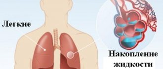
Covid-19 is becoming more insidious and dangerous. Thus, the new delta variant of coronavirus is recognized as dominant throughout the world, and the disease is much more severe. However, people often feel quite well in the first days of the disease. Just a slight fever, a barely noticeable cough and runny nose. But at the same time, hypoxemia develops. And the patient ends up in the hospital with serious lung damage.
− Sergey Nikolaevich, in November 2021, the incidence and mortality rates from Covid-19 in Russia jumped up again. What do you think is the reason for this?
- Several factors play a role here. First, a new strain has emerged - the more aggressive, more virulent delta strain. Secondly, in Russia there is still a low level of vaccination of our population.
Unfortunately, our country is one of the last in terms of global vaccination rates. It is a fact. Russia is not even in the top hundred.
There is only one way out - to get vaccinated. Every person should do this. Without this, it will not be possible to cope with Covid-19.
- The voice of doctors was heard more than once. But many citizens still do not have confidence in vaccines.
“Unfortunately, the voice of doctors is being lost due to the voices of the so-called “anti-vaxxers.” It is also surprising that Russia has an unprecedentedly high level of activity in anti-vaccination campaigns. And in this ranking our country also ranks first. There is no such situation anywhere in the world. Real obscurantism. There is no other way to say it. At the same time, it is impossible to find logical explanations.
− With the emergence of a new strain, new distinctive features of the course of the disease arose that were not previously observed?
- Yes, of course.
Today this information is heard from everywhere. The disease occurs in a faster and more dangerous scenario. In addition, the incubation period has shortened, and severe forms of the disease have become more common among young people and people without concomitant diseases. Children are increasingly appearing in red zones. Therefore, of course, if you compare it with what it was a year ago, there is a difference, and it is serious. − How does Covid-19 affect human lungs?
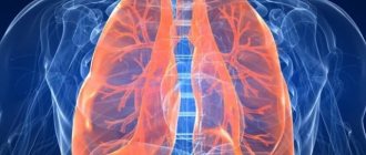
− During the course of the disease, we observe serious damage to the lungs, which is defined by a number of anatomical terms. There is damage, inflammation and blood clots. Unfortunately, it is the lungs that are the most vulnerable and hardest hit system during Covid-19.
The lungs are the “gateway” for infection. Coronavirus infection is an acute respiratory disease that is transmitted by airborne droplets. Accordingly, the infection enters the body through the respiratory tract and epithelium, through cells called alveolocytes.
− Why does our immune system turn against us during illness?
- We don’t fully understand why this happens. But our immune system cannot be considered ideal. At some point, the immune system begins to fight the infection so actively that it attacks its own tissues and organs.
- Is it known why the lungs do not signal us about danger in any way, and the lesion is detected either in the later stages with shortness of breath, or on an x-ray?
- This is not entirely true. As a rule, the body signals this in the form of a cough. But if we talk about shortness of breath, there are indeed some paradoxes observed with Covid-19, including the so-called phenomenon of silent hypoxemia. In this case, the patient may have hypoxia or oxygen starvation, but there is no shortness of breath. Unfortunately, symptoms do not always develop in parallel.
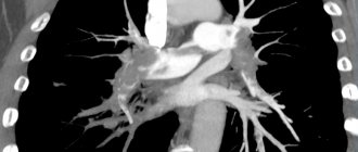
In addition, some patients develop asymptomatic pneumonia, in which serious lung damage is detected during image diagnostics, but the person himself does not feel anything.
We must remember that each person may have an individual response to the disease.
Let's say one patient's lungs are 5% affected, and he immediately feels unwell. And the other will not feel anything, up to a serious defeat of up to 50%, which usually already signals changes, sometimes catastrophic. Why is this happening? There is no exact answer yet. − How act in such a situation ?
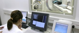
− First of all, diagnosis is important. And here you can’t do without a simple pulse oximeter. There are probably no people left who don’t know what kind of device this is. This small, portable tool is extremely useful. It gives us important information about several parameters, but the most significant of them is saturation, that is, an indicator of oxygen saturation in the blood. This biomarker reflects the development of hypoxemia or respiratory failure. But, of course, we are not limited to a single method. Doctors use a whole range of laboratory, instrumental methods, and image diagnostics. This is the only way to put together a “mosaic” of different elements in order to better understand what is happening to the patient.
− What approaches to restoring damaged lungs exist today?
− The first thing we start with is antiviral therapy. However, it cannot always be used for a particular patient. So, if a person arrives after 5-6 days from the onset of infection, then there is no point in using antiviral therapy. Next, doctors fight the syndromes caused by the disease. This includes anti-inflammatory therapy, therapy aimed at eliminating coagulation disorders and, finally, therapy aimed at correcting hypoxemia or respiratory failure. Of course, each patient may have his own unique characteristics of the course of the disease, so therapy in each specific case will be different.
− Have new diagnostic methods and approaches to therapy emerged?
− If we talk about diagnostics, then the same pulse oximetry is now introduced into all levels of modern medicine. If previously only a pulmonologist carried a pulse oximeter, today every doctor of any specialty carries one. Even people independently purchase this device and use it at home. But there are methods that were given impetus for development by the pandemic situation. For example, ultrasound of the lungs. This technique was known before and was actively used, but right now it is used extremely widely. And I hope that even after Covid-19 is defeated, this method will remain in our practice. However, there are also certain disappointments. As you remember, the first list of antiviral drugs was very extensive. We used drugs that are used in rheumatology, in the treatment of HIV infection and neurological diseases. But today the list of antiviral drugs is very small. At the same time, new drugs will appear very soon that will be included in the list of drugs for the treatment of Covid-19, including those based on monoclonal antibodies. The situation is changing quickly. Two years have passed, but we do not give up the search and are constantly looking for new drugs and more effective methods of treatment.
− How to help patients recover from illness?
− There is no single scenario. It all greatly depends on what problems a person faces after the disease. The well-known post-Covid syndrome includes a number of related problems. A sort of “tangle” that accompanies a person who, at first glance, has coped with the disease. For example, fatigue, shortness of breath, depression, insomnia, cough, etc. are common.
If we talk about serious lung damage, then we act in much the same way as in the treatment of pulmonary fibrosis. As a rule, the defeat is accompanied by the appearance of scars, which are almost impossible to get rid of. Because of these changes, shortness of breath, coughing appear, and the person does not have enough breath for physical work.
On the contrary, some patients may experience cavity formations in the lungs. Essentially, areas where lung tissue is missing. Such disorders are very dangerous and can cause pneumothorax, that is, the accumulation of air or gases in the pleural cavity, bleeding and infection.
− What issues were pressing before Covid-19?
- In fact, there were enough tasks and are still enough to this day. Asthma, chronic obstructive pulmonary disease (COPD), pneumonia. The same pneumonia is similar to coronavirus infection. In both cases, there is damage to the lungs, disorders leading to respiratory failure. However, the treatment methods are completely different. Simple pneumonia is treated with antibiotics, which are not effective against Covid-19.
− If we talk about the educational component, how obvious is the staff shortage?
- There really is a personnel shortage. There are not so many pulmonologists in our country compared to other medical specialties. This is understandable, because most pulmonologists today work in the red zone. This means that there are not enough specialists to treat other diseases - asthma, pulmonary fibrosis and others.
− Do you collaborate with colleagues from other countries?
- Yes. Literally from the very beginning of the pandemic, we have been exchanging experiences with foreign colleagues. Without this, it is impossible to solve the problems facing us. When we first encountered the new coronavirus infection, we had no experience. But we learned from Chinese colleagues, then from European ones. The exchange continues today. We meet at conferences and international forums, where we discuss the main issues and methods of solution.
− What is the current situation in the red zones?
- We started with the fact that today there is another surge in incidence. This means that the workload on doctors has increased again. At the same time, we are talking not only about those who directly work in red zones. But also doctors in clinics, specialists working at ambulance stations, nurses and orderlies. For everyone who works in the healthcare system.
− The only way out is vaccination?
- Yes. You can’t just hope that everyone will get sick and the pandemic will end. After all, this path is associated with numerous losses, including medical personnel.
Link to publication: scientificrussia.ru
Symptoms of myocardial infarction
The main manifestation of a heart attack is very intense, burning, baking, squeezing, pressing, tearing or “dagger” pain behind the sternum
. The pain may spread to the left arm, scapular region, shoulder, neck, and lower jaw.
The duration of the episode is always more than half an hour, the slightest physical stress causes increased pain. An important sign is the lack of analgesic effect of nitrates. Patients may also experience severe weakness, shortness of breath, rapid heartbeat, sweating, and nausea. Most people feel anxiety, doom, and fear of death.
In addition to the typical variant, there are several more atypical
, capable of masquerading as other diseases of the internal organs. Among these are abdominal (epigastric pain, nausea, vomiting), asthmatic (nasty dry cough, suffocation), cerebral (headache, loss of coordination of movements), arrhythmic, erased version (deterioration of sleep, emotional background, feeling of inexplicable discomfort in the chest, sweating ). It is also possible that there are no symptoms—a “silent” heart attack, which accounts for up to 20% of all MIs.
Coronary heart disease: symptoms
What is coronary heart disease?
“Ischemia” translated from Latin means decreased blood flow. Coronary heart disease develops when normal blood flow in the vessels that bring blood to the heart muscle is disrupted. These vessels are called coronary vessels. With the blood flowing through them, the heart muscle receives the oxygen necessary to produce energy. And the heart requires a lot of energy - after all, it performs a colossal job, pumping more than 7 thousand liters of blood per day, and does this constantly throughout our lives. Therefore, a decrease in coronary blood flow is very dangerous - because of it, the full functioning of the heart is disrupted.
Causes of blood flow disorders
- The most common cause is narrowing of one or more blood vessels due to atherosclerosis. In this case, the walls of blood vessels thicken due to the deposition of cholesterol in them - atherosclerotic plaques are formed. They reduce the lumen of the coronary arteries.
- Also, vasoconstriction can be caused by their spasm
Risk factors for developing coronary heart disease
- high blood pressure
- high blood cholesterol
- smoking
- alcohol abuse
- overweight
- low physical activity
- diabetes
- male
- elderly age
- heredity
It should be noted that if we cannot influence the last three of the listed factors, then all the others can be adjusted to one degree or another. This is important both for the prevention of this disease and for improving the prognosis if the disease has already developed.
Variants of manifestation of coronary heart disease
Angina pectoris
Characteristic symptoms:
1. Chest pain. Typical symptoms of pain associated with angina include:
- nature of the pain – squeezing, pressing, pinching
- localization - in the middle of the chest (in the sternum area), pain can spread to the left arm, jaw, under the shoulder blade
- Feeling of heaviness, discomfort in the chest
- Attacks of shortness of breath
The most common symptoms of angina are:
- occur during physical activity or during emotional stress (it is at these moments that the heart muscle’s need for oxygen increases)
- pass with rest or after taking nitroglycerin (or similar drugs - nitromint, isoket)
The appearance of the above symptoms allows one to suspect that a person has angina. To confirm the diagnosis, additional examinations are used, for example, recording an electrocardiogram (ECG) and daily ECG monitoring.
It should be remembered that if the ECG is not performed at the time of the attack, it may be normal. This does not exclude the diagnosis of angina pectoris. To clarify the presence and severity of narrowing of the coronary arteries, the doctor may prescribe coronary angiography. During this study, a contrast agent is injected into the vessels of the heart, which allows you to see them in X-rays and evaluate the presence and degree of narrowing of the lumen.
You should not postpone a visit to the doctor if the symptoms of angina pectoris :
- appeared for the first time (in the first month after the onset of symptoms, the risk of developing myocardial infarction is very high, especially if the correct diagnosis is not made in time and the necessary treatment is not prescribed)
- become more pronounced, become more frequent, occur with less load than before (this indicates the progression of the disease and the risk of complications)
Heart rhythm disturbances
They arise due to changes in the conduction system of the heart against the background of reduced blood supply. Manifests itself in the form of the following symptoms:
- Feeling of heart failure
- Attacks of rapid heartbeat
- Decreased heart rate below normal
Heart failure
This is a condition in which the contractility of the heart decreases and it cannot fully perform its work.
Symptoms:
- Shortness of breath during exercise and when lying down
- Swelling of the legs, more pronounced towards the end of the day
Myocardial infarction
The most dangerous, life-threatening condition that can manifest itself is coronary heart disease. During a heart attack, a blood clot forms on the surface of an atherosclerotic plaque, blocking the lumen of the vessel and disrupting the flow of blood to the heart muscle (myocardium). Due to an acute lack of oxygen, myocardial cells die. This is called a heart attack. The larger the area of damage to the heart muscle, the more severe the patient’s condition.
Symptoms of myocardial infarction:
- Pain in the chest (sternum area), intense, burning. Can spread to the left arm, jaw, under the shoulder blade. Cannot be removed with nitroglycerin.
Pain is the most characteristic symptom of myocardial infarction, but painless forms are also possible (more often in patients with diabetes mellitus and the elderly)
- Sharp weakness
- Cold sweat
- Feeling short of breath, sometimes coughing with foamy sputum
- In some cases:
- Abdominal pain, nausea, vomiting
- Fainting, loss of consciousness (especially in older age)
If symptoms of myocardial infarction appear, you must immediately call an ambulance. The sooner treatment is started, the greater the chance of avoiding massive damage to the heart muscle and life-threatening complications. Patients with a heart attack are hospitalized in specialized departments, where they receive all the necessary care. If treatment is started in a timely manner, the consequences of myocardial infarction may be minimal and the patient has a chance of returning to an active life.
Treatment of coronary artery disease
Effective treatment of the disease requires maximum elimination of risk factors in combination with drug therapy. The medications prescribed by your doctor should be taken regularly. Be sure to monitor blood pressure, pulse, and the presence and severity of symptoms. In some cases, surgical intervention is performed to restore the patency of the affected coronary vessels (stenting, bypass surgery). However, even after surgery, you must take medications, otherwise the disease will progress and new plaques will appear in the vessels.
Coronary heart disease is a serious disease that should not be taken lightly. If its symptoms appear, it is important to consult a doctor in time, who will prescribe the necessary tests and select the optimal treatment. Modern medicine has a large arsenal of means to treat this disease, and with properly prescribed therapy, severe and dangerous complications can be avoided and the opportunity to live a full life can be maintained. This can only be achieved through the joint efforts of the doctor and the patient, a combination of lifestyle changes and regular medication.
Literature:
Clinical recommendations. Stable coronary heart disease. Russian Society of Cardiology 2020
Clinical recommendations. Acute myocardial infarction with ST segment elevation of the electrocardiogram. Russian Society of Cardiology 2020
Martsevich S. Yu. Treatment and prevention of coronary heart disease. //RMJ 2015; 5: 256-258
Diagnosis of myocardial infarction
It is possible to recognize acute myocardial infarction taking into account:
- A characteristic clinical picture
(severe pain in the chest area or the heart area), medical history (duration of a painful attack of more than 30 minutes, lack of results from taking nitroglycerin, presence of previously established coronary heart disease, risk factors) - Changes in the ECG
(negative T wave, pathological Q, QS complexes, ST segment elevation in leads corresponding to the location of the damage to the heart muscle) - Laboratory diagnostic data
(general blood count: increased ESR, leukocytes; biochemical indicators: increased aspartate aminotransferase, alanine aminotransferase, fibrinogen, appearance of CP protein; presence in the blood of cardiac-specific markers of cardiac muscle cell death: myoglobin CPK, LDH, tryponins). - Valuable information is provided by EchoCG, which reveals a local violation of myocardial contractility and thinning.
Stent thrombosis after PCI
In patients with acute coronary syndrome with ST segment elevation (STES), primary percutaneous intervention (PCI) results in a 47% reduction in mortality compared with thrombolytic therapy (4.3% vs 9.2%). In NSTE-ACS, invasive revascularization improves the prognosis in patients at high and intermediate risk of death. The increasing number of PCI with stenting in ACS is accompanied by the risk of complications in the form of stent thrombosis. To illustrate this problem, we present a clinical case. Key words: acute coronary syndrome; OKSpST; ACS without pST; PCI; stenting. In recent years, there has been a continuous increase in the number of primary percutaneous interventions (PCI) for acute coronary syndrome (ACS) [4,5]. However, simultaneously with the increase in the frequency of PCI with stenting, the problem of the development of stent thrombosis and stenosis is growing [1, 6]. The incidence of stent thrombosis is 0.87–2.2% and occurs more often during the first year after surgery [3]. Stent thrombosis is verified if angiographic or pathological confirmation is available. Angiographic confirmation of stent thrombosis based on TIMI flow status is sufficient if there is a new acute onset of resting ischemic symptoms; new ischemic ECG changes, typical increase in markers of myocardial necrosis [7]. Stent thrombosis can develop during the first 24 hours after its installation (acute stent thrombosis), during the 1st month (subacute thrombosis) or from the 1st to 12th month after the intervention (late stent thrombosis). The primary factors contributing to its occurrence are the initial occlusion of the coronary arteries, a long (more than 25 mm) stented segment of the coronary artery, and discontinuation of dual antithrombotic therapy after endovascular intervention [3, 6].
Patient T., 72 years old, was hospitalized with a diagnosis of Unstable angina. Upon admission, he complained of periodically occurring pressing pain in the chest, of moderate intensity, occurring at rest during the last week and relieved by taking nitrates for 5 minutes. Hospitalized by gravity.
From the anamnesis: For a long time, the patient notes increases in blood pressure, maximum blood pressure figures = 180/100 mmHg. usual numbers BP = 110-130/70 mmHg. Since 1980, pressing pain in the chest appeared during exercise, and in 1985 he suffered a myocardial infarction. In 2011, he suffered a hemorrhagic cerebral infarction in the right hemisphere, focal neurological symptoms regressed. Repeatedly underwent PCI on the coronary arteries: TLBAP and LAD stenting in 2001 and 2003, TLBAP and VTK stenting in 2021. After VTK stenting in 2021, the clinical course of angina regressed, chest pain occurred very rarely. Since September 2021, the patient has returned to the angina clinic; a coronary angiography (CAG) was performed at the Federal State Budgetary Institution of the Federal Clinical Clinical Center for Medical and Internal Medicine (FMBA), LAD restenosis was detected, and TLBAP of restenosis was performed, without stenting. Over the past month, the patient's angina symptoms have progressed, tolerance to physical activity has sharply decreased, pain began to occur at rest, and the need for nitrates has increased. Last hospitalization at the Federal State Budgetary Institution National Medical Research Center for Cardiovascular Surgery named after. A.N. Bakulev from 06/13/2018 to 06/18/2018. On June 15, 2018, coronary angiography was performed, it was revealed: the left coronary artery trunk is not changed, previously implanted stents in the s/3 LAD are patent, further restenosis is 90%, a. intermedia stenosis p/3 from the mouth 90%, LMZHV OV stenosis p/3 to 55%, RCA is not changed.
Upon discharge, the patient is recommended to take the following medications: acuside 20/12.5 x 2 times, physiotens 0.4 mg x 2 times, Co-Plavix 100/75 mg in the morning, Preductal OD 80 mg in the morning, atorvostatin 40 mg in the evening, Cardura 4 mg in the evening .
After discharge from June 19, 2018, the patient’s symptoms of angina pectoris and chest pain at rest progressed, and he was forced to use sublingual nitrates several times a day. He sought medical help at Clinical Hospital No. 1 of the UDP and was sent to the ICU with the diagnosis: “IHD: unstable angina. PEAKS. Multivessel coronary lesion."
Objectively: The condition is serious. Position in bed with a low headboard. Body type – normosthenic. Increased nutrition. Height - 168cm, weight - 80kg. BMI-28.3 kg/m2, Overweight.
The skin is of normal color, dry, warm. Normothermia. There is no cyanosis of the lips, no acrocyanosis. There is no swelling. L/U are not enlarged. There are no varicose veins.
CNS: Oriented to place, time, self. Conscious, communicative, adequate. Memory and intelligence are preserved. Pupils D=S. Tongue in the midline. Sensitivity and movement in the limbs are fully preserved. No paresis or paralysis was detected. In the neurological status, focal and meningeal symptoms are not detected.
Respiratory organs: Breathing is independent, nasal, adequate. NPV 18 per minute. SpO2 97% in atmospheric air. Percussion sound is pulmonary with a boxy tint.
Auscultation: vesicular breathing, carried out in all departments. Single dry pneumosclerotic rales on both sides.
Cardiovascular system: The veins of the neck are not swollen. Pulsation of the carotid arteries without any features. The borders of the heart are expanded to the left by 1.5 cm. Heart sounds are muffled and rhythmic. The ECM records sinus rhythm. Heart rate=62, PS=62 per minute, blood pressure 140/76 mmHg. No pathological noises are heard. The pulsation in the arteries of the feet is reduced, more on the right.
Gastrointestinal organs: The tongue is moist. Appetite preserved. The abdomen is soft and painless on palpation in all parts. The liver does not protrude from under the edge of the costal arch and is painless on palpation. The spleen is not palpable. Bowel sounds are heard. The chair is a regular color, decorated.
Urinary organs: Independent urination. There is no dysuria. The effleurage symptom is negative on both sides.
Blood tests for cardiac biomarkers: 06/22/18 12:31 (1st analysis) Troponin-I quantitatively: 28.3 ng/ml; KFK-MB (CK-MB): 27 units/l; Total cholesterol: 2.5 mm/l; LDL: 1.51 mm/l 06/22/2018 23:11 (2nd analysis): Troponin-I quantitatively: 163.8 ng/ml; KFK-MB (CK-MB): 27 units/l; 06/23/2018 06:19: Troponin-I quantitatively: 10235 ng/ml; KFK-MB (CKMB): 755 units/l;
CAG dated 06.22.18: Access: right transradial Study results: LCA. Barrel Not changed. LAD The previously implanted stent in p/3-s/3 is passable, restenosis is 99%, further unchanged. DV Wellhead narrowing 45%, further unchanged. A. intermedia Irregularities of contours in n/3 (small-diameter artery). OS Without hemodynamically significant changes. VTK The previously implanted stent is passable from the orifice, without signs of restenosis, and further without changes. WPV OS Without hemodynamically significant changes. RCA Without hemodynamically significant changes. WMA of RCA Without hemodynamically significant changes. LMWH RCA Without hemodynamically significant changes. Type of blood supply: Balanced.
Progress of the operation: Under local anesthesia sol. Lidocaini 2% - 5.0 the right radial artery was punctured. 10 thousand units were introduced. heparin. A 6F guiding catheter was passed into the ascending aorta and installed at the ostium of the left coronary artery. The coronary guidewire was placed in d/3 of the LAD. Next, after preliminary dilatation with a 2.0x20 mm balloon catheter (12 atm), stenting of the p/3-s/3 LAD was performed. A Resolut stent 2.73x18 mm (14 atm) was implanted. Postdilatation of the implanted stent was performed using a 2.75x15mm (20 atm) balloon catheter. Angiography revealed extravasation of n/3 LAD. A 3.0×23 (14 atm) Aneugraft stent-gravity was implanted in the segment of artery extravasation (n/3 LAD). During control angiography, there is no extravacation. The patient is hemodynamically stable.
. EchoCG dated June 22, 2018: PPT 1 Myocardial mass g LV MM index g/m2 AORTA: 27 mm (N up to 40 mm). AORTIC VALVE: opening amplitude: 18 mm (N > 15 mm); Blood flow speed 1.33 m/sec. Gradient 7.2 mmHg; LEFT ATRIUM: D 45X61 mm (N up to 4.0 cm); V= 81 ml. LEFT VENTRICLE: CDR 4.7 cm (N up to 5.6 cm); DAC cm (N up to 3.7 cm); KDO 124 ml; KSO 47 ml; EF 62%. Relative wall thickness = (norm up to 0.42) IVS THICKNESS 1.4-1.7 cm (N up to 1.1 cm); THICKNESS OF THE ZSLZH 1.3 cm (N up to 1.1 cm). RIGHT ATRIUM: 36 mm. RIGHT VENTRICLE: D 29 mm. MITRAL VALVE: Regurgitation: max. speed of peak E: 0.6 cm/s, peak A 0.9 cm/s; mitral orifice area cm2; diameter of the annulus fibrosus SIGNS OF PULMONARY HYPERTENSION: NO; MPAP 17 mm Hg. PERICARDIAL: NO effusion. Conclusion: Atherosclerosis of the aorta. Calcification of the aortic and mitral valves. Dilatation of the left atrium. Local and global contractility is preserved. Concentric hypertrophy of the LV myocardium.
06/22/18 16:50 The patient was delivered from the cath lab after endovascular intervention - LAD stenting. During the stenting process, extravasation was detected, and therefore a stent-gravt was installed with a positive angiographic effect.
Complaints of intense pressing pain in the heart area, weakness, moderate shortness of breath associated with the inability to breathe deeply due to pain in the heart area.
ECG: sinus rhythm, ST elevation from V1-V, ST depression in II, III, AVF.
Infusion of neoton, isoket, and non-narcotic pain relief was started. It was decided to refrain from carrying out a massive anticoagulant therapy due to the risk of internal bleeding from the site of LAD injury.
At the moment, based on the clinical picture, the characteristic ECG picture, the echocardiographic picture, the clinical picture is interpreted as an acute type IV myocardial infarction in the region of the anterolateral wall of the LV with ST segment elevation.
06/23/18 00:30
A condition with negative dynamics in terms of the resumption of intense chest pain.
Due to severe pain, a decision was made to use narcotic painkillers.
When recording an ECG, pronounced negative dynamics were revealed in terms of expansion of the ischemic zone and an increase in ST segment elevation.
In agreement with endovascular surgeons, a decision was made to repeat CAG.
06/23/18 2:00
The patient was delivered from the cath lab after endovascular intervention - re-stenting of the LAD due to restenosis in the area of the previously implanted stent.
Against the background of revascularization, complaints of pressing pain in the heart area significantly regressed.
The ECG shows positive dynamics in terms of reducing ST elevation from V2-V4.
CAG 06.23.18 01:20: Name of operation: Recanalization, balloon vasodilation with installation of a stent in the anterior interventricular branch. Type of anesthesia: local anesthesia
Research results: LCA. Barrel Not changed. LAD Occluded from the mouth. DV Wellhead narrowing 45%, further unchanged. A. intermedia Irregularities of contours in n/3 (small-diameter artery). OS Without hemodynamically significant changes. VTK The previously implanted stent is passable from the orifice, without signs of restenosis, and further without changes. WPV OS Without hemodynamically significant changes. RCA Without hemodynamically significant changes. WMA of RCA Without hemodynamically significant changes. LMWH RCA Without hemodynamically significant changes. Type of blood supply: Balanced.
Progress of the operation: Under local anesthesia sol. Lidocaini 2% - 5.0 the right radial artery was punctured. 10 thousand units were introduced. heparin. A 6F guiding catheter was passed into the ascending aorta and installed at the ostium of the left coronary artery. Conductor recanalization of n/3 LAD from the mouth was performed. The coronary guidewire was placed in d/3 of the LAD. Next, after preliminary dilatation with a 2.0x20 mm balloon catheter (12 atm), stenting of the LAD from the orifice was performed. A 3.5x30 mm Resolute stent (14 atm) was implanted. A good angiographic result was obtained.
Over the past period, the overall dynamics of the patient’s condition can be characterized as negative due to the development of acute myocardial infarction, accompanied by severe pain, requiring the use of narcotic painkillers, and a decrease in EF to 42%. Following repeated endovascular intervention, the patient's condition stabilized.
During the therapy, the patient's condition remains stable, with satisfactory hemodynamic parameters, no progression of cardiac/respiratory failure, and no cardiac arrhythmias. A repeat echocardiography study is planned. There were no signs of internal bleeding into the pericardial cavity due to damage to the LAD.
06/23/2018 Echocardiography: AORTA: 27 mm (N up to 40 mm). AORTIC VALVE: opening amplitude: 18 mm (N > 15 mm); Blood flow speed 1.33 m/sec. Gradient 7.2 mm Hg; LEFT ATRIUM: D 45X61 mm (N up to 4.0 cm); V= 81 ml. LEFT VENTRICLE: CDR 4.7 cm (N up to 5.6 cm); DAC cm (N up to 3.7 cm); KDO 101 ml; KSO 52ml; EF 49%. Relative wall thickness = (norm up to 0.42) IVS THICKNESS 1.4-1.7 cm (N up to 1.1 cm); THICKNESS OF THE ZSLZH 1.3 cm (N up to 1.1 cm). RIGHT ATRIUM: 36 mm. RIGHT VENTRICLE: D 29 mm. MITRAL VALVE: Regurgitation: max. speed of peak E: 0.6 cm/s, peak A 0.9 cm/s; mitral orifice area cm2; diameter of the annulus fibrosus SIGNS OF PULMONARY HYPERTENSION: NO; MPAP 21 mm Hg. PERICARDIAL: NO effusion. Conclusion: Atherosclerosis of the aorta. Calcification of the aortic and mitral valves. Dilatation of the left atrium. Hypokinesis of the apical segment of the lower wall and IVS. Global contractility is slightly reduced. Concentric hypertrophy of the LV myocardium. For DKG: MR-1-11 st., TR-1 st. Type 1 diastolic dysfunction. Behind the apex of the heart and the lateral wall of the LV, there is separation of the pericardial layers up to 3-4 mm (a small amount of fluid).
Diagnosis: Main diagnosis: IHD: repeated infarction (type IV) in the area of the anterior wall of the LV and the interventricular septum from 06.22.18. Post-infarction cardiosclerosis (MI in 1985) TBAP and LAD stenting in 2001 and 2003, TBAP and VTK stenting in 2021. Selective coronary angiography dated 06/15/2018: the left artery trunk is not changed, previously implanted stents in the s/3 LAD are patent, then restenosis is 90%, a. intermedia stenosis n/3 from the mouth 90%, LMZHV OV stenosis from/3 to 55%, RCA is not changed.
Balloon vasodilation surgery with installation of a 2.73x18 mm Resolut stent in the anterior interventricular branch of the left coronary artery on June 22, 2018, paravasation, installation of a LAD stent graft on June 22, 2018.
LAD stent thrombosis, recanalization, balloon vasodilation with installation of a 3.5x28 mm Xience stent in the anterior interventricular branch on June 23, 2018.
Background: Hypertension stage III, stage 3, risk 4. Diabetes mellitus type 2, moderate severity. Excess body weight. BMI=28.3 kg/m2.
Combined: Multifocal atherosclerosis: Stenosis of the left renal artery, stenting of the left renal artery in 2003, restenosis 95% (according to angiography dated June 15, 2018). Atherosclerosis of the vessels of the lower extremities: Thrombosis of the femoral artery on the left, operation ABBS in 2007. Angiography of the arteries of the lower extremities 06/15/2018: LEFT: OP stenosis 65%, IPA stenosis s/3 55%, BOTH, HBA, SPA and SFA without hemodynamically significant narrowings, RIGHT: SPA, IPA, IPA, BOTH and GBA are patent, SFA is occluded from the mouth, d/3 is filled along the collaterals.
Complications: Killip III AHF; recurrent left ventricular failure; NIV 06/25/18, 06/26/18.
Bilateral hypostatic polysegmental pneumonia. Acute aneurysm in the region of the LV apex. NRS: persistent form of atrial fibrillation-flutter.
Acute kidney injury, contrast-induced nephropathy.
Blockade of the right leg of the ganglion of Gis. NK IIA art. NYHA II FC. Nephroangiosclerosis. Nephropathy of mixed origin. CKD C3A, GFR according to the formula CKD-EPI=46 ml/min/1.73 m2.
Related: CVB. DEP II Art. ONMK from 2011
Cysts of the right kidney. Gastric ulcer, remission. COPD, moderate. Emphysema.
The course of the disease was complicated by acute left ventricular failure on June 23, 2018.
Discussion Thus, the patient had several reasons for the development of stent thrombosis - the presence of ACS itself, a decrease in EF, a long stented LAD segment, and initial subtotal arterial stenosis. The combination of male gender and age over 60 years were also additional predictors of stent thrombosis.
conclusions
1. After invasive revascularization in the scope of PCI with arterial stenting, additional stratification of patients is necessary according to the risk of developing stent thrombosis according to the presence of the most significant factors that increase the risk of its occurrence
2. Strict adherence to recommendations for DAPT in patients with ACS. The default duration of DAPT should be 12 months, regardless of the revascularization method. Ticagrelor is recommended for patients with ACS in the absence of drug contraindications. However, low-income patients require government support to purchase the drug, especially those with a high risk of developing stent thrombosis.
3. Work on maintaining patient compliance with drug therapy remains relevant.
Literature
1. Berezovskaya G. A., Ganyukov V. I., Karpenko M. A. Restenosis and stent thrombosis // Russian Journal of Cardiology. 2012. No. 6. pp. 91–95.
2. Ganyukov V.I. Evidence base for the priority role of primary percutaneous coronary intervention in revascularization of patients with ST-segment elevation myocardial infarction // Complex problems of cardiovascular diseases. 2013. No. 1. pp. 24–34.
3. Ganyukov V. I., Shilov A. A, Bokhan N. S., Moiseenkov G. V., Barbarash L. S. Causes of thrombosis of coronary artery stents // Interventional Cardiology. 2011–2012. No. 27/28. pp. 29–34.
4. Kashtalap V.V., Zavyrylina I.N., Barbarash O.L. Endovascular revascularization in acute coronary syndrome with ST segment elevation in Russia: problems and prospects for further development // Creative Cardiology. 2015. No. 3. pp. 5–15. BULLETIN OF BURYAT STATE UNIVERSITY. MEDICINE AND PHARMACY 2021. Vol. 4 50
5. ESC/EACT recommendations for the treatment of persistent ST-segment elevation myocardial infarction of the European Society of Cardiology. 2021. (escardio.ru).
6. Tereshchenko A. S., Mironov V. M., Merkulov E. V., Levitsky I. V., Samko A. N. Late and very late thrombosis of drug-eluting stents // Atherosclerosis and dyslipidemia. 2014. No. 1. pp. 9–14.
7. FDA Clinical Overview for Panel Packet DES Thrombosis Panel December 7-8, 2006. https://www.fda.gov/ohrms/dockets/ac/06/briefing/2006-4253b
Treatment of myocardial infarction
Myocardial infarction
- an emergency disease in cardiology and serves as an indication for immediate hospitalization in intensive care.
The number one task for doctors is to restore blood flow
in a thrombosed vessel. They achieve this in different ways. The priority and most effective is surgical intervention - angioplasty with stenting of the affected area of the artery or coronary artery bypass grafting. In parallel, medications are used that inhibit the development of a blood clot - disaggregants (aspirin, clopidogrel) and anticoagulants (heparin), and if immediate surgery is not possible, thrombolytics - drugs that dissolve the blood clot (alteplase, prourokinase).
Pain relief
carried out with narcotic analgesics, neuroleptics, intravenous infusions of nitroglycerin.
The size of the necrosis zone can be reduced by reducing myocardial oxygen consumption by using beta-blockers (metoprolol, bisoprolol, atenolol), ACE inhibitors (enalapril, lisinopril, ramipril), and oxygen therapy.
For arrhythmias
antiarrhythmics are used (magnesium, lidocaine, amiodarone), atropine for bradycardia. Anxiolytics and sedatives are prescribed to ensure psycho-emotional peace of the patient.
Strict bed rest
in the early stages of treatment and fractional meals, limited in volume and calorie content.
Differential diagnosis of dyspnea in clinical practice
Mechanisms of dyspnea
Shortness of breath is a manifestation of a discrepancy between the increased demands of gas exchange and the load performed by the respiratory muscles.
Dyspnea can occur during normal gas exchange, but also in the presence of pathology of the respiratory muscles [3]. Shortness of breath is a subjective sensation that is difficult to measure objectively. At the same time, it is possible to determine indirect signs of shortness of breath: changes in frequency (tachypnea), dependence on position (orthopnea, platypnea) and disruption of the respiratory cycle (pathological types of breathing). There are also ways to objectify the patient’s sensations - various scales and questionnaires that are provided to the patient to assess the severity of shortness of breath. However, such instruments are used more in research than in clinical practice. The respiratory regulation system consists of an efferent (directing impulses to the respiratory muscles), an afferent (directing impulses from the receptors to the brain) link and the breathing center itself, which processes information. A disruption in each of these links can lead to shortness of breath. Thus, if external respiration is impaired, the work of the respiratory muscles increases, and with pathology of the respiratory muscles, greater efforts are required. It is believed that impulses from motor efferent neurons, in parallel with the respiratory muscles, are sent to the sensory cortex, which leads to the sensation of shortness of breath. Sensory afferent impulses are enhanced by activation of chemoreceptors by hypoxia, hypercapnia and acidosis. This also results in a feeling of shortness of breath. The same mechanism is activated in response to bronchospasm, increased pulmonary artery pressure, and even hyperinflation. Finally, in severe anxiety disorders, shortness of breath is a consequence of misinterpretation of signals from the afferent circuit, as well as increased breathing rates that exceed physiological needs [4–6].
Pathophysiological causes of shortness of breath are usually divided into pulmonary and extrapulmonary. The first includes the occurrence of shortness of breath in obstructive diseases (bronchial obstruction, stenosis of the upper respiratory tract), restrictive lung pathology (infiltrative processes, pulmonary fibrosis, after resection of a lung lobe) and vascular diseases of the lungs (PE, pulmonary hypertension, intrapulmonary shunts). Extrapulmonary mechanisms include extrapulmonary restriction (morbid obesity, diaphragmatic paralysis, neuromuscular diseases, severe kyphoscoliosis), cardiovascular diseases (systolic and diastolic myocardial dysfunction, valvular defects) and other causes, including anemia, severe acidosis of any origin, third trimester pregnancy and respiratory regulation disorders (panic attack with hyperventilation syndrome, alveolar hyperventilation) (Tables 1, 2).
Shortness of breath due to lung diseases
In clinical practice, shortness of breath most often occurs in diseases with the development of obstructive disorders of the external respiration function, i.e., with increased resistance to the flow of inhalation or exhalation. With restrictive respiratory disorders due to infiltration, fibrosis or edema, the compliance of the lungs decreases. Detection of severe broncho-obstructive syndrome usually does not cause significant difficulties due to the typical medical history and characteristic auscultatory pattern (dry wheezing, including during forced exhalation). At the same time, with a combination of obstructive and restrictive lung lesions, the auscultatory picture can be quite poor. The same is true for mild persistent bronchial asthma in trained individuals. The key to identifying bronchial obstruction as the cause of shortness of breath in such patients is to conduct a pulmonary function test (always using a bronchodilator) and body plethysmography in unclear cases.
Dyspnea due to chronic pulmonary pathology is characterized by aggravation during exercise, as well as a lack of connection between shortness of breath and body position. The exception is orthopnea in patients with severe exacerbation of bronchial asthma or chronic obstructive pulmonary disease (COPD), platypnea (increased shortness of breath in an upright position due to the development of hypoxemia) in patients with the presence of intrapulmonary shunts with blood discharge from right to left (with vascular malformations, portopulmonary hypertension ), as well as after pneumonectomy. It is believed that the development of platypnea may be associated with the opening of the oval window after pneumonectomy. In a standing position, the flow of blood from right to left increases, which leads to shortness of breath [7].
Shortness of breath due to heart disease
Dyspnea is one of the main manifestations of left ventricular failure, both systolic and diastolic. Shortness of breath is associated with increased pressure in the chambers of the heart and, as a consequence, pulmonary venous hypertension. Hypervolemia, another manifestation of heart failure, plays a significant role in increasing the pressure in the chambers. Dyspnea in heart failure increases with exertion, and in case of decompensation, at rest. In the latter case, shortness of breath increases in the supine position (orthopnea), including after falling asleep (cardiac asthma). Decompensated left-sided heart failure is characterized by signs of hypervolemia (moist rales in the lungs, pleural effusions, bulging of the external jugular vein, edema). In some cases, with decompensation due to swelling of the bronchial wall, bronchial obstruction with characteristic manifestations may develop (wheezing, dry rales, changes in pulmonary function tests). The presence of signs of fluid overload and known heart pathology (history of myocardial infarction, valve defects, long-term history of arterial hypertension, atrial fibrillation) allows diagnosing heart failure as the cause of shortness of breath without much difficulty. It is much more difficult to identify heart failure as the cause of dyspnea in the absence of signs of volume overload, which is especially true in diastolic heart failure. In this situation, determining the level of brain-type natriuretic peptide (BNP) may be useful.
The concentration of MNUP increases in parallel with the increase in overload of the ventricular myocardium (right or left) with volume or pressure, i.e., the filling pressure of the chambers. Values of BNP (BNP) more than 400 pg/ml, and its n-terminal precursor (NT-proBNP) - more than 1600 mg/dl - indicate a cardiac cause of shortness of breath. BNP values less than 100 pg/ml, and NT-proBNP values less than 300 pg/ml are likely to exclude it. On the other hand, MNUP also reflects an increase in pressure in the right chambers, thus its content in the blood can increase with pulmonary hypertension, PE and cor pulmonale. In morbidly obese patients, especially women, BNP levels, on the contrary, may be significantly reduced even in the presence of heart failure [8].
A difficult clinical task is the differential diagnosis between shortness of breath in heart failure with preserved ejection fraction, without yet signs of severe fluid overload, and shortness of breath as an equivalent of angina pectoris. One gets the impression that the latter is overdiagnosed in domestic clinical practice. Key to the differential diagnosis in this case are the characteristics of dyspnea (longer in heart failure), the results of stress testing, and the response to loop diuretic therapy. It should be noted that nitrates reduce shortness of breath in both cases. Therefore, in these patients, a positive response to nitroglycerin cannot be considered as a differential diagnostic sign.
Another cause of short attacks of shortness of breath may be heart rhythm disturbances, for example, frequent ventricular extrasystole, especially of the bigeminy or trigeminy type, with an initially low pulse and short paroxysms of atrial fibrillation. Rhythm disturbances are not always detected when recording a standard 12-lead ECG. Daily Holter ECG monitoring may be required to clarify the nature of rhythm disturbances and their correspondence in time to symptoms.
Another cause of short-term episodes of shortness of breath may be pulmonary arterial hypertension (primary, as part of systemic connective tissue diseases), which is characterized by “crises” - increases in pressure in the pulmonary vessels, accompanied by shortness of breath.
Despite this, in most cases, the differential diagnosis of “cardiac” and “pulmonary” dyspnea does not cause great difficulties. The exception is patients with concomitant diseases of the heart and lungs, in whom it is necessary to identify the prevailing mechanism.
Other causes of shortness of breath
Shortness of breath during moderate exertion is quite common in anemia and thyrotoxicosis, conditions with high cardiac output. In this case, the severity of shortness of breath depends on the initial state of the cardiovascular system.
Dyspnea and tachypnea, even at rest, accompany metabolic acidosis of any origin. In clinical practice, most often this is diabetic ketoacidosis, acidosis due to renal failure (including renal tubular acidosis with hyperkalemia in patients with diabetic nephropathy and a moderate decrease in filtration during spironolactone therapy), as well as acidosis due to poisoning with salicylates and antifreeze. An increase in progesterone concentrations, characteristic of the third trimester of pregnancy, also contributes to the development of shortness of breath with light exertion.
Dyspnea during exertion is also caused by diseases that cause extrapulmonary restrictive disorders, including severe kyphoscoliosis, pleural effusion, significant thickening of the pleura and pathology of the diaphragm.
Finally, shortness of breath as part of hyperventilation syndrome is a common manifestation of anxiety disorders and a number of neuroses and neurosis-like conditions, in which clinical manifestations can be quite pronounced.
Clinical approach to a patient with complaints of shortness of breath
When analyzing complaints and medical history, special attention should be paid to the description of the patient’s feeling of shortness of breath, the speed of its development and the effect on the severity of shortness of breath of changes in body position, the addition of infections and changes in external factors, such as temperature and humidity. The range of diseases leading to the sudden onset of shortness of breath and its gradual development varies. Moreover, a sharp increase in long-term shortness of breath may indicate both the progression of the main process and the addition of a second disease. Among the diseases leading to the sudden development of severe shortness of breath, the most common in clinical practice are pneumonia, decompensated or acute heart failure (including with the development of silent myocardial infarction of the status asthmaticus type), pulmonary embolism, broncho-obstructive syndrome (exacerbation of bronchial asthma or COPD ), pneumothorax (including spontaneous), foreign body aspiration, hyperventilation syndrome and metabolic acidosis (most often ketoacidosis) [9]. Most of these diseases, with a typical clinical picture, do not cause significant difficulties for diagnosis, with the exception of PE, in which most often the only symptoms are shortness of breath, tachycardia, chest pain and decreased oxygen saturation at rest. It should be noted that cyanosis and hemoptysis occur in a minority of patients with PE [10]. The same applies to the classic ECG change Q1S3T3 (the most common ECG change in PE is nonspecific ST–T changes along the anterior wall of the left ventricle) [11]. Most diseases that lead to the development of severe shortness of breath require hospitalization and inpatient treatment.
In outpatient practice, we more often encounter cases of chronic shortness of breath, when the differential diagnosis is made between cardiac, pulmonary, cardiopulmonary and “non-cardiac and non-pulmonary” causes of shortness of breath. The occurrence of shortness of breath in a horizontal position is most typical for heart failure, but also occurs in bronchial asthma associated with gastroesophageal reflux and morbid obesity. Night attacks of shortness of breath and suffocation suggest the presence of heart failure or bronchial asthma. When collecting anamnesis, it is necessary to pay special attention to cardiovascular risk factors and the patient’s professional path (Fig. 1).
Shortness of breath when talking indicates a significant decrease in the vital capacity of the lungs (with pulmonary edema, late stages of interstitial diseases) or hyperstimulation of the respiratory center (panic attack, acidosis). The participation of accessory muscles during breathing indicates severe bronchial obstruction and/or a significant decrease in lung elasticity. A careful examination may reveal signs of certain diseases associated with shortness of breath. Thus, swelling of the neck veins in a sitting position indicates an increase in pressure in the right atrium, i.e., the presence of right ventricular heart failure. Thickening of the nail phalanges like Hippocrates' fingers may indicate the presence of interstitial lung diseases as a cause of shortness of breath; Raynaud's syndrome is associated with pulmonary hypertension in systemic scleroderma and other systemic connective tissue diseases. Paradoxical movement of the abdominal wall (inward movement during inspiration while lying down) indicates damage to the diaphragm, usually bilateral.
In many cases, a thorough analysis of complaints, anamnesis and examination of the patient is sufficient to make a diagnosis. If the cause of shortness of breath is not clear, the next step is chest x-ray (CH), which can identify cardiomegaly as a common manifestation of heart failure, as well as infiltrative changes in the lungs, hyperinflation as a manifestation of broncho-obstructive diseases and pleural effusion. Most patients also need to undergo electrocardiography and pulmonary function testing if ventilation disorders are suspected. In many cases, the definition of MNLP, as discussed above, provides significant assistance. Among other causes of chronic shortness of breath in clinical practice, the most common are anemia, thyrotoxicosis, obesity or exercise, chest pathology and neuromuscular diseases [9]. Therefore, performing a clinical blood test as well as TSH can provide the information necessary to make a diagnosis.
In the absence of a clear clinical picture, as well as the presence of concomitant diseases of the heart and lungs, it is necessary to conduct a stress test with gas analysis and spiroergometry. This technique allows you to determine indicators of pulmonary gas exchange during exercise: oxygen consumption, carbon dioxide production, as well as minute pulmonary ventilation. Since in lung diseases, exercise tolerance is limited by disturbances in respiratory mechanics (obstructive or restrictive), shortness of breath occurs as a result of achieving maximum voluntary ventilation (MVV). The difference between MVV and VEmax measured at peak load is called respiratory reserve and is normally 50–80% of MVV. In patients with chronic lung diseases, VEmax during exercise approaches MVV to a much greater extent. This means that exercise tolerance in such patients has “pulmonary limits”, respiratory reserve <50%.
In cardiac disease, shortness of breath occurs due to decreased contractile reserve, so the limitation of peak oxygen uptake (VO2peak) and ventilatory threshold (VT) are caused by inadequate transport of oxygen to the periphery, and the ventilatory reserve remains normal (> 50%). There are other respiratory parameters for differentiating dyspnea, each of which has more or less good sensitivity and specificity. Through a comprehensive analysis of these parameters, spiroergometry allows one to draw conclusions about the factors limiting physical performance [12, 13].
Shortness of breath is a common complaint that prompts medical attention. The use of a stepwise approach, based on the analysis of complaints, the clinical picture and the use of additional methods in individual cases, makes it possible to identify the cause of shortness of breath in most patients at the outpatient level.
Literature
- Elliott MW, Adams L, Cockcroft A et al. The language of breathlessness. Use of verbal descriptors by patients with cardiopulmonary disease // Am. Rev. Respirat. Disease. 1991. Vol. 144. P. 826–832.
- Chuchalin A.G. Dyspnea: pathophysiological and clinical aspects // Pulmonology: scientific and practical journal. 2004. No. 5. P. 6–16.
- Tobin MJ Dyspnea. Pathophysiologic basis, clinical presentation, and management // Arch. Intern. Med. 1990. Vol. 150. P. 1604–1613.
- Banzett RB, Pedersen SH, Schwartzstein RM, Lansing RW The affective dimension of laboratory dyspnea: air hunger is more unpleasant than work/effort // Am. J. Respira. Crit. Care Med. 2008. Vol. 177(12). P. 1384–1390.
- American thoracic society consensus. Dyspnea. Mechanisms, assessment, and management: a consensus statement. American Thoracic Society // Am. J. Respira. Crit. Care Med. 1999. Vol. 159(1). P. 321–340.
- Mahler D. A., Harver A., Lentine T. et al. Descriptors of breathlessness in cardiorespiratory diseases // Am. J. Respira. Crit. Care Med. 1996. Vol. 154(5). P. 1357–1363.
- Amao E., Val E., Michel F. Platypnea-orthodeoxia syndrome // Rev. Clin. Esp. 2013. Vol. 213(2). P. 120–121.
- Morrison LK, Harrison A, Krishnaswamy P et al. Utility of a rapid B-natriuretic peptide assay in differentiating congestive heart failure from lung disease in patients presenting with dyspnea // J. Am. Coll. Cardiol. 2002. Vol. 39(2). P. 202–209.
- Ponka D., Kirlew M. Top 10 differential diagnoses in family medicine: Dyspnea // Can. Fam. Physician. 2007. Vol. 53(8). P.1333.
- Worsley DF, Alavi A. Comprehensive analysis of the results of the PIOPED Study. Prospective Investigation of Pulmonary Embolism Diagnosis Study // J. Nucl. Med. 1995. Vol. 36(12). P. 2380–2387.
- Rodger M., Makropoulos D., Turek M. et al. Diagnostic value of the electrocardiogram in suspected pulmonary embolism // Am. J. Cardiol. 2000. Vol. 86(7). P. 807–9. A10
- Toma N., Bicescu G., Dragoi R. et al. Cardiopulmonary exercise testing in differential diagnosis of dyspnea // Maedica (Buchar). 2010. Vol. 5(3). P. 214–218.
- Arena R., Sietsema KE Cardiopulmonary Exercise Testing in the Clinical Evaluation of Patients With Heart and Lung Disease // Circulation. 2011. Vol.123. P. 668–680.
Prevention of myocardial infarction
Prevention of coronary artery disease and its extreme expression - myocardial infarction - comes down to influencing modifiable risk factors, namely: physical activity, nutrition, body weight, bad habits, blood glucose and cholesterol levels, blood pressure.
After a heart attack, it is important to regularly independently measure blood pressure and heart rate, systematically undergo ECG and EchoCG diagnostics, and take daily medications to prevent the progression of heart dysfunction and recurrence of the disease.
How to recognize myocardial infarction: symptoms and signs of the disease
The main sign of myocardial infarction is severe chest pain, which can radiate to the left shoulder, neck, ear and interscapular area. The intensity of pain is closely related to the area of damage to the heart muscle. On average, an angina attack lasts from 15 to 60 minutes. In patients with diabetes mellitus, cardiac pathology can be painless.
Additional symptoms of myocardial infarction include arrhythmia, systolic murmurs, weakness, anxiety, and profuse sweating. With large-scale lesions of the heart tissue, a nonproductive cough and shortness of breath appear. Symptoms of myocardial infarction also include sudden cardiac arrest.
Necrosis of an area of muscle tissue leads to fever, which lasts from 3 to 10 days. The patient continues to have signs of heart failure and blood pressure drops. An atypical course of a heart attack occurs in elderly people with severe symptoms of cardiosclerosis and circulatory disorders.