Publications in the media
Variant angina is angina characterized by the appearance of pain at rest and accompanied by a transient rise in the ST segment. This type of angina is caused by transient spasm of the coronary arteries, so it usually occurs unrelated to physical activity. Statistical data. The prevalence is unknown, but the disease appears to be quite rare.
Etiology and pathogenesis • The tone of the coronary vessels depends on the balance of vasodilator and vasoconstrictor factors. Vasodilating factors include nitric oxide (NO), the so-called endogenous relaxing factor. In the presence of atherosclerosis and hypercholesterolemia, the production of this factor by the endothelium apparently decreases, or it breaks down to a greater extent, i.e. endothelial vasodilator function decreases. This leads to an increase in the activity of vasoconstrictor agents, which contributes to the development of spasm of the coronary arteries. Severe spasm causes transmural ischemia, which is characterized by dyskinesia of the left ventricular wall, detected by echocardiography, and ST segment elevation on the ECG • Variant angina can occur with stable angina pectoris in 50% of patients. Its appearance is often noted in patients in the acute period of myocardial infarction, as well as after coronary artery bypass surgery and percutaneous transluminal coronary angioplasty.
Clinical manifestations • Typical anginal pain behind the sternum, occurring more often at night or in the early morning hours, the duration of the attack can be more than 15 minutes. Sublingual administration of nitroglycerin in most cases stops an attack of variant angina. The appearance of pain at night or early in the morning without connection with external factors is typical • At the height of pain, the appearance of ventricular arrhythmias or AV blockades is possible. Fainting due to ventricular arrhythmias or AV block may be diagnostic of variant angina • A characteristic associated symptom is migraine, which occurs in 25% of patients. In 25% of patients, variant angina is combined with Raynaud's phenomenon • The disease can occur in waves - after several attacks, a long period of remission is possible, and then resumption of attacks of variant angina.
Instrumental data • If it is possible to record an ECG during a painful attack, an elevation of the ST segment is recorded (usually in several leads at once), and its return to the baseline after relief of the pain syndrome • Daily ECG monitoring can also reveal episodes of ST segment elevation • ECG during a test with physical load provokes angina pectoris with ST segment elevation in 30% of patients in the active phase of the disease • Provocative tests: cold, test with hyperventilation, pharmacological tests with dopamine, acetylcholine. A cold test can detect an attack of angina and ECG changes in 10% of patients (place the hand up to the middle of the forearm in water at a temperature of +4 ° C for 3–5 minutes; the test is considered positive if ischemic changes appear on the ECG during immersion or over the next 10 minutes ) • Coronary angiography allows you to identify transient local spasm of the coronary artery, usually located at the site of atherosclerotic lesion (and regardless of the degree of its severity).
TREATMENT
Drug therapy • Sublingual nitroglycerin is used to relieve an attack of variant angina. In case of exacerbation of the disease (increased frequency of attacks), it is possible to use long-acting nitrates: isosorbide mononitrate is prescribed in a dose of 10-40 mg 2-4 times / day, and retard forms - 40-120 mg 1-2 times / day • Slow calcium blockers may be recommended channels - long-acting preparations of nifedipine (10–30 mg/day), verapamil (480 mg/day), diltiazem (360 mg/day). A combination of nifedipine and verapamil, nifedipine and diltiazem is possible, as well as a triple combination: long-acting nitrates + 2 slow calcium channel blockers • There has been a positive effect of using a-blockers, amiodarone, guanethidine, clonidine in case of variant angina • b-blockers can prolong an attack of variant angina, therefore, they are not indicated for this category of patients • For patients with variant angina, as with other forms of coronary artery disease, the use of acetylsalicylic acid is indicated for the prevention of MI.
Surgery. If severe atherosclerotic narrowing of the arteries is detected by coronary angiography, coronary bypass grafting or balloon dilatation is recommended. However, there is evidence that rates of operative mortality and postoperative MI in patients with variant angina are higher than in patients without variant angina.
Forecast. Spontaneous remission (disappearance of attacks) occurs quite often, sometimes lasting for years. A number of patients develop MI within 3 months. To a large extent, the prognosis of patients with variant angina is influenced by the severity of atherosclerosis of the coronary arteries.
Synonyms. Prinzmetal's angina • Vasospastic angina • Spontaneous angina.
ICD-10 • I20.8 Other forms of angina
Angina pectoris
Atherosclerosis
Diabetes
Stroke
24811 May 20
IMPORTANT!
The information in this section cannot be used for self-diagnosis and self-treatment.
In case of pain or other exacerbation of the disease, diagnostic tests should be prescribed only by the attending physician. To make a diagnosis and properly prescribe treatment, you should contact your doctor. Angina pectoris: causes, symptoms, diagnosis and treatment methods.
Definition
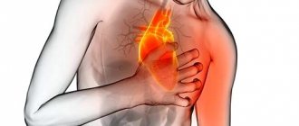
Angina is a clinical syndrome manifested by a feeling of tightness or squeezing pressing pain in the chest, which is most often localized behind the sternum and can radiate (“give”) to the left arm, neck, lower jaw, and to the epigastric region (epigastric region).
Pain occurs during physical activity or exposure to other factors that increase the heart's need for oxygen, and lasts from 1 to 15 minutes. It goes away with rest (when you stop exercising) or 1-3 minutes after taking nitroglycerin.
Translated from ancient Greek, angina means “narrow (weak, cramped) heart.”
Causes of angina
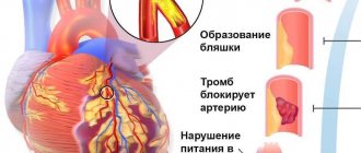
The development of angina is based on three mechanisms:
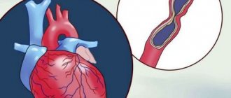
- atherosclerotic lesions of the coronary arteries;
- transient vascular thrombus formation;
- decreased coronary blood flow due to spasm or increased tone of the coronary arteries.
Angina pectoris makes itself felt during physical activity or stressful situations when there is a narrowing of the lumen of the coronary artery by 50-70%.
The severity of angina depends on the degree of coronary artery stenosis, its location and extent, the number and number of affected arteries. An atherosclerotic plaque can block a vessel completely or partially. With an increase in blood pressure, the inner layer of the coronary arteries (endothelium), damaged by the atherosclerotic process, can be easily damaged, blood penetrates into the plaque, the blood clotting process is activated and a blood clot is formed, which can partially or completely block the vessel.
The formation of a blood clot, especially against the background of a vessel spasm, can lead to its complete or partial blockage.
When myocardial tissue is damaged, pain mediators (serotonin, histamine, bradykinin, etc.) are released, which affect pain receptors.
There are modifiable (those that can be influenced) and non-modifiable factors that can provoke the development of angina. Modifiable diseases include dyslipoproteinemia (violation of the normal ratio of blood lipids), arterial hypertension, diabetes mellitus, smoking, low physical activity, obesity, and stress. Non-modifiable factors - male gender, age, family history of cardiovascular diseases (myocardial infarction or ischemic stroke in close relatives - up to 65 years (for women) and up to 55 years (for men)).
Classification of the disease
The most widespread classification of angina pectoris, developed
on the basis of WHO expert recommendations
:
- Stable angina pectoris (indicating functional class from I to IV).
- Class I – the patient tolerates physical activity well, angina attacks occur only during high-intensity exercise; Class II - slight limitation of usual physical activity, angina attacks occur when walking on level ground for a distance of more than 500 m, when climbing more than one floor; Class III – pronounced limitation of normal physical activity, attacks occur when walking at a normal pace on level ground for a distance of 100-150 m, or when climbing one floor; Class IV - angina occurs with little physical activity, walking on level ground at a distance of less than 100 m.
- Unstable angina:
- new-onset angina (less than 1 month from the onset of attacks);
- progressive angina (increasing attacks in frequency, duration, intensity with expanding localization and irradiation);
- early post-infarction (within 2 weeks after acute myocardial infarction) or post-operative angina.
- Spontaneous (vasospastic, variant, Prinzmetal) angina.
In practice, doctors also use the clinical classification of stable angina
:
- stable exertional angina (indicating the functional class);
- vasospastic angina;
- microvascular angina.
Symptoms of angina
Pain from angina can sometimes be perceived not as true pain, but as a feeling of discomfort, a feeling of heaviness, squeezing, tightness, distension, burning or lack of air.
Most often, the pain is localized behind the sternum or along the left edge of the sternum. It can radiate to the neck, lower jaw, teeth, interscapular space, and less often to the elbow or wrist joints, mastoid processes.
Pain with angina pectoris usually lasts from 1 to 15 minutes.
Pain occurs at the peak of physical or emotional stress. Stressful situations, due to increased activity of the sympathoadrenal system, lead to an increase in heart rate, increased blood pressure and myocardial contractility, which means the myocardial need for oxygen increases. After taking nitroglycerin or stopping the exercise, the pain stops, and the pain attack stops faster in a sitting or standing position.
As angina progresses, a moment comes when attacks occur even with minimal exertion, and then even under conditions of physical rest.
Angina at rest joins angina pectoris and is combined with it. In such cases, attacks occur during times of increased oxygen consumption by the heart muscle, for example, during REM sleep, when the heart begins to beat faster.
In some patients, an attack of angina may occur in a horizontal position due to increased blood flow to the heart.
Vasospastic angina develops independently of physical and emotional stress, is caused by spasm of the coronary arteries, and usually occurs at a younger age than exertional angina due to atherosclerosis of the coronary arteries. In patients with vasospastic angina, many typical risk factors for atherosclerosis cannot be identified. The disease may be accompanied by threatening disturbances in heart rhythm, leading to the development of myocardial infarction and/or sudden death.
A feature of vasospastic angina is very severe attacks, usually localized in a typical location. They occur at night or early in the morning, and also when exposed to cold on exposed areas of the body.
Microvascular angina is characterized by attacks that occur some time after physical activity, during emotional stress and at rest; they are poorly controlled by nitroglycerin. The cause of microvascular angina is considered to be dysfunction of small coronary arteries (100-200 μm in diameter) in the prearteriolar segment of the coronary bed. In more than 70% of cases, microvascular angina coexists with classical angina in patients with atherosclerotic stenoses.
Diagnosis of angina pectoris
The diagnosis of angina is established on the basis of a combination of complaints (pain characteristic of angina) and information received from the patient about the course of the disease.
For all patients with suspected coronary heart disease, the following questions are asked:
- current or past smoking;
- the presence of cardiovascular diseases and/or deaths from cardiovascular diseases in the patient’s immediate relatives (father, mother, siblings);
- previous cases of seeking medical help and the presence of previously registered electrocardiograms, studies and conclusions;
- the presence of concomitant diseases in order to assess additional risks;
- currently taken medications.
To clarify the diagnosis and existing complications, data from laboratory and instrumental examination methods are used.
All patients with suspected angina are recommended to have a lipid profile tested to detect dyslipoproteinemia:
- triglycerides (Triglycerides);
How to distinguish the types of angina from each other
The main symptom of any type of this condition is pressing, squeezing pain in the left side of the chest, which can “radiate” to the left side of the face, neck, shoulder or shoulder blade.
Stable exertional angina
An attack of pain in the heart that occurs as a result of a decrease in arterial blood flow to the myocardium during exercise or stress. A decrease in blood supply to the heart muscle leads to a sharp decrease in the amount of oxygen supplied to the heart, which causes pain.
Signs of stable angina pectoris
- An attack is triggered by physical or emotional stress.
- Duration - from two to five minutes.
- The pain goes away after the action of the factor that caused it ceases or after taking nitrates.
Sometimes atypical manifestations are possible - shortness of breath (increased breathing rate), a feeling of suffocation, a burning sensation or heat in the chest.
The severity depends on the functional class (FC):
- 1st FC - attacks occur only with excessive stress;
- 2nd FC - attacks occur during normal exertion: fast walking, climbing stairs (more than two flights), after a heavy meal, stress;
- 3rd FC - attacks occur with minor exertion: walking at an average pace, climbing 1-2 flights of stairs;
- 4th FC - attacks occur at rest.
New or acute angina
After the first attack occurs, and up to three months after it, they speak of new-onset angina. During this period, the doctor draws conclusions about the severity of the pathology (determines the functional class) and the prognosis - the possibility of transition to unstable angina. In an acute attack, symptoms can range from mild to severe.
Unstable angina
Progressive exertional angina, or unstable angina, is said to occur when:
- the number of attacks increases;
- attacks appear at rest or after a heavy meal;
- the duration of attacks increases (up to 15-20 minutes or more);
- response to nitroglycerin decreases.
Progressive angina may precede myocardial infarction.
Vasospastic or variant angina
This type is spoken of when an attack occurs for no apparent reason - when it cannot be associated with the action of provoking factors. The cause of myocardial ischemia is a sudden spasm of the coronary arteries. More often, attacks of variant (vasospastic) angina occur in older women and occur at night or in the morning. Taking nitroglycerin is ineffective.
Microvascular angina
The clinical picture is no different from exertional angina. This diagnosis is made only after functional and instrumental research methods - echocardiography (ultrasound of the heart), coronary angiography or CT angiography. Based on the results of these studies, one can judge the condition of the coronary vessels. The main difference between microvascular angina and other types is the absence of damage to the coronary arteries.
Giving up bad habits, dosed physical activity and other prevention methods help prevent or delay the onset of any type of angina. Once diagnosed, angina requires treatment regardless of type
Forms of pathology
Cardiologists distinguish three types of the disease: primary, stable and progressive. The first and second forms can transform into the third over time. A more extensive classification of angina pectoris is based on the patient's tolerance to physical activity. The intensity of the maximum permissible human activity allows us to distinguish four functional classes.
| Functional class (FC) | Description |
| I | Daily loads are tolerated without consequences. The attack develops against the background of unusual activity - running, climbing a large number of flights of stairs, lifting heavy objects. |
| II | Minor physical activity provokes the development of symptoms. Causes may include leisurely walking, climbing stairs to a height of more than one floor, emotional stress |
| III | The patient cannot tolerate minimal physical activity. An attack occurs when walking a distance of 50-100 meters. Climbing stairs is difficult |
| IV | Activity is significantly limited. Angina pectoris occurs when performing basic actions or while at rest |
Angina pectoris (angina pectoris) - symptoms and treatment
After collecting complaints, anamnesis of life and illness, and examination, additional laboratory and instrumental examinations are prescribed.
Laboratory tests are mainly aimed at diagnosing dyslipidemia, which contributes to the formation of atherosclerotic plaques. For this purpose, a blood lipid spectrum analysis is prescribed, which includes total cholesterol and its fractions.
Instrumental methods primarily include electrocardiogram (ECG) . Many patients go to the cardiologist with the question “is everything normal with my heart” with only one ECG film. But, unfortunately, these indicators are of informative value only during an attack. In most cases, it is impossible to determine angina pectoris from one ECG film taken at rest, since it can only show rhythm disturbances, hypertrophy (enlargement) of various parts of the heart, conduction disturbances, or the presence of a previous myocardial infarction.
There is also an ultrasound examination of the heart , which shows its structure and can reveal possible abnormalities. Echocardiography is intended to determine the size of the heart and its chambers, the presence of valve defects (stenosis and insufficiency), neoplasms and previous infarction. But this study, like an ECG, is not informative enough if it was carried out outside an attack of angina, at rest.
Holter ECG monitoring (outpatient monitoring) is much more informative than the above studies. It is advisable to perform it in order to identify signs of myocardial ischemia during daily activities. To do this, the patient walks, eats and sleeps with the device attached to it for about a day, i.e., performs normal daily physical activity. When an attack occurs, the person being examined records the incident in a diary and then informs the doctor about when and under what conditions the symptoms arose. The sensitivity of this method in diagnosing coronary heart disease is 44-81%.
Since angina attacks practically do not occur at rest, there are examination methods using physical activity. By analogy with an electrocardiogram, a bicycle ergometry and treadmill test : an ECG is taken while the patient performs physical activity on a bicycle or treadmill. The sensitivity of this method is approximately 68%.
A more sensitive method is stress echocardiography - this is the same ultrasound examination of the heart, only performed after physical activity. As your heart rate increases during running, your myocardium consumes more oxygen and nutrients. When a vessel narrows, a sufficient amount does not enter the muscle, resulting in an attack of chest pain and disturbances in contraction. As a result, hypokinesia develops, which can be seen with ultrasound. This study has greater sensitivity (80-85%) and specificity (84-86%)
If it is impossible to perform an ECG and EchoCG during exercise, you can use transesophageal electrical pacing (TEES) or pharmacological tests . These methods are based on increasing the myocardial oxygen demand by increasing heart rate without significantly changing blood pressure.
There are also two less common research methods: stress myocardial perfusion scintigraphy and multislice cardiac computed tomography.
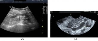
The main method for assessing the condition of the arteries, in which vessel narrowing can be visually recognized, is coronary angiography (CAG) - a radiopaque examination method that reliably determines the location and degree of narrowing of the arteries, as well as the type of blood supply, signs of thrombosis, ulceration, calcification and spasm of the coronary artery.
According to the timing of completion, this examination is divided into emergency (within 6 hours), urgent (within 6-12 hours) and planned.
Emergency coronary angiography is performed in case of unstable angina or myocardial infarction, when every minute is important.
Indications for planned coronary angiography include:
- objective signs of myocardial ischemia;
- transient ischemic changes detected during an ECG at rest or during 24-hour ECG monitoring;
- positive exercise test with bicycle ergometry, treadmill test, TES, stress echocardiography or myocardial scintigraphy;
- the occurrence of attacks of angina pectoris II-IV FC or angina pectoris at rest;
- condition of early post-infarction angina;
- life history of dangerous ventricular arrhythmias with a high risk of clinical death;
- planning surgery on the heart valve apparatus in people over 40 years of age;
- carrying out differential diagnosis with non-coronary myocardial diseases (including atypical pain syndrome);
- social indications in the presence of minimal and unclear signs of myocardial ischemia, provided that the patient’s profession is associated with a risk to the lives of other people (pilots, drivers), combat duty, etc.;
- undergone heart transplantation (the study is carried out every year, sometimes in combination with intravascular ultrasound).
There are currently no absolute contraindications for prescribing CAG.
An intermediate place is occupied by emergency coronary angiography. It is carried out in the event of a deterioration in the condition of a patient who is being treated in a hospital due to the progression of angina pectoris, when attacks of rest angina pectoris occur, there is no effect from maximum therapy, as well as in case of deterioration in condition after endovascular surgery or coronary artery bypass grafting.[1][10]
Diagnosis of pathology
The patient is examined by a cardiologist. The most effective way to diagnose exertional angina is an ECG obtained during an attack. Ischemia is induced by stress testing. Conducting them in a clinical setting eliminates the possibility of complications developing in the patient. Holter monitoring can detect myocardial ischemia and heart rhythm disturbances.
EchoCG is aimed at assessing myocardial contractility. The use of this technique allows for differential diagnosis and exclusion of other pathologies of the cardiovascular system from the patient’s medical history. Laboratory tests - general and biochemical blood tests - provide an opportunity to detect signs of atherosclerotic damage to the patient’s blood vessels.
What is angina?
In Russia, the classification given in the Clinical Guidelines of the Ministry of Health for Coronary Heart Disease of 2021 has been adopted. According to this classification, angina occurs:
- voltage, stable, indicating functional class (FC);
- vasospastic (other names - spontaneous, variant, Prinzmetal's angina);
- microvascular.
In the International Classification of Diseases, 10th revision (ICD-10), in addition to the above-mentioned options, there is also unstable angina, which can be either new or progressive. This type is usually mentioned in cases of progression of coronary insufficiency. The greater the degree of stenosis of the coronary arteries, the more severe the attacks will be
Syndrome X in cardiology (“microvascular angina”)
In approximately 10–20% of patients who undergo diagnostic coronary angiography due to acute or chronic cardiac ischemic syndrome, the coronary arteries are intact. Even if we assume that in some of them the symptoms of ischemia may be due to other cardiac and non-cardiac causes, then at least one out of ten patients with typical angina pectoris does not have hemodynamically significant stenoses of the coronary arteries. The presence of typical angina with unchanged coronary arteries was first described by N. Kemp in 1973 [1]. This syndrome is called “ X(X) syndrome” .
Cardiac syndrome X is a pathological condition characterized by the presence of signs of myocardial ischemia in the absence of atherosclerosis of the coronary arteries and spasm of the epicardial coronary arteries on coronary angiography (signs of myocardial ischemia: typical attacks of angina and ST segment depression ≥ 1.5 mm (0.15 mV) duration more than 1 minute established during 48-hour ECG monitoring). Thus, cardiac syndrome X is diagnosed in patients:
• with typical chest pain;
• with positive stress tests;
• with angiographically normal epicardial coronary arteries and no clinical or angiographic evidence of coronary artery spasm;
• with the absence of systemic arterial hypertension with and without left ventricular hypertrophy, as well as the absence of disturbances in the systolic function of the left ventricle at rest.
In rare cases, patients with syndrome X develop left bundle branch block with subsequent development of dilated cardiomyopathy. It should be noted that in the absence of changes in the coronary arteries during angiography, there is often occlusive pathology of the distal vessels (microvascular angina).
Some authors use the term “microvascular angina,” meaning that patients with typical angina have a normal coronary angiogram and reduced coronary reserve.
Syndrome X is usually classified as one of the clinical forms of coronary artery disease, since the concept of “myocardial ischemia” includes all cases of imbalance in oxygen supply and myocardial demand for it, regardless of the reasons causing it.
It should be noted that the capabilities of the angiography method in assessing the state of the coronary bed, in particular microvascular, are limited. Therefore, the concept of “angiographically unchanged coronary arteries” is very arbitrary and indicates only the absence of atherosclerotic plaques narrowing the lumen of the vessels in the epicardial coronary arteries. The anatomical features of the small coronary arteries remain “angiographically invisible.” Causes of cardiac syndrome X:
The etiology of cardiac syndrome X remains unclear and only some pathophysiological mechanisms leading to the development of typical clinical and instrumental manifestations of the disease have been established:
• increased sympathetic activation; • endothelial dysfunction; • structural changes at the microcirculation level; • metabolic changes (hyperkalemia, hyperinsulinemia, “oxidative stress”, etc.); • increased sensitivity to intracardiac pain; • chronic inflammation; • increased stiffness of arteries, etc.
There are a number of hypotheses that determine the pathogenesis of syndrome X. According to the first of them, the disease is caused by myocardial ischemia due to functional or anatomical disorders of microcirculation in intramuscular (intramural) prearterioles and arterioles, i.e. in vessels that cannot be visualized by coronary angiography. The second hypothesis assumes the presence of metabolic disorders leading to disruption of the synthesis of energy substrates in the heart muscle. The third hypothesis suggests that syndrome X occurs with increased sensitivity to painful stimuli (decreased pain threshold at the level of the thalamus) from various organs, including the heart.
Despite intensive research over the past 35 years regarding the pathogenesis of coronary syndrome X, many important questions remain unanswered.
Among patients with cardiac syndrome X, middle-aged people, mostly women, predominate. Less than 50% of patients with cardiac syndrome X have typical exertional angina, and the majority have atypical chest pain. Symptoms of cardiac syndrome X:
The main complaint includes episodes of angina-type chest pain that occurs during physical activity or is provoked by cold or emotional stress; with typical irradiation, in some cases the pain is longer lasting than with ischemic heart disease, and is not always relieved by taking nitroglycerin (in most patients the drug worsens the condition). Symptoms accompanying cardiac syndrome X resemble vegetative-vascular dystonia. Cardiac syndrome X is often found in people who are suspicious, with a high level of anxiety, against the background of depressive and phobic disorders. Suspicion of these conditions requires consultation with a psychiatrist. The diagnostic criteria for cardiac syndrome X include: • typical chest pain and significant depression of the ST segment during physical activity (including on the treadmill and bicycle ergometer); • transient ischemic depression of the ST segment ≥ 1.5 mm (0.15 mV) lasting more than 1 minute during 48-hour ECG monitoring; positive dipyridamole test; • positive ergometrine (ergotavin) test, a decrease in cardiac output against its background; • absence of atherosclerosis of the coronary arteries during coronary angiography; • increased lactate content during ischemia when analyzing blood from the coronary sinus area; • ischemic disorders during stress myocardial scintigraphy with 201 Tl.
Syndrome X resembles stable angina. However, clinical manifestations in patients with syndrome X are very variable, and in addition to exertional angina, attacks of resting angina can also be observed.
When diagnosing cardiac syndrome X, the following should also be excluded: • patients with spasm of the coronary arteries (vasospastic angina), • patients in whom non-cardiac causes of chest pain are documented by objective methods, for example:
- musculoskeletal causes (osteochondrosis of the cervical spine, etc.); — neuropsychic causes (anxiety-depressive syndrome, etc.); - gastrointestinal causes (esophageal spasm, gastroesophageal reflux, gastric or duodenal ulcer, cholecystitis, pancreatitis, etc.); - pulmonary causes (pneumonia, tuberculosis in the lungs, pleural overlays, etc.); - latent infections (syphilis) and rheumatological diseases.
Treatment of cardiac syndrome X:
Treatment for the group of patients with syndrome X remains not fully developed. The choice of treatment is often difficult for both treating physicians and the patients themselves. The success of treatment usually depends on the identification of the pathological mechanism of the disease and is ultimately determined by the participation of the patient himself. An integrated approach to the treatment of patients with cardiac syndrome X is often necessary.
There are various approaches to drug treatment: antianginal drugs, ACE inhibitors, angiotensin II receptor antagonists, statins, psychotropic drugs, etc. Antianginal drugs such as calcium channel blockers (nifedipine, diltiazem, verapamil, amlodipine) and β-adrenergic blockers (atenolol, metoprolol , bisoprolol, nebivolol, etc.) are necessary for patients with documented myocardial ischemia or impaired myocardial perfusion. Sublingual nitrates are effective in 50% of patients with cardiac syndrome X. There is evidence regarding the effectiveness of nicorandil, which has a bradycardic effect, the α1-blocker prazosin, L-arginine, ACE inhibitors (perindopril and enalapril), cytoprotectors (trimetazidine).
General advice on changing quality of life and treating risk factors, especially aggressive lipid-lowering therapy with statins (reducing total cholesterol to 4.5 mmol/l, LDL cholesterol less than 2.5 mmol/l), should be considered as vital components of any chosen treatment strategies.
Physical training. With cardiac syndrome X, tolerance to physical activity decreases, physical detraining and inability to perform exercise are observed due to a low pain threshold. Physical training increases the pain threshold, normalizes endothelial function and “delays” the appearance of pain during exercise in this category of patients.
Forecast.
The prognosis of patients with cardiac syndrome “X” is usually favorable. Complications typical for patients with coronary artery disease with stenotic atherosclerosis of the coronary arteries (in particular, myocardial infarction) are extremely rare. Survival with long-term follow-up is 95–97%, but in the majority of patients, repeated attacks of angina over many years have a negative impact on the quality of life. While not associated with increased mortality or an increased risk of cardiovascular “events,” cardiac syndrome X often seriously impairs patients' quality of life and poses a significant burden to the health care system. It must be emphasized that the prognosis is favorable in the absence of endothelial dysfunction. In these cases, the patient should be informed about the benign course of the disease. When excluding patients with left bundle branch block and patients with secondary microvascular angina due to serious systemic diseases such as amyloidosis or multiple myeloma, the prognosis of patients with cardiac syndrome X is favorable both in terms of survival and preservation of left ventricular function, but in some patients clinical manifestations of the disease persist for quite a long time.