Cardiologist
Sokolov
Denis Vladimirovich
16 years of experience
Cardiologist of the first category, candidate of medical sciences, member of the Asute Cardiovascular Care Association (ASSA)
Make an appointment
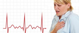
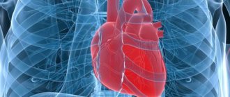
Angina pectoris is a pathology that manifests itself in representatives of different age groups in the form of attacks of myocardial ischemia. Symptoms arise against the background of increased physical or emotional stress that the patient faces. People suffering from angina pectoris experience pain in the heart area and shortness of breath. Autonomic reactions develop as a result of exposure to unfavorable external factors on the body.
Reasons for the development of pathology
Symptoms of unstable angina develop against the background of a gradual narrowing of the lumen of the coronary arteries. This phenomenon causes necrosis of cardiomyocytes. Lipid deposits on the coronary vessels lead to the formation of atherosclerotic plaques. The patient suffers from decreased blood flow in the coronary arteries.
A significant amount of lipid deposits can cause blood clots. Damaged vessels become overly susceptible to substances released by platelets. This phenomenon provokes further narrowing of the arterial lumen.
Interview and examination of the patient
An important role in the differential diagnosis of angina pectoris is played by questioning and examination of the patient. When compiling a medical history, the cardiologist studies all the symptoms that concern the patient, risk factors, for example, the presence of bad habits, obesity, etc., as well as possible genetic predisposition. During the examination, the doctor listens to heart sounds, looks for signs of damage to the main arteries, as well as signs of lipid metabolism disorders - one of the main causes of the formation of cholesterol plaques. Externally, they manifest themselves in the appearance of xanthoma and xanthelasma - skin neoplasms of a characteristic yellow color.
Forms of pathology
The classification of unstable angina used by cardiologists takes into account the timing of the onset of primary symptoms of the disease.
| Form of angina | Description |
| First appeared | Pain syndrome develops against the background of many years of asymptomatic disease. Pain occurs during periods of physical activity of the patient. The frequency and intensity of attacks increase over time |
| Progressive | Over the course of a month, there is a sharp increase in the number of attacks. The pain syndrome develops at rest. The course of the disease is complicated by arrhythmia and functional disorder of the left ventricle |
| Spontaneous | It is characterized by single attacks that are not associated with the patient’s physical activity. The duration of the acute phase is at least 15 minutes. ECG reveals signs of ischemia, but cardiomyocyte necrosis does not develop |
| Variant | Unstable angina occurs when the patient is at rest. The attacks last more than 10 minutes. Cardialgia appears regularly, at the same time interval. Between attacks, the patient can perform any physical activity without consequences |
| Post-infarction | Develops several days after myocardial infarction. It is characterized by an extensive area of necrosis with an unfavorable course. Unstable angina of this form requires emergency care. |
Canadian classification
To determine the severity of symptoms of angina pectoris, the Canadian Society of Cardiology has developed a special classification in the form of a table, which includes the following functional classes of angina pectoris:
Functional class 1
When performing normal physical activity for a person, he feels good. Pain only appears during intense and prolonged work, such as weightlifting or long-distance running.
Functional class 2
Pain occurs even during normal walking, when a person walks more than 200 meters. Also, angina pectoris develops if the patient climbs stairs above the 2nd floor, goes outside in very cold weather, or overeats.
Functional class 3
The attack begins when walking from 100 to 200 meters, or when climbing to the 2nd floor.
Functional class 4
Doing any physical work causes pain. An attack can develop even in a completely calm state.
Signs of pathology
The main symptom of unstable angina is intense pressing pain behind the sternum. The attack can last 10-15 minutes. The pain radiates to the left arm, shoulder, shoulder blade, neck and jaw. Taking nitroglycerin by the patient does not completely relieve symptoms. Over time, the frequency and intensity of attacks increase.
Manifestations of unstable angina develop against the background of:
- physical activity;
- stressful conditions;
- changeable weather;
- excessive food consumption.
Patients may experience interruptions in heart rate that occur with minimal physical activity (moving around the apartment, performing daily household chores). Later, shortness of breath joins the listed symptoms, and the person faces a lack of air.
Pharmacological tests
Pharmacological tests are an alternative to stress tests and are recommended for patients who, for various reasons, cannot perform physical activity. The principle of their action is the introduction of special drugs, due to which the myocardial need for oxygen increases, thereby simulating activity. Depending on the drug, this need arises as a result of an increase in heart rate or redistribution of blood in favor of vessels not affected by atherosclerosis.
Diagnostic procedures
The absence of specific symptoms does not allow the cardiologist to confirm the diagnosis during a physical examination. The doctor carries out differential diagnosis to exclude heart attack and non-ischemic heart pathologies from the patient’s history. Clinical guidelines suggest the following tests to confirm unstable angina:
- electrocardiography;
- general and biochemical blood tests;
- echocardiography;
- coronary angiography.
Cardiologists detect changes in the condition of the coronary arteries and decreased left ventricular function. Based on these data, the diagnosis is confirmed.
Diagnosis of angina pectoris
An important task in the treatment of cardiovascular diseases is the differential diagnosis of angina pectoris. In most cases, angina pectoris is a symptom of coronary heart disease that develops as a result of cholesterol deposition on the inner walls of blood vessels, so the efforts of cardiologists should be aimed at diagnosing the latter.
If you feel discomfort and pain in the chest or left shoulder after heavy exercise or stress, immediately contact a cardiologist. Self-medication can worsen the course of the disease.
Remember, only a specialist using modern diagnostic tools can identify coronary heart disease, prescribe treatment and prevent a tragic outcome!
Therapeutic measures
Conservative treatment involves prescribing several groups of drugs to the patient. Antianginal drugs relieve symptoms of angina pectoris. In acute attacks accompanied by intense pain, intravenous infusion of painkillers is performed.
Thrombolytics reduce the likelihood of blood clots. A similar goal is pursued when prescribing direct anticoagulants to a patient.
Lipid-lowering drugs remain an integral component in the drug treatment of unstable angina. They help normalize cholesterol levels in the patient’s blood. Constant use of such drugs reduces the likelihood of complications and recurrent attacks.
If conservative treatment is ineffective, the patient is prescribed surgery. The type of surgical intervention is determined by a cardiologist based on coronary angiography data. Imaging will allow us to understand the extent of damage to the coronary arteries. The most common methods of surgical treatment of angina remain coronary bypass surgery and coronary angioplasty.
Statistical data
Pathologies of the cardiovascular system remain the most common cause of death among Muscovites – 55% of cases in 2019. This figure is three times higher than the European average. The total number of Russian residents suffering from heart disease exceeds 16 million people. A quarter of them have congenital pathologies of the cardiovascular system. Prevention of attacks of myocardial ischemia remains one of the key tasks of cardiologists when monitoring patients at risk.
Treatment
The goals of treatment are to improve the prognosis (prevent heart attack) and eliminate symptoms of the disease. Non-medicinal (sports, diet), medicinal (tablets and drip infusions) and surgical treatment methods are used.
At the EXPERT Clinic, patients have the opportunity to receive a full consultation with a cardiologist on lifestyle changes and modification of risk factors. If necessary, treatment in a day hospital under the supervision of experienced medical personnel is possible.
Possible complications
Untreated angina can cause:
- sudden ventricular fibrillation with fatal outcome;
- acute myocardial infarction;
- acute failure of the heart muscle with pulmonary edema;
- thromboembolism of the pulmonary arteries.
Therefore, an increase in attacks and the appearance of unusual symptoms are important reasons for urgent medical attention.
When referred for inpatient treatment, refusal is considered as the patient’s responsibility for his own life.
Publications in the media
Stable angina pectoris is one of the main manifestations of coronary artery disease. The main and most typical manifestation of angina pectoris is chest pain that occurs during physical activity, emotional stress, when going out into the cold, walking against the wind, or at rest after a heavy meal.
Statistical data. Every year, angina pectoris is recorded in 0.2–0.6% of the population, with its predominance in men aged 55–64 years (0.8% of cases). It occurs in 30,000–40,000 adults per 1 million population per year, and its prevalence depends on gender and age • In the age group of the population 45–54 years, angina pectoris is observed in 2–5% of men and 0.5–1% of women , in the group of 65-74 years - in 11-20% of men and 10-14% of women (due to a decrease in the protective effect of estrogens in menopause) • Before MI, angina pectoris is observed in 20% of patients, after MI - in 50% of patients.
Etiology • In most cases, angina pectoris occurs due to atherosclerosis of the coronary arteries. Although the correlation between the degree of atherosclerotic narrowing, its extent and the severity of the clinical manifestations of angina is insignificant, it is believed that the coronary arteries must be narrowed by at least 50–75% before a discrepancy between the myocardial oxygen demand and its delivery appears and the clinical picture of the disease arises • Other causes (relative coronary insufficiency) •• Aortic stenosis •• Hypertrophic cardiomyopathy •• Primary pulmonary arterial hypertension •• Severe arterial hypertension •• Aortic valve insufficiency.
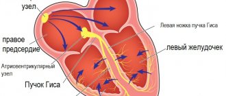
Pathogenesis • As a result of a discrepancy (imbalance) between the myocardial need for oxygen and its delivery through the coronary arteries due to atherosclerotic narrowing of the lumen of the coronary arteries, the following occur: •• Myocardial ischemia (clinically manifested by chest pain) •• Violations of the contractile function of the corresponding part of the heart muscle •• Changes biochemical and electrical processes in the heart muscle. In the absence of a sufficient amount of oxygen, cells switch to an anaerobic type of oxidation: glucose breaks down to lactate, intracellular pH decreases and energy reserves in cardiomyocytes are depleted • Subendocardial layers are primarily affected • The function of cardiomyocyte membranes is disrupted, which leads to a decrease in the intracellular concentration of potassium ions and an increase in intracellular concentration of sodium ions • Depending on the duration of myocardial ischemia, changes can be reversible or irreversible (myocardial necrosis, i.e. infarction) • Sequences of pathological changes during myocardial ischemia: impaired myocardial relaxation (impaired diastolic function) - impaired myocardial contraction (impaired systolic function ) - ECG changes - pain syndrome.
Canadian Cardiovascular Society Classification Pain does not occur when walking or climbing stairs. Seizures occur with severe, rapid or prolonged strain at work • Class II - “mild limitation of usual activities.” Pain occurs when walking or quickly climbing stairs, walking uphill, walking or climbing stairs after eating, in the cold, against the wind, during emotional stress, or within a few hours of waking up. Walking more than 100–200 m on level ground or climbing more than 1 flight of stairs at a normal pace and under normal conditions • Class III - “significant limitation of usual physical activity.” Walking on level ground or climbing one flight of stairs at a normal pace under normal conditions provokes an attack of angina pectoris • Class IV - “impossibility of any physical activity without discomfort.” Seizures may occur at rest
CLINICAL MANIFESTATIONS
Complaints. Characteristics of the pain syndrome • Localization of pain - retrosternal • Conditions for the occurrence of pain - physical activity, strong emotions, large meals, cold, walking against the wind, smoking. Young people often have the so-called phenomenon of “going through pain” (the “warm-up” phenomenon) - a decrease or disappearance of pain while increasing or maintaining the load (due to the opening of vascular collaterals) • Duration of pain - from 1 to 15 minutes, has an increasing character (“ crescendo"). If the pain continues for more than 15 minutes, the development of MI should be assumed • Conditions for stopping pain - stopping physical activity, taking nitroglycerin • The nature of pain during angina (squeezing, pressing, bursting, etc.), as well as the fear of death, are very subjective and not have serious diagnostic significance, since they largely depend on the physical and intellectual perception of the patient • Irradiation of pain - both to the left and right parts of the chest and neck. Classic irradiation - to the left arm, lower jaw.
Associated symptoms are nausea, vomiting, increased sweating, fatigue, shortness of breath, increased heart rate, increased (sometimes decreased) blood pressure.
Angina equivalents: shortness of breath (due to impaired diastolic relaxation) and severe fatigue during exercise (due to decreased cardiac output due to impaired systolic myocardial function with insufficient oxygen supply to skeletal muscles). In any case, symptoms should decrease when exposure to the provoking factor (physical activity, hypothermia, smoking) or nitroglycerin is stopped.
Physical data • During an attack of angina pectoris - pallor of the skin, immobility (patients “freeze” in one position, since any movement increases the pain), sweating, tachycardia (less often bradycardia), increased blood pressure (less often, its decrease) • Extrasystoles, “rhythm” can be heard gallop”, systolic murmur resulting from mitral valve insufficiency as a result of dysfunction of the papillary muscles • An ECG recorded during an attack of angina can detect changes in the terminal part of the ventricular complex (T wave and ST segment), as well as cardiac arrhythmias.
Laboratory data - supporting value; They can only determine the presence of dyslipidemia, identify concomitant diseases and a number of risk factors (DM), or exclude other causes of pain (inflammatory diseases, blood diseases, thyroid diseases).
Instrumental data
• ECG during an attack of angina: repolarization disturbances in the form of changes in T waves and ST segment displacement up (subendocardial ischemia) or down from the isoline (transmural ischemia) or heart rhythm disturbances.
• Daily ECG monitoring allows you to identify the presence of painful and non-painful episodes of myocardial ischemia in the usual conditions for patients, as well as possible heart rhythm disturbances throughout the day.
• Bicycle ergometry or treadmill (stress test with simultaneous recording of ECG and blood pressure). Sensitivity - 50-80%, specificity - 80-95%. The criterion for a positive stress test during bicycle ergometry is ECG changes in the form of horizontal ST segment depression of more than 1 mm lasting more than 0.08 s. In addition, stress tests can reveal signs associated with an unfavorable prognosis for patients with angina pectoris: •• typical pain syndrome •• ST segment depression of more than 2 mm •• persistence of ST segment depression for more than 6 minutes after cessation of exercise •• appearance of segment depression ST when the heart rate (HR) is less than 120 per minute •• the presence of ST depression in several leads, ST segment elevation in all leads, with the exception of aVR •• absence of a rise in blood pressure or its decrease in response to physical activity •• the occurrence of cardiac arrhythmias ( especially ventricular tachycardia).
• EchoCG at rest allows you to determine the contractility of the myocardium and conduct a differential diagnosis of pain syndrome (heart defects, pulmonary hypertension, cardiomyopathies, pericarditis, mitral valve prolapse, left ventricular hypertrophy in arterial hypertension).
• Stress echocardiography (echocardiography assessment of the mobility of left ventricular segments with an increase in heart rate as a result of dobutamine administration, transesophageal pacemaker or under the influence of physical activity) is a more accurate method for detecting coronary artery insufficiency. Changes in local myocardial contractility precede other manifestations of ischemia (ECG changes, pain). The sensitivity of the method is 65–90%, specificity is 90–95%. Unlike bicycle ergometry, stress echocardiography can detect coronary artery insufficiency when one vessel is affected. Indications for stress echocardiography are: •• atypical angina pectoris (the presence of angina equivalents or an unclear description of the pain syndrome by the patient) •• difficulty or impossibility of performing stress tests •• uninformativeness of bicycle ergometry in a typical clinical picture of angina pectoris •• absence of changes on the ECG during stress tests due to for blocks of the His bundle branches, signs of left ventricular hypertrophy, signs of Wolff–Parkinson–White syndrome in a typical clinical picture of angina pectoris •• positive stress test during bicycle ergometry in young women (since the likelihood of coronary artery disease is low).
• Coronary angiography is the “gold standard” in the diagnosis of coronary artery disease, since it allows us to identify the presence, location and degree of narrowing of the coronary arteries. Indications (recommendations of the European Society of Cardiology; 1997): •• exertional angina pectoris above functional class III in the absence of the effect of drug therapy •• exertional angina pectoris I–II functional class after MI •• exertional angina pectoris with His bundle branch block in combination with signs of ischemia according to data myocardial scintigraphy •• severe ventricular arrhythmias •• stable angina in patients undergoing vascular surgery (aorta, femoral, carotid arteries) •• myocardial revascularization (balloon dilatation, coronary artery bypass grafting) •• clarification of the diagnosis based on clinical or professional (for example, pilots) considerations.
• Myocardial scintigraphy is a method of visualizing the myocardium that allows identifying areas of ischemia. The method is very informative when it is impossible to evaluate the ECG due to blockades of the His bundle branches.
Diagnostics. In typical cases, stable angina pectoris is diagnosed based on a detailed history, a detailed physical examination of the patient, a resting ECG recording, and subsequent critical analysis of the data obtained. It is believed that these types of examinations (history, examination, auscultation, ECG) are sufficient to diagnose angina pectoris with its classic manifestation in 75% of cases. If there is any doubt about the diagnosis, 24-hour ECG monitoring, stress tests (veloergometry, stress echocardiography) are performed consistently, and if appropriate conditions are present, myocardial scintigraphy is performed. At the final stage of diagnosis, coronary angiography is necessary.
Differential diagnosis. It should be borne in mind that chest pain syndrome can be a manifestation of a number of diseases. We should not forget that there may be several causes of chest pain at the same time • Cardiovascular disease •• MI •• Angina pectoris •• Other causes ••• possibly of ischemic origin: aortic stenosis, aortic valve insufficiency, hypertrophic cardiomyopathy, arterial hypertension, pulmonary hypertension , severe anemia ••• non-ischemic: aortic dissection, pericarditis, mitral valve prolapse • Gastrointestinal diseases •• Esophageal diseases - esophageal spasm, esophageal reflux, esophageal rupture •• Stomach diseases - peptic ulcer • Diseases of the chest wall and spine •• Anterior thoracic syndrome walls •• Syndrome of the anterior scalene muscle •• Costal chondritis (Tietze syndrome) •• Damage to the ribs •• Herpes zoster • Lung diseases •• Pneumothorax •• Pneumonia involving the pleura •• PE with or without pulmonary infarction • Pleural diseases.
TREATMENT. The goals are to improve the prognosis (prevention of MI and sudden cardiac death) and reduce the severity (elimination) of symptoms of the disease. Non-drug, medicinal (drug) and surgical treatment methods are used.
• Non-drug treatment - impact on risk factors for coronary artery disease: dietary measures to reduce dyslipidemia and reduce body weight, smoking cessation, sufficient physical activity in the absence of contraindications. Normalization of blood pressure levels and correction of carbohydrate metabolism disorders are also necessary.
• Drug therapy - three main groups of drugs are used: nitrates, b-blockers and slow calcium channel blockers. Additionally, antiplatelet agents are prescribed.
Nitrates. When nitrates are administered, systemic venodilation occurs, leading to a decrease in blood flow to the heart (reduction in preload), a decrease in pressure in the chambers of the heart and a decrease in myocardial tension. Nitrates also cause a decrease in blood pressure, reduce resistance to blood flow and afterload. In addition, the expansion of large coronary arteries and an increase in collateral blood flow are important. This group of drugs is divided into short-acting nitrates (nitroglycerin) and long-acting nitrates (isosorbide dinitrate and isosorbide mononitrate).
• To relieve an attack of angina, nitroglycerin is used (tablet forms sublingually in a dose of 0.3–0.6 mg and aerosol forms - spray - are also used in a dose of 0.4 mg sublingually). Short-acting nitrates relieve pain in 1–5 minutes. Repeated doses of nitroglycerin to relieve an attack of angina can be used at 5-minute intervals. Nitroglycerin in tablets for sublingual use loses its activity after 2 months from the moment the tube is opened due to the volatility of nitroglycerin, so regular replacement of the drug is necessary.
• To prevent angina attacks that occur more often than once a week, long-acting nitrates are used (isosorbide dinitrate and isosorbide mononitrate) • Isosorbide dinitrate at a dose of 10-20 mg 2-4 times a day (sometimes up to 6) 30-40 minutes before expected physical activity. Retard forms of isosorbide dinitrate - at a dose of 40-120 mg 1-2 times / day before the expected physical activity • Isosorbide mononitrate at a dose of 10-40 mg 2-4 times / day, and retard forms - at a dose of 40-120 mg 1-2 r/day also 30–40 minutes before the expected physical activity.
• Tolerance to nitrates (loss of sensitivity, addiction). Regular daily use of nitrates for 1–2 weeks or more can lead to a decrease or disappearance of the antianginal effect •• The reason is a decrease in the formation of nitric oxide, acceleration of its inactivation due to increased activity of phosphodiesterases and increased formation of endothelin-1, which has a vasoconstrictor effect •• Prevention - asymmetric (eccentric) administration of nitrates (for example, 8 a.m. and 3 p.m. for isosorbide dinitrate or only 8 a.m. for isosorbide mononitrate). In this way, a nitrate-free period lasting more than 6–8 hours is provided to restore the sensitivity of the SMC of the vascular wall to the action of nitrates. As a rule, a nitrate-free period is recommended for patients during periods of minimal physical activity and a minimal number of painful attacks (individually in each case) •• Other methods of preventing nitrate tolerance include the use of sulfhydryl group donors (acetylcysteine, methionine), ACE inhibitors (captopril, etc. ), angiotensin II receptor blockers, diuretics, hydralazine, however, the incidence of tolerance to nitrates with their use decreases to a small extent.
Molsidomine is similar in action to nitrates (a nitrocontaining vasodilator). After absorption, molsidomine is converted into an active substance that is converted into nitric oxide, which ultimately leads to relaxation of vascular smooth muscle. Molsidomine is used in a dose of 2–4 mg 2–3 times/day or 8 mg 1–2 times/day (long-acting forms).
b-Adrenergic blockers. The antianginal effect is due to a decrease in myocardial oxygen demand due to a decrease in heart rate and a decrease in myocardial contractility. For the treatment of angina pectoris the following is used:
• non-selective b-adrenergic blockers (act on b1- and b2-adrenergic receptors) - for the treatment of angina, propranolol is used in a dose of 10-40 mg 4 times / day, nadolol in a dose of 20-160 mg 1 time / day;
• cardioselective b-adrenergic blockers (act primarily on b1-adrenergic receptors of the heart) - atenolol at a dose of 25–200 mg/day, metoprolol 25–200 mg/day (in 2 doses), betaxolol (10–20 mg/day), bisoprolol ( 5–20 mg/day).
• Recently, b-blockers have been used that cause peripheral vasodilation, such as carvedilol.
Slow calcium channel blockers. The antianginal effect consists of moderate vasodilation (including coronary arteries), reducing myocardial oxygen demand (in representatives of the verapamil and diltiazem subgroups). Used: verapamil - 80-120 mg 2-3 times / day, diltiazem - 30-90 mg 2-3 times / day.
Prevention of MI and sudden cardiac death
• Clinical studies have shown that the use of acetylsalicylic acid at a dose of 75–325 mg/day significantly reduces the risk of developing MI and sudden cardiac death. Patients with angina pectoris should be prescribed acetylsalicylic acid in the absence of contraindications - peptic ulcer, liver disease, increased bleeding, intolerance to the drug.
• The prognosis of patients with stable angina pectoris is also positively influenced by reducing the concentration of total cholesterol and LDL cholesterol using lipid-lowering drugs (simvastatin, pravastatin). Currently, optimal levels are considered to be no more than 5 mmol/l (190 mg%) for total cholesterol and no more than 3 mmol/l (115 mg%) for LDL cholesterol.
Surgery. When determining the tactics of surgical treatment of stable angina pectoris, it is necessary to take into account a number of factors: the number of affected coronary arteries, left ventricular ejection fraction, and the presence of concomitant diabetes. Thus, with one or two vessel lesions with a normal left ventricular ejection fraction, myocardial revascularization is usually started with percutaneous transluminal coronary angioplasty and stenting. In the presence of two or three-vessel disease and a decrease in the left ventricular ejection fraction of less than 45% or the presence of concomitant diabetes, it is more advisable to perform coronary artery bypass grafting (see also Atherosclerosis of the coronary arteries).
• Percutaneous angioplasty (balloon dilatation) - expansion of a section of the coronary artery narrowed by an atherosclerotic process with a miniature balloon under high pressure under visual control during angiography. The success of the procedure is achieved in 95% of cases. Complications are possible during angioplasty: •• mortality is 0.2% with single-vessel disease and 0.5% with multi-vessel disease, MI occurs in 1% of cases, the need for coronary artery bypass grafting appears in 1% of cases; •• late complications include restenosis (in 35–40% of patients within 6 months after dilatation), as well as the appearance of angina (in 25% of patients within 6–12 months).
• In parallel with the expansion of the lumen of the coronary artery, stenting has recently been used - implantation of stents (thin wire frames that prevent restenosis) at the site of narrowing.
• Coronary artery bypass grafting is the creation of an anastomosis between the aorta (or internal mammary artery) and the coronary artery below (distal) the site of narrowing to restore effective blood supply to the myocardium. A section of the saphenous vein of the thigh, the left and right internal mammary arteries, the right gastroepiploic artery, and the inferior epigastric artery are used as a graft. Indications for coronary artery bypass grafting (recommendations of the European Society of Cardiology; 1997) •• Left ventricular ejection fraction less than 30% •• Lesion of the trunk of the left coronary artery •• The only unaffected coronary artery •• Left ventricular dysfunction in combination with three-vessel disease, especially with damage to the anterior interventricular branches of the left coronary artery in the proximal part •• When performing coronary bypass surgery, complications are also possible - MI in 4-5% of cases (up to 10%). Mortality is 1% for single-vessel disease and 4–5% for multi-vessel disease. Late complications of coronary artery bypass grafting include restenosis (with the use of vein grafts in 10–20% of cases during the first year and 2% each year for 5–7 years). When arterial grafts are used, shunts remain open in 90% of patients for 10 years. Within 3 years, angina returns in 25% of patients.
The prognosis for stable angina pectoris with adequate treatment and monitoring of patients is relatively favorable: mortality is 2–3% per year, fatal MI develops in 2–3% of patients. A less favorable prognosis is for patients with a decrease in the ejection fraction of the left ventricle, a high functional class of stable angina pectoris, elderly patients, patients with multivessel disease of the coronary arteries, stenosis of the main trunk of the left coronary artery, proximal stenosis of the anterior interventricular branch of the left coronary artery.
Age characteristics • Children. The most common cause of symptoms of angina pectoris in children is hereditary dyslipidemia • Elderly - high sensitivity to the side effects of drugs (for example, severe depression when prescribing b-blockers) • Pregnancy - after clarifying the diagnosis, careful observation by an obstetrician and cardiologist is necessary, an increase in the need for oxygen during pregnancy increases the symptoms of angina pectoris.
Prevention • Stop smoking, diet low in cholesterol and fat, regularly perform a set of special exercises • Lipid-lowering drugs.
Synonyms • Angina pectoris • Angina pectoris • Heberden's disease.
ICD-10 • I20.8 Other forms of angina