To decipher any electrocardiogram, doctors use a term such as “metabolic changes in the myocardium.” It is believed that it represents a generalized concept expressing the presence of deviations from the isoline of cardiogram segments.
As a rule, these deviations indicate disturbances in metabolic processes in the heart muscle and insufficient supply of nutrition to its cells (cardiomycytes), which means that subsequently a malfunction may occur and the contractility of the heart muscle may be impaired.
Interesting! There are cases when such changes are shown by the cardiogram of a healthy person, and they do not mean anything “terrible”. This is explained by the fact that they can be the consequences of increased workload, severe stress, or excessive alcohol consumption on the eve of the examination. To get rid of them, it is enough to return to a normal lifestyle.
But, most often, metabolic disorders in the myocardium are reflected not only in the electrocardiogram; they are accompanied by patient complaints about general well-being and problems associated with blood circulation (for example, heart rhythm disturbances). Such cases require special attention and appropriate therapy.
Process description
Metabolic changes in the myocardium are a pathological condition accompanied by metabolic disorders:
- proteins,
- carbohydrates,
- fats,
- vitamins,
- electrolytes (potassium, calcium, magnesium).
Such changes are not considered a separate disease, but occur against the background of other pathologies.
Impaired nutrition of myocardial cells leads to a decrease in its contractility, as well as loss of electrical automaticity. Therefore, the condition is often accompanied by various arrhythmias.
Causes
There are many reasons for metabolic changes in the myocardium. Among them are routine conditions (overwork, stress, consumption of energy drinks). In these cases, metabolic disorders in cardiomyocytes do not require urgent medical intervention, as they disappear on their own within a few hours after the factor is eliminated.
The most common causes of metabolic disorders are heart disease or noncardiac pathologies:
- mycardiofibrosis;
- cardiomyopathy provoked by the action of drugs (for example, cardiac glycosides) or toxic substances and allergens;
- myocardial dystrophy;
- cardiac amyloidosis;
- "gouty" heart;
- alcoholic cardiomyopathy;
- endocrine diseases (thyrotoxicosis);
- general metabolic disorders;
- hypovitaminosis;
- malabsorption, atrophic gastritis, pancreatitis with impaired exocrine activity;
- chronic anemia;
- obesity of any degree;
- strict protein-free diets for weight loss, vegetarianism;
- indolent infections;
- pathology of renal excretory function;
- hepatitis, liver failure.
What is myocardium
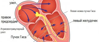
What does the concept of “myocardium” mean and do its changes always indicate any pathology? The concept refers to the middle muscular layer of the heart, which makes up the bulk of the organ.
The main task of this layer is to conduct electrical impulses along neuromuscular fibers. Due to various disorders, muscle tissue ceases to fully perform its functions, which entails a change in heart rhythm and the formation of specific cells that are not able to pass these impulses through themselves. In medical practice, these areas are called necrotic zones.
There are focal or isolated lesions, characterized by disruption of myocardial tissue in individual areas, as well as diffuse lesions, when necrosis is distributed evenly throughout the entire region of the heart. In place of the scars, overgrown connective tissue is formed, which doctors call electrically inert, that is, unable to pass electrical signals through itself. This leads to muscle hypertrophy, the development of heart failure and many other problems.
Expert advice
Often, hypokalemia (a decrease in the level of potassium in the blood), which leads to disruption of the heart muscle, occurs as a result of uncontrolled or incorrect use of loop diuretics (Lasix, Furosemide). I always focus the attention of my patients on the fact that this therapy increases the removal of electrolytes from the body. Therefore, it is necessary to artificially replenish their volume, in particular by taking Panangin or Caldium.
Features of ECG in various diseases
Changes in the myocardium occur under the influence of various internal and external provoking factors. Depending on the disease, electrocardiography indicators may be as follows:
- myocarditis - in this case, the shape and size of the teeth in all leads changes, the rhythm of heart beats is disrupted. A laboratory blood test reveals an inflammatory process;
- necrosis of the heart muscle - characterized by a distorted Q wave;
- myocardial ischemia – here there is an altered amplitude, shape and polarity of the T wave in the leads associated with the necrosis zone;
- myocardial dystrophy - manifests itself on the cardiogram, like myocarditis. A laboratory blood test helps make an accurate diagnosis.
Diagnosis and differential diagnosis of pathologies, which are characterized by changes in the structure of the myocardium, are carried out exclusively in a clinical setting using the necessary laboratory and instrumental methods.
Inflammatory
During electrocardiography, a specialist can see on the cardiogram a decrease in the waves in all leads and a disturbance in the heart rate. What do such violations mean?
Inflammatory changes develop against the background of such diseases:
- rheumatic organ damage caused by streptococcal diseases such as scarlet fever, tonsillitis, tonsillitis;
- diphtheria, typhus;
- viral diseases - influenza, measles, rubella and others;
- rheumatism;
- autoimmune pathologies, including lupus erythematosus and other disorders.
Inflammatory changes are most often provoked by viral and bacterial agents
Important! Rheumatism of the heart muscle is considered a severe inflammatory pathology leading to irreversible changes in the organ.
Dystrophic
This type of change occurs under the influence of a deficiency of certain microelements necessary for the normal functioning of cardiomyocytes. This entails a violation of the contractility of the myocardium, leading to the formation of lesions in the form of scars and connective tissue in the heart area. This occurs due to the following reasons:
- heart and kidney failure, resulting in the accumulation of toxins that poison the tissues of the entire body, including the myocardium;
- impaired glucose absorption, hormonal imbalance, decreased metabolic processes under the influence of endocrine diseases such as thyroid disease, adrenal dysfunction, diabetes mellitus and others;
- oxygen starvation of the muscle layer due to anemia. In this case, cardiomyocytes receive oxygen in insufficient quantities, which acts as a factor provoking dystrophic changes in the myocardium;
- The chronic course of certain diseases - malaria, influenza, tuberculosis - can cause the appearance of dead areas;
- vitamin deficiencies, frequent stress, regular physical activity;
- dehydration of the body under the influence of various reasons;
- poisoning, including toxic or chemical damage (medicines, alcohol, food poisoning).
Dystrophic changes are often reversible. If the problem is detected in a timely manner and the underlying disease is treated, it is possible to restore the damaged tissue.
Metabolic
Metabolic disorders occur when intracellular potassium-sodium metabolic processes fail. These may be moderate changes or the formation of large areas of damage. In turn, a lack or excess of potassium and sodium leads to a deficiency of energy to ensure the contractile function of the heart.
Myocardial infarction on ECG
On the cardiogram, the doctor notes the following changes:
- myocardial ischemia. During an examination for this disorder, the specialist sees on the ECG a pathological change in the T wave (frequency and shape) in the leads corresponding to the affected areas of the myocardium;
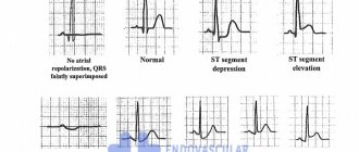
- myocardial infarction. Here, mirror changes are noted on the electrocardiogram, repolarization of the left ventricle, as evidenced by a shift of the ST segment above the isoline;
- necrotic processes in the myocardium. This disease is characterized by a distorted Q wave;
- irreversible damage to the wall of the muscle layer (transmural necrosis), which is characterized by the complete absence of the R wave.
Treatment for myocarditis and other diseases should be comprehensive, since the lack of therapy often entails many negative consequences and even the death of the patient.
Scarring
What do cicatricial changes in the myocardium mean? This type of pathology can be focal or diffuse in nature and occur against the background of the following conditions:
- long-term course of coronary heart disease. Hypoxia provokes accelerated growth of connective tissue, which leads to its growth in the thickness of the myocardium;
- transfer of myocardial infarction. More often, changes here are focal in nature;
- extensive transmural myocardial infarction – here necrosis covers a large area of the heart muscle, so it is worth highlighting this condition separately;
- surgical manipulations in the myocardium. Intervention in the tissue of a vital organ leads to the formation of scars, followed by their replacement with connective tissue.
Important! It is worth noting that many people suffer from several forms of changes at once. For example, patients with coronary artery disease who have suffered a myocardial infarction have signs of focal and at the same time diffuse cardiosclerosis.
Symptoms
Signs of metabolic disorders in the myocardium are often absent, so pathology is found by chance, thanks to characteristic changes on the ECG. Sometimes cardiac discomfort “eclipses” the signs of the underlying disease.
For example, I observed how, with an increase in the secretion of thyroid hormones, patients were more concerned about tremors in the hands, insomnia, and constant thirst. Therefore, they may simply not pay attention to periodic unpleasant sensations behind the sternum.
Among the symptoms of metabolic disorders in the heart muscle, I would note:
- overwork and decreased performance;
- sleep disturbance;
- palpitations, disturbance of normal rhythm;
- shortness of breath;
- periodic headaches and dizziness;
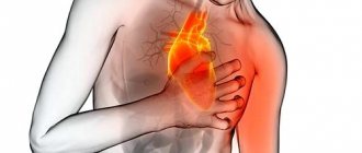
- pain in the left half of the chest, stabbing or squeezing in nature;
- pallor of the skin, cyanosis of the nasolabial triangle;
- short-term loss of consciousness, which may recur periodically.
It is very difficult to call such a clinical picture strictly specific. Similar conditions can occur with many somatic, neurological, infectious diseases and even with ordinary stress. I strongly advise you not to put off consulting a doctor until later. Early detection means faster recovery.
Key symptoms to watch out for
Symptoms will vary depending on which disease is the root cause of the changes. But there are also four common symptoms that are common to everyone. Particular attention should be paid when there is:
- pain in the chest area;
- frequent heart rhythm disturbances;
- the appearance of shortness of breath during physical activity;
- increased fatigue.
All of them can be “informants” of developing problems, which can only be identified by doing an electrocardiogram of the heart.
Diagnostic criteria
To identify metabolic changes in the myocardium, it is necessary to exclude hypoxic-ischemic processes in it.
The following signs speak in favor of metabolic disorders:
- clinical picture (pain, arrhythmias);
- muffled heart sounds during auscultation, the appearance of a slight systolic murmur, expansion of the boundaries of the organ during percussion (tapping);
- ECG signs (see table below);
- absence of manifestations of ischemia during Holter monitoring and exercise tests;
- slight enlargement of the heart, revealed by X-ray examination;
- normal state of blood vessels and valves during echocardiography against the background of decreased myocardial contractile function.
I draw your attention to the fact that the listed criteria should be considered comprehensively.
Characteristic changes on the ECG
The simplest screening method for diagnosing metabolic disorders in the myocardium is an electrocardiogram. The table shows common metabolic dysfunctions and their signs on film.
| Pathological condition | ECG signs |
| Increased calcium concentration in the blood |
|
| Decreased calcium levels in the body |
|
| Hypokalemia |
|
| Hyperkalemia |
|
| Thyrotoxicosis |
|
| Chronic alcoholism |
|
Remember that deciphering an ECG is not an easy task and requires in-depth physical analysis. This should only be carried out by a qualified person with the appropriate skills.
Types of changes
Violations of myocardial structure can be diagnosed in patients with various cardiac pathologies, as well as in absolutely healthy people, by chance during a routine examination. In this case, different degrees of inability of cardiac cells to contract and relax, their atrophy and replacement with connective tissue are detected. As already mentioned, a distinction is made between diffuse and focal changes. Diffuse on the ECG are accompanied by registration of multiple areas of accumulation of cardiomyocytes in all leads. Focal lesions are diagnosed more often in the first and second leads and are single in nature. Here, scar formation occurs in localized areas of the organ.
Norm and pathology of myocardial tissue
Clinical case
Patient P., 45 years old, was admitted to the emergency department. Delivered by ambulance from the store where she lost consciousness. Complains of weakness, irritability, trembling hands, palpitations. From the anamnesis: a year ago, thyrotoxicosis was diagnosed; he does not take the prescribed therapy. Fainting occurs 1-2 times a month, periodic headaches and dizziness, and increased blood pressure are also disturbing. She did not seek medical help. Objectively: the thyroid gland is enlarged, pressure: 150/90 mm Hg, heart rate 100 beats/min, tremor in the fingers, BMI 18, heart sounds are muffled, the boundaries are expanded by 0.5-0.7 mm. Preliminary diagnosis: “Thyrotoxicosis, thyrotoxic myocardial dystrophy.”
Dynamic ECG, Echo-CG, exercise tests, blood test for TSH and free T4, ultrasound of the thyroid gland, clinical blood and urine tests were performed. The diagnosis was confirmed, after which the patient was sent for treatment to an endocrinologist. After a two-week hospital stay, her condition improved. The patient was discharged for outpatient observation at her place of residence.
Which doctors were prescribed to you for metabolic changes in the myocardium? Share your experience in the comments.