Peripartum cardiomyopathy (Meadows Cardiomyopathy, Peripartum disease)
Conservative therapy
After confirmation of the diagnosis, patients are necessarily hospitalized in a cardiology hospital.
In case of severe circulatory failure, transfer to the intensive care unit for intra-aortic balloon counterpulsation is indicated. Despite the rapidly progressive course, almost half of the patients experience spontaneous recovery. Non-drug treatment includes limiting fluid intake to 2 liters per day and table salt to 3-4 grams per day. When prescribing drug therapy to pregnant women, it is necessary to take into account the potential undesirable effect of certain medications on the fetus. Recommended:
- Dopamine receptor agonists.
The drugs act on the main pathogenetic link of PPCM. By suppressing the production of prolactin, they reduce the formation of its cardiotoxic fragment (16 kDa). Bromocriptine is used in pregnant women; after childbirth, it is possible to switch to cabergoline or quinagolide. - ACE inhibitors.
Essential medications that slow the progression of heart failure. - Beta blockers.
Prescribed as an alternative to ACE inhibitors for pregnant women, as well as in the presence of tachyarrhythmias. - Vasodilators.
Vasodilator medications are used to relieve vasospasm when systolic pressure is more than 110 mmHg. Art. - Diuretics.
To reduce congestion in the lungs and remove fluid, thiazide and loop diuretics are used. - Cardiac glycosides.
To maintain adequate cardiac output during arterial hypotension, digoxin and levosimendan are indicated. - Anticoagulants.
Since dilatation of the cardiac cavities is accompanied by a high risk of thrombus formation, patients are recommended to take anticoagulants (warfarin, rivaroxaban). Only heparin is allowed for pregnant women.
Surgery
In case of severe CHF (left ventricular ejection fraction reaches less than 40%), which develops in pregnant women, termination of pregnancy by cesarean section is required. Extremely severe peripartum cardiomyopathy, resistant to conservative therapy, is an indication for heart transplantation. For persistent tachyarrhythmias accompanied by severe hemodynamic disturbances (critical drop in blood pressure), implantation of a cardioverter-defibrillator is used.
Cardiomyopathies
Diabetes
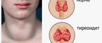
Thyrotoxicosis
21607 November 25
IMPORTANT!
The information in this section cannot be used for self-diagnosis and self-treatment.
In case of pain or other exacerbation of the disease, diagnostic tests should be prescribed only by the attending physician. To make a diagnosis and properly prescribe treatment, you should contact your doctor. Cardiomyopathy: causes, symptoms, diagnosis and treatment methods.
Definition
Cardiomyopathies (CM) are a group of heterogeneous (dissimilar) diseases characterized by pathology of the heart muscle with its structural and/or functional disorders that are not caused by coronary heart disease, hypertension, valvular defects and congenital diseases.
Causes of cardiomyopathies
It is believed that cardiomyopathy occurs as a result of a combination of various factors: genetic (gene mutations), autoimmune inflammation (in some bacterial and viral diseases), organ damage in a generalized (multiple organ) disease.
Classification of cardiomyopathies
According to the World Health Organization classification, cardiomyopathies are divided into idiopathic, specific and unclassified myocardial diseases.
Idiopathic cardiomyopathies:
- dilatational;
- hypertrophic (obstructive or non-obstructive);
- restrictive;
- arrhythmogenic dysplasia of the right ventricle;
- periportal (postpartum).
Specific cardiomyopathies (associated with a specific systemic or non-systemic disease):
- infectious;
- metabolic:
- endocrine (for hypo- and hyperthyroidism, myxedema, acromegaly, pheochromocytoma, diabetes mellitus, obesity),
- for storage diseases (amyloidosis, hemochromatosis, sarcoidosis, leukemia, mucopolysaccharidosis, glycogenosis, lipidosis),
- with a deficiency of microelements, nutrients and electrolyte imbalance;
Unclassified myocardial diseases include endomyocardial fibroelastosis, or the childhood form of cardiomyopathy, and idiopathic Fiedler's myocarditis.
Symptoms of cardiomyopathy Hypertrophic cardiomyopathy
Clinical manifestations vary from asymptomatic forms to severe disturbances in the functioning of the heart - up to sudden death, which is most often recorded in childhood or adolescence during physical activity or immediately after it.
Patients are concerned about the following symptoms:
- shortness of breath during exercise (in some patients at rest and at night), caused by venous stagnation of blood in the lungs due to diastolic dysfunction of the enlarged left ventricle of the heart;
- cardialgia associated with a discrepancy between coronary blood flow and myocardial weight;
- dizziness and syncope (fainting); in children they are observed during physical activity and emotional stress;
- rapid heartbeat due to heart rhythm disturbances;
- fast fatiguability.
Dilated cardiomyopathy
The disease is characterized by dilation (stretching) of the left ventricle with impaired pumping function of the heart in the absence of other diseases (arterial hypertension, valvular pathology, coronary heart disease) that could cause such disorders. The main clinical symptoms usually appear gradually, and in some patients are absent altogether, despite the presence of ventricular dilatation for months or even years. By the time of diagnosis, 73% of patients have symptoms of congestive heart failure. Patient complaints are usually associated with manifestations of congestive heart failure: heart pain, shortness of breath, general weakness and fatigue, peripheral edema, rapid heartbeat, heaviness in the right hypochondrium and epigastrium, symptomatic angina.
Restrictive cardiomyopathy
With the disease, the walls of the ventricles become rigid, inextensible (not necessarily thickened), which prevents the heart from filling the heart normally with blood between beats. Symptoms are determined by the severity of heart failure, and the disease can be asymptomatic for a long time. Patients are concerned about weakness, increased fatigue, shortness of breath even with slight physical exertion, cough, which may worsen in a lying position, swelling, abdominal enlargement, and in children, delay in physical development.
Arrhythmogenic right ventricular cardiomyopathy
The disease is characterized by the progressive replacement of right ventricular heart cells with adipose or fibrofatty tissue, leading to atrophy and thinning of the ventricular wall, and its dilatation.
For a long time, arrhythmogenic cardiomyopathy is asymptomatic, although the organic damage underlying the disease slowly progresses. Clinical signs of the disease (palpitations, paroxysmal tachycardia, dizziness and/or fainting) usually appear in adolescence. The leading clinical manifestations are life-threatening arrhythmias and sudden cessation of blood circulation, which occurs during exercise or strenuous sports activity.
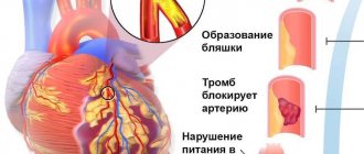
Ischemic cardiomyopathy
The disease is caused by diffuse morphofunctional disorders that develop as a result of chronic and episodes of acute myocardial ischemia.
The development of ischemic cardiomyopathy can result from extensive myocardial infarction or numerous small focal infarctions.
In typical cases, the clinical picture includes exertional angina, cardiomegaly (enlarged heart), chronic heart failure (and some patients have no clinical and ECG signs of angina).
Symptoms are identical to those of heart failure in patients with dilated cardiomyopathy.
Hypertensive cardiomyopathy
The disease develops against a background of uncontrolled high blood pressure over a long period of time.
The intensity of symptoms depends on the severity of the disease and the nature of its course. The most common complaints of patients include pain and tightness in the chest, shortness of breath, cough, lack of appetite, and peripheral edema.
Metabolic cardiomyopathy
Metabolic cardiomyopathy is a non-inflammatory lesion of the myocardium, which is based on a metabolic disorder leading to myocardial dystrophy and insufficient contractile function of the heart.
Clinical manifestations are varied and not specific, and the initial stage of the disease can occur without any symptoms at all, but over time, in the absence of proper treatment, severe heart failure develops.
Inflammatory cardiomyopathy
The disease is a combination of myocarditis and myocardial dysfunction. Patients complain of chest pain, often radiating to the left arm, attacks of rapid heartbeat, shortness of breath during exercise and then at rest, peripheral edema, and fatigue.
Diagnosis of cardiomyopathy
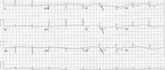
Diagnosis of cardiomyopathy is based on collecting anamnesis and laboratory and instrumental examinations of the patient. The main diagnostic criterion for cardiomyopathy is the presence of myocardial dysfunction, detected by ultrasound examination of the heart.
Diagnosis of dilated cardiomyopathy
DCM is diagnosed both by examining your medical history (symptoms and family history) and by a physical examination and clinical tests, such as blood tests, ECG, chest x-ray, echocardiography (ultrasound of the heart), stress test, catheterization heart, CT and MRI.
Other tests to identify the cause of DCM are rarely done, because When performing a myocardial biopsy, a tissue sample taken from the heart must be examined under a microscope to determine the cause of these symptoms.
Relatives of patients with dilated cardiomyopathy should be regularly screened for DCM. Genetic testing will also be useful to identify abnormal genes.
What pathologies can provoke the secondary form?
Secondary cardiomyopathy appears against the background of pathologies associated with the functioning of the human cardiovascular system.
The list of these diseases includes:
- IHD, coronary heart disease;
- Infections leading to destructive changes in the heart muscle;
- Endocrine diseases;
- Arterial hypertension;
- Shifts in metabolism and accumulation of substances;
- Electrolyte balance;
- In connective fibers;
- Amyloidosis;
- Neuromuscular pathology;
- Severe poisoning;
- During pregnancy.
Coronary heart disease is distinguished by a number of characteristic symptoms. Against the background of atherosclerosis, the lumen of the coronary arteries narrows, and a lack of oxygen (O2) occurs in the myocardium. This leads to the destruction of cardiomyocytes and replacement with connective fibers.
Myocardial infections also cause disorders in the heart muscle, inflammatory processes, edema, and damage to cardiomyocytes. Acute myocarditis develops, with the replacement of cardiac fibers by connective tissue, resulting in cardiomyopathy.
Endocrine diseases and hormonal disorders disrupt the functioning of the heart as a result of the influence of hormones and hyperstimulation of functions. These complications can cause:
- Thyroid dysfunction;
- Adrenal diseases;
- Diabetes.
Arterial hypertension with a sustained increase in blood pressure is also included in the list, leading to a pathogenic effect on the myocardium. Under these conditions, the load on the heart significantly increases, as a result, the structure of the cardiac fibers is disrupted, with loss of elasticity and the development of cardiomyopathy.
Shifts in metabolism lead to the accumulation of substances that are foreign to the myocardium and cause irreversible effects on the functioning of the heart muscle. Among the main reasons:
- Pathologies of glycogen accumulation;
- Phytanic acid in Refsum syndrome, genetic origin;
- Sphingolipids in Farbi's disease, with a genetic nature;
- Hemochromatosis with accumulation of iron in the fibers.
Problems with maintaining electrolyte balance are accompanied by changes in the amount of ions in the blood, which affects the functioning of the heart and the structure of myocardial fibers. Potassium, phosphorus, chlorine, magnesium, and calcium ions are washed out during severe diarrhea and various kidney diseases.
Pathological processes of connective tissue in the heart area cause systemic damage and disrupt the structure of myocardial fibers. Inflammation of these fibers leads to cardiosclerosis and gradual replacement of the heart muscle with connective tissue with the development of cardiomyopathy.
The list of diseases that provoke the pathological process includes:
- Scleroderma;
- Dermatomyositis;
- Lupus erythematosus;
- Rheumatoid arthritis.
Amyloidosis is characterized by the deposition of amyloid and causes disruption of the heart muscle. Moreover, the accumulation of this complex of proteins and polysaccharides occurs directly in the myocardium, which leads to the development of pathology.
Neuromuscular pathology causes disturbances in the transmission of impulses by nerve fibers with subsequent deviations in the functioning of the myocardium. In this case, asystole or extrasystole is observed, with a pronounced failure in the contraction of the heart muscle. In particularly severe conditions, the muscle tone of the heart decreases sharply. This leads to the development of a severe form of cardiomyopathy.
Poisoning with poisons, alcohol, surrogates, toxins directly affects the functioning of the heart and causes emergency conditions incompatible with human life. Toxic compounds directly affect the damage of cardiomyocytes, while the passage of the nerve impulse is blocked, and acute heart failure develops.
Severe poisoning with the development of cardiomyopathy is caused by various substances :
- Alcohol products and surrogates;
- Medicines, drugs (amphetamines group and others);
- Toxic compounds, heavy metals, including arsenic, lead, mercury and others.
Chronic consumption of these products causes irreversible changes in the heart muscle, pain in the heart area, and myocardial infarction. The high risk of developing emergency conditions leads to the fact that a person is constantly on the verge of life and death.
Radiation activity directly affects the human body and also refers to damaging factors that cause disturbances in the functioning of the heart. In some cases, such as in pregnant women, problems with the myocardium are temporary. But with chronic alcoholism, drug addiction, and the use of chemical substances, the disease continues to progress and the patient cannot be cured.
Reversible conditions include cardiomyopathy in pregnant women . It occurs more often in the last trimester or after childbirth due to stress. During this period, hemodynamics change dramatically, the circulatory system adapts to provide adequate nutrition to the fetus. At the same time, the volume of circulating blood changes, the level of blood pressure increases or decreases. These changes lead to the development of a reversible form of the disease.
Treatment of cardiomyopathy is prescribed by a doctor in accordance with the indications and depends on the cause of the disease. If the source of severe pathology cannot be determined, the disease is classified as a primary form and a course of maintenance therapy is prescribed.
Rate this article
Article rating 4.45 out of 5. Votes: 11.
Latest articles
Sexual transmission of infections
Sexually transmitted infections or STIs are dangerous diseases that can undermine health and lead to dire consequences. Most of the diseases in this group are…
Hormonal disorders
A woman’s well-being largely depends on the endocrine system. Any hormonal imbalance can cause a sharp deterioration in health, weakening the immune system...
Cervical dysplasia
Cervical intraepithelial neoplasia or cervical dysplasia is a precancerous condition expressed by pathological changes in cells. In most...
Treatment of dilated cardiomyopathy
Treatment of DCM is the treatment of chronic heart failure (CHF) in its purest form, since etiotropic treatment is possible only with a known etiology of DCM. Another important goal is to reduce the size of the heart by reducing the level of hormones in the blood that cause it to enlarge and ultimately worsen symptoms. Patients usually take various medications to treat heart failure. Cardiologists also recommend lifestyle changes.
What drugs are used to treat DCM?
To improve heart function, most people take medications such as beta blockers, ACE inhibitors and/or various diuretics. If you have an arrhythmia, your doctor may give you medicine to control your heart rhythm. Blood thinners may be used to prevent blood clots from forming. Discuss with your doctor which treatment is best for you.
What is used in surgery to treat DCM?
People with severe dilated cardiomyopathy may need one of the following surgeries:
- Cardiac resynchronization therapy is a three-chamber cardiac stimulation (one electrode in the right atrium, two in the ventricles).
- The operation of wrapping the heart with an elastic mesh frame prevents the progression of CHF; in the initial stages of DCM, it can lead to the reverse development of the disease.
- Mechanical heart ventricles (a micropump installed in the cavity of the left ventricle) - can be used for temporary hemodynamic support, but the functions of the heart itself have been shown to improve after several months of operation of the device.
- Heart transplantation is currently no longer considered the treatment of choice for DCM, due to the possibility of alternative procedures listed above.
The prognosis for DCM remains extremely serious - in the case of the idiopathic form of the disease, the annual mortality rate is from 5 to 45%; for forms with a known etiology, the mortality rate may be lower. Advances in treatment have led to a significant increase in survival rate for this form of cardiomyopathy.