Material and methods
The criterion for inclusion in the study was the presence of obliterating atherosclerosis in patients, clinically manifested as critical ischemia of the lower extremities, stage 3-4 according to Pokrovsky. In this case, the necrotic process should not spread beyond the toes. Patients who met these criteria were randomly divided into two groups: the first - control (30 patients) and the second, in which Actovegin was used (30 patients). Due to random distribution, both cohorts were comparable in gender, age, comorbidity and severity of the underlying disease, which made it possible, along with the use of modern methods of medical statistics, to obtain reliable research results.
The average age of the patients was 69.5±7.2 years. There were 34 men, 26 women. Taking into account the advanced age of the patients, concomitant pathology was present within the following range: multifocal atherosclerosis (AT) in 100% of cases, hypertension - 82.4, coronary heart disease (CHD) in 88.1% of patients, chronic heart failure (CHF) suffered from 85%, chronic renal failure (CRF) was detected in 23.7% (Fig. 1).
Treatment of patients in the control group consisted of prescribing a course of standard conservative therapy used in general surgical hospitals for critical ischemia of the lower extremities. This complex includes drugs that improve the rheological properties of blood, antispasmodics, drugs for the treatment of atherosclerosis (usually from the group of statins). The regimen was as follows: pentoxifylline 15 ml, rheopolyglucin 400.0 ml, antiplatelet agents (in some cases with double antiplatelet therapy), low molecular weight heparins in prophylactic doses, statins. In the second group, Actovegin was added to the above complex at a dose of 1200 mg intravenously, by drip for 15 days. Then a similar dosage in tablet form for the next 15 days.
The effectiveness of treatment was monitored by assessing the clinical picture (pain-free walking distance), the level of microcirculation - oxygen tension in tissues (polarography, TSM-400, Denmark) and laser Doppler imaging parameters using an LDI device from AIMAGO (Switzerland). For double control, morphological methods were also used: cytological and histological studies, the material for which was taken after surgical removal of dry necrosis, or in the area of the demarcation line.
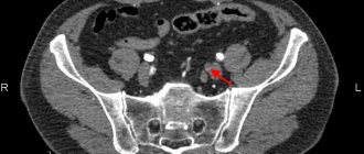
The reliability of the results of studying microcirculation was achieved using the regional tissue perfusion index (RTI tpO²). The latter is the ratio of the absolute value of tissue oxygen saturation at the desired point (the first interdigital space on the foot) to that at the control (ulnar fossa) point (Fig. 2).
The results were assessed before treatment, after a course of parenteral administration of Actovegin (on day 15), upon completion of the drug (on day 30), and also on day 60 from the start of treatment.The study of skin microcirculation in our patients was carried out using laser Doppler imaging using an LDI device from AIMAGO (Switzerland). LDI technology (LaserDoppler Imaging) in Russian literature - laser Doppler imaging, allows for non-contact observation of microblood flow over an area of 100 cm², with a penetration depth of up to 2 mm, simultaneous video recording of the study and the possibility of measurements at several nearby points at once. Moreover, the technical study is so simple that it can be performed by nursing staff, since reproducibility practically does not depend on the qualifications of the researcher. The resulting image was analyzed in real time. The obtained data were entered into the device’s memory, which made it possible, if necessary, to re-view the video recording of the measurement. At the same time, the number of relative perfusion units (APU) was taken into account, reflecting the amount of blood flowing per unit of time in one cubic unit of tissue volume.
Neuroprotection is defined as creating conditions for the neuron to adapt to new functional conditions and reduce damage to brain tissue. Currently, the universality of the mechanisms of cell damage in various pathological processes has been proven. The final link in these cases is a violation of redox processes, a violation of metabolism and energy supply to cells [16]. The use of pleiotropic drugs is considered a promising direction in neuroprotection. The pleiotropic neuroprotective effect involves a simultaneous modulating effect on various damaging pathological mechanisms - excitotoxicity, inflammation, apoptosis, oxidative stress, etc. [20].
Today, there are several pharmacological groups of drugs with neurometabolic action [15]. These include Actovegin; the features of clinical action and the mechanisms of its implementation have not yet received enough attention, which served as the basis for writing the proposed review.
Actovegin belongs to the group of drugs that activate metabolism without direct vasodilating effects [17]. It is a highly purified deproteinized hemodialysate obtained by ultrafiltration from the blood of young calves, which includes more than 200 biological substances [1]. Actovegin mainly consists of substances with low molecular weight, including amino acids, biogenic amines and polyamines, sphingolipids, hexoses, lactate, succinate, choline, vitamins, adenosine monophosphate and inositol phosphooligosaccharides [20]. The drug also contains macroelements - magnesium, sodium, calcium, phosphorus and neuroactive microelements - silicon and copper. One of the most important macronutrients is magnesium, which is part of many proteins and enzymes. In addition, magnesium is part of glutathione peroxidase, which takes part in the metabolism of H2O2, leading to the formation of glutathione [6, 8, 9].
Preclinical trials have shown that at the molecular level, Actovegin improves the uptake and utilization of oxygen and glucose [2, 10, 18]. Glucose transporters play an important role in neuronal homeostasis and glucose utilization, and changes in glucose homeostasis in the CNS lead to disruption of neuronal activity and cognitive functions, which is especially dangerous for the hippocampus, an area of the brain associated with learning and memory processes [20]. In an in vitro
P. Buchmayer et al. [18] it was shown that actovegin has a neuroprotective effect: when it is added to a neuron culture, the number of “living” neurons and synaptic contacts on their processes increases. Under ischemic conditions, actovegin improves metabolic balance by increasing glucose uptake and oxygen consumption by tissues [17, 18]. We are talking about the antioxidant and antiapoptotic mechanisms of action of actovegin, which underlie their neuroprotective properties, as well as its ability to influence neuroplasticity, neurogenesis, and the trophic function of the nervous system. It is the multicomponent nature of Actovegin that determines its pleiotropic neuroprotective mechanism of action and clinical effectiveness [20].
Before moving on to the presentation of clinical studies related to cerebrovascular diseases (CVD), we recall that one of the most common manifestations is impaired cognitive function. They include deficits in attention, concentration, decreased ability to quickly navigate in a changing environment; memory impairment, especially for current events; slowness of thinking processes, rapid exhaustion during intense mental work; narrowing the range of interests [15].
V. Jansen et al. [17] conducted a double-blind, placebo-controlled study of the therapeutic effect of Actovegin-Forte in the form of tablets (containing 200 mg of the active substance) on mnestic-intellectual abilities in patients with CVD. The study included 120 patients whose age was 60-72 years (average - 67 years); the average duration of the disease was 2.5 years. All patients were randomized into 3 groups: group 1 included 40 patients treated with Actovegin-Forte, 3 tablets 3 times a day; patients of the 2nd group (also 40 people) received placebo in the form of pills (2 pills 3 times a day or 3 pills 3 times a day); Group 3 (40 patients) took Actovegin-Forte 2 tablets 3 times a day. The duration of therapy was 12 weeks. The patients' condition was assessed before the start of therapy and then after 4, 8 and 12 weeks of treatment. When examining patients, a number of neuropsychological tests were used to assess mnestic-intellectual functions: a test to assess vocabulary; short syndromic test; Pauli test; Benton test; mosaic test; light flicker frequency analysis test. It was found that in the groups of patients treated with Actovegin, success in completing the task in the jigsaw test increased compared to placebo. By the end of the 12-week course of therapy with Actovegin, in comparison with placebo, the patients’ lack of concentration of thinking, attention and volitional disorders disappeared; mnestic-intellectual abilities in general have increased. The authors assessed the effectiveness of 12-week therapy with Actovegin-Forte at 92%, which served as a basis for recommending this drug for long-term outpatient treatment of elderly patients with CVD.
H. Letzel et al. [5] conducted a multicenter study of the effectiveness of Actovegin in 1549 elderly patients (average age 74.1 years) with impaired cerebral functions. Patients included in the study were prescribed Actovegin according to the standard regimen: 10 ml of Actovegin solution intravenously for 2 weeks, then 4 weeks of oral administration of Actovegin tablets (2 tablets 3 times a day). Patients were examined before the start of therapy, after 2 and 4 weeks of oral administration of the drug. After 4 weeks from the start of Actovegin therapy, in 80% of cases, an improvement in the general condition was noted: a decrease in the severity (or cessation) of headaches, dizziness, anxiety and fear, improvement in memory and concentration (according to psychological testing). In 10.9%, when using the drug parenterally, undesirable effects were observed (feelings of heat and nausea). Poor tolerability of the drug was noted in a small percentage (0.9%) of cases. The authors emphasize the importance of combination therapy - starting with intravenous Actovegin to achieve a quick and good response and then continuing it with oral pills. This therapy has been recommended for elderly patients with organic syndrome.
A.A. Fedorovich et al. [12, 13, 19] describe a number of studies that confirmed the effect of Actovegin on the vasomotor and metabolic activity of smooth muscle cells of the microvascular bed of the skin in humans. The authors infused 250 ml of Actovegin solution into the cubital vein of the left arm in doses of 1.0 g of dry matter and 2.0 g of dry matter at a rate of 2-2.5 ml/min for 120-130 minutes. On average, the increase in the amplitude of the endothelial (Ae) rhythm under the influence of the first dose of Actovegin was 75%, the contribution of the endothelium to the modulation of microblood flow increased by 30%, and the functional contribution to the overall level of tissue perfusion increased by 65%. When tested with a higher dose of Actovegin, the most pronounced increase in the activity of the microvascular endothelium was shown: the increase in AE in this group averaged 191%, and the contribution of the endothelium to the overall level of tissue perfusion was 163%. The maximum peak of drug activity after intravenous administration was observed in the interval of 2-6 hours. 2 hours after the end of the infusion, a slight but significant decrease in diastolic blood pressure and mean blood pressure was noted. These changes were noted against the background of a significant increase in all tone-forming links of microcirculation modulation (endothelial, neurogenic and myogenic), with the most significant increase observed in endothelial activity. The increase in the functional contribution of the endothelium to total tissue perfusion averaged 79%. The authors concluded that Actovegin not only has a pronounced metabolic effect, increasing the functional activity of the microvascular endothelium, but also affects the vasomotor function of microvessels, resulting in a significant improvement in the functional state of the smooth muscle apparatus of the microvessels.
Taking into account persistent hemodynamic cerebral insufficiency, Actovegin may be the drug of choice for the treatment of chronic CVD in patients with myocardial damage [11, 14]. Neuroendocrine imbalance in the form of activation of local neurohormonal systems and severe endothelial dysfunction initiates vascular remodeling processes: the thickness of the arterial walls increases and the lumen decreases. Changes in the microcirculatory bed in atherosclerosis of the cerebral arteries: reduction of the bed as a consequence of hypoxia and microvascular convolutes compensating for this hypoxia. Low cardiac output, asymptomatic cerebral microembolization, fluctuations in blood pressure with episodes of hypotension, and metabolic changes can cause cerebral disorders. Narrowing of the lumen of intracranial arteries, along with dysfunction of the endothelium and rigidity of the vascular wall, causes a chronic reduction in cerebral blood flow, the appearance of diffuse or focal changes in the brain substance and clinical signs of CVD. All of the above components of chronic cerebrovascular disorders are a direct indication for the prescription of the drug Actovegin. It is also important to emphasize that Actovegin has a satisfactory efficacy-safety ratio.
A number of clinical studies in recent years have confirmed the positive effect of Actovegin on cognitive function. As established by S.G. Bugrova [3], this applies primarily to mild and moderate cognitive impairment in vascular diseases. In the work of E.A. Derevyannykh et al. [4] revealed a broader effect of Actovegin not only on cognitive functions, but also on EEG and indicators of blood supply to the brain, as well as neurological manifestations of chronic cerebrovascular insufficiency. A.A. Skoromets and V.V. Kovalchuk [7], when comparing several drugs (actovegin, instenon, berlition, rheopolyglucin, gliatilin) in the process of rehabilitation of patients with ischemic and hemorrhagic stroke, found that in ischemic stroke all of the listed drugs have a positive effect, while in hemorrhagic stroke - only actovegin .
results
Changes in the level of skin perfusion that arose as a result of the study in patients are indicated in Table. 1.
Table 1. Oxygen regime of tissues before and after treatment with Actovegin
| Observation days | Before treatment | 15 days | 30 days | 60 days |
| Control group | 0,41±0,03 | 0,46±0,02 | 0,46±0,09 | 0,42±0,08 |
| Actovegin group | 0,43±0,04 | 0,51±0,03 | 0,52±0,02 | 0,54±0,09 |
Before treatment, both groups were comparable in terms of the initial level of microcirculation. In the control cohort of patients, despite standard therapy, no statistically significant improvement in transcutaneous oxygen tension was obtained, which may indirectly indicate that these medications do not affect the microcirculation.
In the Actovegin group, skin perfusion improved by 35% by day 15 of observation, at the end of the parenteral course. This result was maintained at the achieved level by days 30 and 60 with the help of an extended course of taking the drug in tablet form (Table 2)
Table 2. Effect of atovegin on microcirculation of the skin of the foot
| Time | First group (n-30) (APU) | Second group (n-30) (APU) |
| Before treatment | 42±3.1 | 43±2.4 |
| 15 days | 45±2.8 | 62±1.7 |
| 30 days | 48±1.7 | 75±2.1 |
| 60 days | 53±1.4 | 89±2.6 |
After morphological studies in the control group, a tendency towards intensification and growth of inflammation in the demarcation zone was revealed, followed by the development of necrobiotic and then necrotic changes in this area. Which was macroscopically expressed in the proximal spread of the area of dry necrosis (gangrene).
By day 15, compared with the initial data according to histological examination, activation of reparative processes was observed in the form of increased activity and an increase in the number of macrophages and fibroblasts (Fig. 4, Fig. 5).
During the same time period, cytological data indicate a significant decrease in the activity of the inflammatory process in the form of a 3-4-fold decrease in leukocyte infiltration by neutrophils (Fig. 6, Fig. 7).
In the Actovegin group, significant changes were noted during the period of parenteral administration of the drug, which was a manifestation of the activation of the corresponding metabolic links that are affected by the drug at the initial stage of treatment. In subsequent periods, consolidation of the result at the achieved level was observed.
Actovegin in the treatment of diseases of the nervous system
Treatment of patients with various neurological diseases with Actovegin has been discussed in a number of reviews [5,8,13,17]. Actovegin is a highly purified hemodialysate obtained by ultrafiltration from the blood of calves. The drug contains organic low-molecular compounds, which eliminates the development of prion diseases, such as Creutzfeldt-Jakob disease. The technology for producing Actovegin excludes the presence of protein components that have antigenic and pyrogenic properties. Actovegin contains amino acids, oligopeptides, nucleosides, intermediate products of carbohydrate and fat metabolism, antioxidant enzymes, electrolytes, and trace elements. There are no exact data on the pharmacokinetics of Actovegin, since it is a multicomponent drug and its composition includes substances originally contained in the human body. Experimental studies have shown that the effect of Actovegin on glucose metabolism begins after 5 minutes, and the peak of action appears after 120 minutes. after intravenous administration of the drug. The antihypoxic effect of Actovegin is associated with its ability to increase the absorption of oxygen by tissues, which increases the resistance of cells to hypoxemia. As a result of normalization of the supply of oxygen and glucose to tissues, the formation of marcroergic phosphates (ATP, ADP) increases and cellular energy imbalance decreases. An increase in oxygen absorption by the vascular wall with the administration of Actovegin leads to the normalization of endothelium-dependent reactions and a decrease in peripheral vascular resistance. The antioxidant effect of Actovegin is ensured by the presence of superoxide dismutase in the drug, which is confirmed by atomic emission spectrometry, as well as magnesium ions, which increase the activity of glutathione synthetase, which converts glutathione into glutamine [6]. It has been shown that Actovegin helps to increase the rate of redox processes in hepatocytes, reduces ultrastructural and functional damage to cardiomyocyte mitochondria, and increases the reduced level of glucose metabolism in chronic alcoholism. Improving the energy capabilities of cells of various organs and tissues, including the nervous system, is associated with Actovegin’s ability to improve the transfer of glucose into the cell by activating its transporters (GluT) and the insulin-like action of one of the components of the drug - inositol phosphooligosaccharide. Due to the fact that Actovegin modulates the activity of intracellular glucose transport [21], lipolysis is activated. The possibility of using Actovegin for the treatment of diabetes mellitus and metabolic syndrome is being considered [12,15]. Simultaneous administration of the antihypertensive drug b-blocker bisoprolol and Actovegin leads to an increase in cerebral perfusion in patients with metabolic syndrome. Actovegin is recommended for widespread use in a wide variety of fields of medicine, for example in the form of a 20% gel, 5% ointment or cream in the treatment of dermatological pathologies. The main task of the external form of the drug is to accelerate tissue healing. However, neurological diseases are the main indication for its use. In neurology, Actovegin is used in the form of intravenous, intramuscular solutions or tablets for oral administration. In intensive care units, Actovegin at a dose of 800–1200 mg is prescribed for the prevention of reperfusion syndrome in patients with acute myocardial infarction, after thrombolytic therapy and balloon angioplasty, in the treatment of various types of shock, after circulatory arrest and asphyxia, and in severe heart failure [14] . The prospects of using Actovegin in the early stages of the formation of persistent vegetative states caused by cerebral hypoxia are considered. At the same time, they proceed from the idea that the leading factor in the preservation of neuroglial structures in conditions of hypoxia and ischemia is the maintenance of stable cerebral blood flow, oxygenation and the creation of conditions for activating the utilization of oxygen and glucose in order to ensure the normal functioning of the Krebs cycle. Currently, Actovegin is widely used for the treatment of vascular diseases of the brain and cognitive impairment of varying severity [17]. In a study of the effectiveness of Actovegin’s effect on cognitive functions in elderly and senile people, it was revealed that after 2 weeks of starting drug therapy, a statistically significant improvement in memory, attention and thinking was noted. Clinical improvement in cognitive function, according to neuropsychological testing, is accompanied by normalization of the electroencephalogram and an increase in the amplitude of the evoked cognitive potential P300, and even a single administration of the drug led to an improvement in electrophysiological indicators of brain function [25,26]. Several double-blind, placebo-controlled, randomized studies have studied the effect of Actovegin in patients with mild or moderate dementia. After oral administration of Actovegin at a dose of 2 tablets 3 times a day for 8 weeks, the speed of mental processes significantly increased [24]. A study involving 120 patients with cerebrovascular insufficiency and cognitive deficit showed that during long-term treatment of discirculatory encephalopathy with cognitive impairment syndrome, preference should be given to oral administration of Actovegin [19]. Parenteral administration of Actovegin in this group of patients showed improvement in cognitive functions, primarily memory and attention, reduction in the severity of affective disorders and improvement in social adaptation. It should be especially noted that the effectiveness of the drug increases with increasing time of the course of infusion therapy, not only in vascular dementia, but also in Alzheimer's disease [2,23]. A course of infusion therapy with Actovegin leads to a more pronounced and faster improvement in the condition of patients with cognitive impairment, so it is recommended to start treatment with the parenteral form of the drug, and then switch to taking tablets [3]. The possibility of using Actovegin, given its effect on glucose utilization, in patients with type 2 diabetes with diabetic encephalopathy for the treatment of cognitive impairment is of great interest. In a study of 60 patients with type 2 diabetes mellitus (DM) who had cognitive impairment of varying severity, intravenous administration at a dose of 400 mg for 3 weeks led to an improvement in the sum of scores on the MMSE scale (25.50 ± 2.52 points before treatment and 26 .47±2.50 points after treatment, p<0.05), with memory improving to the greatest extent (Fig. 1) [11]. The improvement in cognitive functions was not accompanied by a significant decrease in depression and anxiety, as well as changes in cerebral hemodynamics according to duplex scanning. Thus, it can be assumed that the clinical effectiveness of Actovegin in patients with type 2 diabetes with cognitive impairment is primarily due to an improvement in cerebral metabolism. In a pilot study carried out on 10 patients with severe traumatic brain injury (TBI), data were obtained on the advisability of using Actovegin in this group of patients. However, the effectiveness of the drug in the treatment of TBI needs to be confirmed by the results of double-blind placebo-controlled randomized studies [4]. There is much less data on the possibility of treating diseases of the peripheral nervous system with Actovegin. However, a vascular component is present in many mononeuropathies, including radiculopathies, multiple mononeuropathies and polyneuropathies. Works on the treatment of elderly patients with discogenic radiculopathies who have intense pain syndrome with Actovegin, Xefocam and indomethacin are presented. When using Actovegin, patients experienced a more rapid regression of pain syndrome compared to groups of patients receiving only Xefocam or indomethacin [16]. In addition, as a follow-up study showed, patients in the group of patients receiving combination therapy with Actovegin were 43% less likely to have a need for re-hospitalization due to relapse of pain. The severity of neurological deficit was significantly lower in the group of patients receiving Actovegin than in other groups. In addition, in the group of patients treated with Actovegin, a significant improvement in well-being, motor activity and sleep was noted. It was concluded that the inclusion of Actovegin in the complex therapy of elderly patients with discogenic radiculopathy allows for faster relief of pain, accelerates regression of neurological disorders and improves the well-being of patients. Significantly more studies evaluating the effectiveness of Actovegin have been carried out in diabetic lesions of peripheral nerves - diabetic distal symmetric sensory-motor polyneuropathy (DPN) [5,18,22], which is predetermined by the mechanisms of pharmacological action of the drug. Reduced blood flow in endoneural vessels caused by microangiopathy underlies the formation of DPN. One can agree with the point of view that “diabetes begins as a metabolic disease and ends as a vascular pathology” [1]. According to the modern point of view, the development of DPN is associated with oxidative stress resulting from disorders of glucose metabolism, as well as the weakness of one’s own antioxidant systems [20]. Metabolic disorders, causing changes in the content of phosphate energy substrates in the cytoplasm of cells, lead to the development of the phenomenon of “pseudohypoxia”. Thickening of the vascular wall of endoneurial vessels and changes in the rheological properties of blood in diabetes form true hypoxia. This understanding of the metabolic and vascular processes underlying the development of DPN makes it advisable to use Actovegin, which affects the development of hypoxia and glucose metabolism, for the treatment of DPN. W. Jansen and E. Beck studied the effect of Actovegin in patients with DPN in a controlled study: one group of 35 patients received placebo, another group of 35 patients received Actovegin at a dose of 600 mg (2 tablets) 3 times a day for 24 weeks [22]. The criteria for assessing the effectiveness of the drug were clinical characteristics of polyneuropathy (tendon reflexes, superficial and deep sensitivity, intensity of pain) and EMG indicators of peripheral nerve function (velocity of propagation of excitation (RPV), as well as the distance that patients could walk without pain. Improvement in the condition of patients in Actovegin treatment group was observed in the majority of patients 8 weeks after the start of treatment, and the optimal effect was achieved after 16 weeks of treatment.Significant improvement compared with the placebo group was shown in almost all clinical indicators: pain-free walking distance, tendon reflexes, superficial and deep sensitivity (p <0.01).SRV significantly (p<0.001) increased in the Actovegin group compared to the placebo group. Patients in the Actovegin group felt better and had fewer complaints about disturbances in psycho-emotional status, which correlated with an improvement in their physical condition. In the work of Yavorskaya V.A. et al. [18] in an open study, Actovegin was used for the treatment of DPN in 24 patients with type 1 and 2 diabetes in the form of daily infusions for 20 days. The effectiveness criteria were clinical indicators, data from peripheral blood flow studies, and the results of an EMG examination. An improvement in the clinical condition of patients was noted in the form of a decrease in pain, improved sensitivity and tendon reflexes, and an increase in muscle strength. Rheovasography showed an improvement in blood flow in the legs, and an EMG examination showed an increase in the amplitude of the M-response and SRV when stimulating the nerves of the legs. The use of Actovegin in the complex treatment of 33 patients with diabetic foot syndrome of varying severity according to the Wagner classification showed that the connection of the drug to traditional treatment contributed to the rapid relief of pain and the acceleration of the processes of granulation and epithelization of ulcerative defects with their healing [7]. In the study by Morgoeva F.E. et al. The effectiveness of intravenous monotherapy with Actovegin in patients with type 2 diabetes was studied [5]. The group of 30 patients who received Actovegin once a day intravenously at a dose of 400 mg, in 200 ml of physiological solution for 3 weeks (15 infusions) included patients with diabetes duration of at least 10 years at the age of 58.94 ± 1.29 years (9 men and 21 women). The presence of DPN was determined based on neurological examination, EMG results, quantitative sensory and autonomic testing. The group included patients who had stages 2a and 2b of DPN according to the PJ Dyck classification and an HbA1C level of no more than 10%. The assessment of positive neuropathic symptoms (unpleasant sensations of patients) was carried out using the TSS scale (Total Symptom Score - a general score of symptoms of neuropathy) with an analysis of the severity of pain, burning, numbness and paresthesia. Negative neuropathic symptoms (neurological deficits) were assessed using the NISLL scale (Neuropathy Impairment Score - a score of neuropathic disorders for the legs) with testing of muscle strength, reflexes and sensitivity of various modalities. The study of the functional state of the peripheral nerves of the legs was carried out using EMG stimulation with testing of the motor (n. peroneus) and sensory (n. suralis) nerves. Quantitative assessment of superficial and deep sensitivity, as well as autonomic cardiac innervation was carried out using a CASE-IV device (Medical Electronics, USA) with determination of thresholds of cold, temperature, pain and vibration sensitivity. The level of oxidative stress was assessed by the content of malondialdehyde in blood serum and erythrocyte hemolysate. The state of microcirculation was studied in the nail bed of the second finger of the hand using a GY-0.04 computer capillaroscope (Russia). Digital image analysis made it possible to determine the speed of blood flow, the number of “sludge phenomena,” and the amount of perivascular edema. The patients were examined before and after the course of treatment with Actovegin. Before treatment, in the group of patients with diabetes with DPN, a significant increase in the level of lipid peroxidation in plasma and erythrocyte membranes was observed compared with a group of 15 healthy volunteers of the same age, which indicated the severity of oxidative stress. Treatment with Actovegin led to a decrease in the level of malondialdehyde (MDA) in plasma and erythrocyte membranes, followed by its normalization. Thus, Actovegin had an undoubted antioxidant effect, acting on the pathogenetic mechanisms of the development of DPN. The state of the rheological properties of blood was assessed by computer capillaroscopy before and after treatment with Actovegin. After treatment with Actovegin, a significant improvement was noted in the main characteristics of capillary blood flow, not only reflecting the rheological properties of the blood, but also the state of permeability of the capillary wall (Fig. 2). Before treatment, the severity of positive neuropathic symptoms on the TSS scale was significant (7.79 points). After treatment, there was a significant decrease in both the sum of scores on the TSS scale and the scores of each sensory symptom separately (p<0.001) (Table 1). The decrease in the severity of painful sensations in patients after treatment with Actovegin was not associated with psycho-emotional disorders, since no dynamics in the severity of depression and anxiety were noted after treatment (Table 2). When assessed before treatment, the severity of negative neuropathic symptoms was insignificant (sum of points on the NISLL scale – 2.27 points), and disturbances of sensitivity and reflexes were mainly detected. After treatment with Actovegin, a significant decrease in the severity of neurological deficit was noted (p<0.05), but not in individual groups of symptoms (muscle strength, reflexes, sensitivity) (Fig. 3). When studying the functional state of the motor nerve, no significant changes in the main electrophysiological characteristics were revealed after treatment with Actovegin. When studying the functional state of the sensory nerve, a significant increase in the amplitude of the sensory response was revealed (3.87 ± 2.43 μV before treatment and 6.19 ± 3.16 μV after treatment, p < 0.05) with the SRT unchanged (Table 3). A study of temperature and pain sensitivity thresholds using quantitative sensory testing showed that after treatment with Actovegin, there is a decrease in sensitivity thresholds associated with the state of thin nerve fibers (p < 0.05). There was no significant increase in the threshold of vibration sensitivity (thick fibers), although an EMG examination revealed a significant increase in the amplitude of the sensory response. Heart rate variability, according to the study of R-R intervals at rest and during the deep breathing test, did not significantly change after treatment with Actovegin, although a tendency towards normalization was noted. Thus, the study showed that anti -ovegin treatment, reducing the severity of oxidative stress and improving the condition of the microcirculation system, leads to patients with type 2 diabetes to regression of the clinical manifestations of DPN, which is confirmed by an improvement in the functional state of the peripheral nerves (EMG and quantitative sensory testing). Despite the fact that all studies of the effectiveness of Actovegin in DPN showed the feasibility of its use, there is a need to conduct a large -scale controlled study with a large number of patients in the treatment group and the placebo group. In 2007–2008 Under the patronage of the pharmaceutical, a multicenter randomized double -centered placebo -controlled study was carried out with the planned inclusion of 550 patients (“Actovegin against placebo in patients with diabetic polyneuropathy”). Currently, the results are statistically processed and will be allegedly published at the end of 2008. It is hoped that this study will finally solve the issue of the effectiveness and safety of the use of Actovegin for the treatment of DPN. From a pathophysiological point of view, there is no doubt that the Actovegin with an anti -hyponsant and antioxidant effect can be used for a wide range of neurological diseases of the central and peripheral nervous system, in the pathogenesis of which hypoxia, ischemia and oxidative stress play the role of hypoxia. The drug can be used for vascular diseases of the brain, for cognitive disorders caused by vascular or vascular -deigenerative factors. Diseases of the peripheral nervous system, for example, ischemic neuropathies, tunnel and radicular syndromes, in the genesis of which the vascular factor plays an important role, is also a possible goal for the use of actovengin.
Literature 1. Efimov A.S. Diabetic angiopathies // M. "Medicine" - 1989. 2. Kammerer S. Early initiation of therapy in patients with dementia // Russian medical journal. – 2003 – No. 10 – p.583–585. 3. Kunz G., Schumann G. The use of Actovegin in moderate dementia: results of a multicenter double-blind placebo-controlled randomized trial // Neurological Journal - 2004 - No. 1 - pp. 40–44. 4. Mihalovich N., Hack J. Antihypoxants in emergency treatment of traumatic brain injuries // Russ. honey. magazine – 2004 – No. 10 – p.621–625. 5. Morgoeva F.E., Ametov A., Strokov I.A. Diabetic encephalopathy and polyneuropathy: therapeutic possibilities of Actovegin // Russ. medical journal – 2005 – No. 6 – p.302–304. 6. Nordvik B. Mechanism of action and clinical use of the drug Actovegin // In collection. “Actovegin. New aspects of clinical application” – M. – 2002 – p. 18–24. 7. Obolensky V.N. Complex treatment of patients with diabetic foot syndrome // In collection. “Experience in the clinical use of Actovegin in endocrinology” – M., 2005 – pp. 39–46. 8. Piradov M.A., Rumyantseva S.A. RMJ – 2005 – No. 15 – p.980–983. 9. Rumyantseva S.A. Pharmacological characteristics and mechanism of action of Actovegin // In collection. “Actovegin. New aspects of clinical application” – M. – 2002 – p. 3–9. 10. Rumyantseva S.A., Benevolenskaya N.G., Evseev V.N. Antihypoxants in resuscitation and neurology // Russ. medical journal – 2004 – No. 22 – pp. 302–304. 11. Strokov I.A., Morgoeva F.E., Strokov K.I. and others. Therapeutic correction of diabetic polyneuropathy and encephalopathy with Actovegin // Russ. medical journal – 2006 – No. 9 – p.698–703. 12. Sych Yu.P., Zilov A.V. Possibilities of using Actovegin in the treatment of diabetes mellitus // Problems of endocrinology – 2003 – No. 3 – pp. 51–53. 13. Chugunov A.V., Kamchatnov P.R., Kabanov A.A. and others. Possibilities of metabolic therapy in patients with chronic cerebrovascular disorders // Consilium medicum – 2006 – No. 2. 14. Shilov A.M. Antihypoxants and antioxidants in cardiological practice // Russ. honey. journal – 2004 – No. 2 – pp. 112–114. 15. Shishkova V.N. Prospects for using the drug Actovegin in patients with metabolic syndrome and prediabetes. Modern ideas about disorders of carbohydrate metabolism // RMJ – 2007 – No. 27. 16. Shmyrev V.I., Bobrova T.A. Actovegin and Xefocam in combination therapy of vertebrogenic pain syndromes in the elderly // Treatment of nervous diseases - 2002 - No. 1 - pp. 37–39. 17. Shmyrev V.I., Ostroumova O.D., Bobrova T.A. Possibilities of the drug Actovegin in the prevention and treatment of dementia // Russian medical journal. – 2003 – No. 4 – pp. 216–220. 18. Yavorskaya V.A., Egorkina O.V., Mashkin O.N. and others. Clinical experience with the use of Actovegin in diabetic polyneuropathy // In the collection. “Experience in the clinical use of Actovegin in endocrinology” – M. – 2005 – art. 27–30. 19. Jansen V., Bruckner G.V. Treatment of chronic cerebrovascular insufficiency using Actovegin forte tablets (double-blind placebo-controlled study) // Russ. medical journal – 2002 – No. 12–13 – pp. 543–546. 20. Brownly M. Biochemistry and molecular cell biology of diabetic complications // Nature – 2001 – Vol.414 – P.813–820. 21. Jacob S., Dietze GJ, Machicao F. et al. Improvement of glucose metabolism in patients with type II diabetes after treatment with hemodialysate // Arzneimittelforschung – 1996 – No. 3 – P.269–272. 22. Jansen W., Beck E. Treatment of diabetic polyneuropathy. Controlled double-blind study // In collection. “Experience in the clinical use of Actovegin in endocrinology” – M., 2005 – pp. 11–20. 23. Kinzler E., Lehmann E., Groth J. et al. Actovegin in der Behandlung geriatrischer Patienten mit hirnorganischem psychosyndrom // Munch Med Wochenschr – 1988 – Vol. 130 – P.644–646. 24. Oswald WD, Steger W, Oswald B et al. Die Verbesserung fluider kognitiver Leistungen als Indikator fur die klinische Wirksamkeit einer nootropen Substanz.Eine placebokontrollierte Doppelblind–Studie mit Actovegin // Z. Gerontopsychol.–psychiatrie. – 1991 – Vol.4 – P.209–220. 25. Saletu B., Grunberger J., Linzmayer L. et al. EEG brain mapping and psychometry in age-associated memory impairment after acute and 2-week infusions with the hemoderivative Actovegin: double-blind, placebo-controlled trials // Neurophychobiol. – 1990/1991 – Vol.24 – P.135–148. 26. Semlitsch HV, Anderer P, Saletu B et al. Topographic mapping of cognitive event-related potentials in a double-blind, placebo-controlled study with the hemoderivative Actovegin in age-associated memory impairment // Neurophychobiol. – 1990/1991 – Vol.24 – P.49–56.
List of used literature
- Karpov Andrey Vladimirovich “Complex treatment of patients with critical ischemia of the lower extremities.” Abstract for the degree of Doctor of Medical Sciences. St. Petersburg, 2007;
- Morozov Mikhail Yurievich “Evaluation of the effectiveness of mydocalm, emoxipin and actovegin in the complex treatment of patients with obliterating atherosclerosis of the lower extremities.” Abstract for the academic degree of Candidate of Medical Sciences. Saransk, 2007;
- Mainugin S. V., Fomin A. A., Krasavin V. A., Krasavin V. G., Averin S. V., Kamensky S. Yu., Khudoyarov T. A. “First experience in using laser Doppler imaging technology when choosing the level of amputation of the lower limb in patients with critical ischemia // Thrombosis, hemostasis and rheology”. – 2012, No. 3;
- Fomin A. A., Mainugin S. V., Krasavin V. A., Kazmiruk N. A., Kamensky S. Yu., Khudoyarov T. A. “Laser Doppler visualization in the practice of the department of purulent surgery // Thrombosis, hemostasis and rheology" - 2012, No. 4;
- Brecht M., de Grool H. “Protection from hypoxic injury in cultured hepatocytes hy glycine, alanine and serine. Amino Acids." 1994. Vol.6. P. 25-35.
diabetes ischemia lower limb ischemia surgery
Diabetic polyneuropathy: effectiveness of Actovegin
I.A. LINES
1 Doctor of Medical Sciences, Professor,
A.S.
FOKINA 2, O.S.
FEDOROVA
3,
V.A.
GOLOVACHEV 1 1
Department of Nervous Diseases of the First Moscow State Medical University named after.
THEM. Sechenov 2
Department of Endocrinology, First Moscow State Medical University named after.
THEM. Sechenov 3
Federal State Budgetary Institution "Federal Bureau of Medical and Social Expertise", Moscow Diabetes mellitus (DM) is widespread throughout the world, for example, in the USA, DM was detected in 12% of the population aged 60 years and older, and in Europe in this age group - in 8–10%.
The prevalence of diabetes is constantly growing, outstripping the most pessimistic forecasts of the beginning of the century, and there is also a “rejuvenation” of type 2 diabetes. In diabetes, as a result of metabolic disturbances due to hyperglycemia and pathology of blood flow in the vessels of the microcirculatory system, various lesions are formed in most organs and systems of the body. The “classical” complications of diabetes include nephropathy, retinopathy and distal symmetric sensory-motor polyneuropathy (DPN), and DPN is most often detected in patients with diabetes [1]. Lesions of peripheral nerves in patients with diabetes are diverse, which is reflected in the classification of diabetic neuropathies [2]. The most common type of diabetic neuropathy is DPN (30–50% of patients with diabetes), other diabetic neuropathies (diabetic painful fine fiber polyneuropathies, diabetic amyotrophy, diabetic thoracoabdominal neuropathy, diabetic motor neuropathy, diabetic mononeuropathies) are observed much less frequently (1–3% of patients SD). S. Tesfaye et al. proposes the term “typical” diabetic polyneuropathy for DPN, and the term “atypical” for other variants of diabetic polyneuropathy (thin fiber) [3, 4]. This is hardly justified, since the term “typical” implies the most common variant of the disease, and the term “atypical” implies an unusual course, symptoms, and severity of the same disease, i.e., having similar pathophysiological mechanisms of development. At the same time, fine-fiber diabetic polyneuropathies have fundamentally different clinical manifestations, a different onset, different characteristics of objective research methods, the state of peripheral nerve function and, apparently, other mechanisms of formation [2, 5, 6]. Thus, we are talking about forms of diabetic polyneuropathies that are fundamentally different from DPN. Therefore, practitioners should not use thyremin “atypical diabetic polyneuropathy” at this time. It is more appropriate to keep the previous qualifying names: acute painful diabetic polyneuropathy (Ellenberg syndrome) and acute painful diabetic polyneuropathy associated with the normalization of carbohydrate metabolism.
DPN is most often encountered in the clinical practice of neurologists and endocrinologists; it develops slowly, has a subclinical phase, and in type 2 diabetes can be its manifest manifestation. Many patients with even quite severe damage to the peripheral nerves do not complain, and polyneuropathy can only be detected during a neurological examination and electrophysiological examination. DPN refers to the so-called. neuropathies that depend on the length of the nerves, so its phenotype is quite typical: positive and negative neuropathic symptoms always predominate in the legs, in the distal parts (fingers, feet, legs), and due to damage primarily to thin fibers, sensory disorders always dominate over motor ones. In the later stages of DPN, generalized damage to peripheral nerves is manifested by simultaneous damage to somatic (motor and sensory) and autonomic nerve fibers. According to the classification of DPN stages proposed by PJ Dyck [7], states with the absence of neuropathy are distinguished - N0, the subclinical phase of DPN, in which there are no positive and severe negative neuropathic symptoms, however, upon examination (EMG, quantitative sensory and autonomic testing) a dysfunction of somatic or autonomic nerves - N1, and stage N1 is also established in cases where there is any one symptom indicating trouble in the peripheral nerves (only a decrease in Achilles reflexes or only a decrease in one sensory modality). In the presence of typical patient complaints (pain, burning, numbness, paresthesia) and/or severe sensory neurological deficit, the stage of symptomatic DPN is determined - N2a, and if symptoms of involvement of motor fibers are detected in the clinic (the patient cannot stand or walk on his heels) - stage N2b . In the event that damage to the peripheral nerves leads to disruption of the social and work adaptation of patients (severe pain syndrome, diabetic foot, severe orthostatic hypotension), we are talking about stage N3. The diagnosis of DPN can only be made in patients with type 1 or type 2 diabetes established on the basis of international criteria. The average time for the development of DPN is about 9 years, therefore the early development of polyneuropathy should force one to exclude another nature of the formation of peripheral nerve pathology in patients. In approximately 10% of patients with diabetes, polyneuropathies of non-diabetic etiology are detected [8]. Diagnosis of DPN is based on identifying typical positive neuropathic symptoms (pain, burning, numbness, paresthesia), as well as positive neuropathic symptoms (decreased sensitivity of various modalities (primarily pain, temperature and vibration), decreased or absent tendon reflexes in the legs, primarily Achilles turn). Additional symptoms of damage to peripheral sympathetic fibers on the feet may be: dry skin, calluses, changes in temperature (increase) and skin color (redness). To objectively confirm damage to somatic nerve fibers, EMG is used, which reveals a decrease in excitation conduction velocity (ECV) along the motor (peroneal and tibial nerves) and sensory (sural) nerves of the legs, a decrease in the amplitude of motor and sensory responses, and an increase in residual latency. To identify damage to autonomic fibers, the study of heart rate variability using RR intervals at rest, during deep breathing, and during an orthostatic test is used; a study of the power spectrum of high and low frequencies of the ECG is also used during daily monitoring. To identify damage to the autonomic nerves of the extremities, a study of cutaneous evoked sympathetic responses is used. In DPN, there may be no responses or a significant prolongation of latency [6]. The most accurate way to judge the presence of DPN in patients with diabetes is the so-called method developed at the Mayo Clinic. compositional score – NIS LL (Neuropathy Impairment Score Low Limb – score of neuropathic symptoms in the legs) + 7 electrophysiological tests [11]. In recent years, the earliest method for detecting pathology of peripheral nerves in patients with diabetes is considered to be confocal microscopy of thin corneal nerves and biopsy of the skin of the foot with examination of the condition of thin epidermal fibers. Risk factors for the development of DPN include poor glycemic control, type 1 and duration of diabetes, excess weight, and arterial hypertension, age, smoking, dyslipidemia, and low insulin and C-peptide levels may also play a role. Intensive therapy of patients with type 1 diabetes with insulin can reduce the risk of developing vascular disorders and DPN, although it cannot completely eliminate its occurrence or lead to a serious regression of symptoms [14]. A review of prospective studies of the effect of glycemia on DPN emphasizes that long-term maintenance of normoglycemia in patients with type 1 diabetes can improve the function of somatic and autonomic nerves. In type 2 diabetes, no convincing data have been obtained on the possibility of reducing the risk of developing DPN with intensive insulin therapy. At the same time, there is no doubt that good control of diabetes is a priority in preventing the development of DPN and ensuring maximum effectiveness of its pathogenetic therapy.
In 2001, M. Brownlee, in an article published in the journal Nature, summarized experimental and clinical data on the molecular and biochemical basis of the pathogenesis of DPN. In 2005, M. Brownlee summarized the results of experimental and clinical studies of the pathogenesis of late complications of diabetes in the Benting Lecture [9, 10]. According to modern concepts, in the formation of the pathological process in nerve fibers, endothelium and the vascular wall in DPN, the leading role is played by the blockade of the hexoseamine pathway of glucose utilization with the accumulation of intermediate products of glucose metabolism, in particular, glucose-6-phosphate and glyceraldehyde-3-phosphate. An increase in the concentration of metabolic intermediate products triggers the activation of protein kinase C and the formation of a large number of AGEs (Advanced Glycated end products), which leads to disruption of endothelium-dependent reactions and the function of nerve cell structures, for example, axoplasmic flow. The reason for the blockade of glucose metabolism is the activation of special polymerases (PARP), which occurs in response to the destruction of mitochondrial DNA by superoxide. Oxidative stress, the cause of which in diabetes is the formation of a large number of free radicals against the background of the weakness of one’s own antioxidant system (decreased content of antioxidant enzymes), is responsible for impaired glucose metabolism in the cytoplasm of cells. This theory is supported by facts confirming the dependence of the timing of development of DPN in patients with diabetes on the polymorphism of certain genes. An association was found between the timing of development of DPN and the polymorphism of the mitochondrial and endothelial superoxide dismutase genes and the PARP gene, which is in good agreement with the idea of the leading role of mitochondrial superoxide in the formation of late complications of diabetes [12].
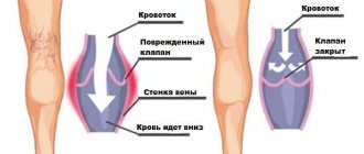
Damage to endoneurial vessels in diabetes occurs as a result of metabolic disorders, but later begins to play an independent role in the formation of pathology of peripheral nerves. It has been shown that there is a correlation between the number of nerve fibers in the peripheral nerve and the wall thickness of the endoneurial vessels in diabetes and, thus, the vascular component is fundamental for the development of DPN [13]. In the apt expression of A.S. Efimova, “diabetes begins as a metabolic disease and ends as a vascular pathology” [27], which must be taken into account when prescribing pathogenetic therapy to patients with DPN.
Treatment of DPN is based on the use of drugs that act on the pathogenetic mechanisms of its formation, symptomatic treatment, for example, the use of pain medications (anticonvulsants, antidepressants, local anesthetics, opioids), prevention of late complications and, if possible, exclusion of risk factors for their development. Pathogenetic therapy of DPN is determined by modern ideas about the mechanisms of formation of late complications of diabetes, emphasizing the relationship of metabolic and vascular factors, which has been repeatedly considered in the literature. In this regard, the possibilities of using: 1) aldose reductase blockers to reduce the utilization of glucose through the polyol pathway are discussed; 2) γ-linolenic acid preparations to normalize the metabolism of essential fatty acids and the synthesis of prostaglandins; 3) vasodilators and prostaglandin analogues, which help reduce hypoxia and increase endoneurial blood flow; 4) protein kinase C inhibitors to improve endothelium-dependent reactions of the vascular wall; 5) nerve growth factors; 6) inhibitors of the formation and accumulation of advanced glycation end products AGEs, 7) antioxidants, thiamine. Currently, the main drugs that have proven their effectiveness in DPN are antioxidants, Actovegin and thiamine.
The main antioxidant drug used to treat DPN is α-lipoic (thioctic) acid (ALA), which is a powerful natural fat-soluble antioxidant. The effectiveness of ALC in DPN has been shown in a number of clinical studies conducted within the framework of evidence-based medicine, and confirmed by a meta-analysis of four studies on the effectiveness of ALC in DPN [15].
Another opportunity to influence the pathogenetic mechanisms of DPN formation is to influence the activity of the transketolase enzyme. This enzyme is able to reduce the pathological accumulation of intermediate products of glucose metabolism. Enzyme activity directly depends on the concentration of thiamine. In 2003, the hypothesis about the ability of thiamine to reduce the severity of metabolic disorders caused by hyperglycemia was confirmed in an experimental study that showed the ability of thiamine to activate transketolase and prevent vascular damage to the retina in diabetes [16, 17].
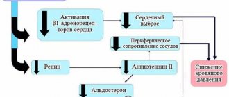
In recent years, data have been obtained on the effectiveness of treatment of DPN with Actovegin. The drug Actovegin from Takeda (Japan) contains highly purified hemodialysate obtained by ultrafiltration from the blood of calves. The drug contains organic low-molecular compounds, which eliminates the development of prion diseases, such as Creutzfeldt-Jakob disease, when Actovegin is administered into a human vein. The technology for producing Actovegin excludes the presence of protein components that have antigenic and pyrogenic properties. Actovegin is a mixture of natural substances of inorganic (electrolytes - sodium, potassium, calcium, magnesium, chlorides, nitrogen compounds) and organic (acetate, lactate, amino acids, nucleosides, glycosphingolipids, intermediate products of carbohydrate and fat metabolism, antioxidant enzymes). There is no exact data on the pharmacokinetics of Actovegin, since it is a multicomponent drug, and its composition includes substances originally contained in the human body. The production method of Actovegin and the discussion of studies on the mechanisms of its action are discussed in detail in reviews devoted to the almost half-century history of this drug [18, 23]. It is known from experimental work that Actovegin’s activation of glucose uptake into cells begins 5 minutes after intravenous administration of the drug and the peak of its action is detected after 120 minutes. It is currently believed that the insulin-like effect of Actovegin and stimulation of glucose metabolism are associated with inositol phosphooligosaccharides contained in the drug. Due to the fact that Actovegin modulates the activity of intracellular glucose transport, lipolysis is activated [19]. The antihypoxic effect of Actovegin is associated with its ability to increase the absorption of oxygen by tissues, which increases the resistance of cells to hypoxemia [20, 21]. As a result of the supply of oxygen to tissues, the formation of macroergic phosphates (ATP, ADP) increases, oxidative phosphorylation enzymes are activated, the synthesis of carbohydrates and proteins and the breakdown of anaerobic glycolysis products (lactate) are accelerated, and cellular energy imbalance is reduced. An increase in oxygen absorption by the vascular wall with the administration of Actovegin leads to normalization of endothelium-dependent reactions and a decrease in peripheral vascular resistance. As a result, the formation of high-energy phosphates in cellular structures increases. The improvement in blood flow in the microcirculation system under the influence of Actovegin, associated with the normalization of endothelium-dependent reactions (vasodilation) and a decrease in peripheral vascular resistance, reflects an increase in oxygen metabolism of the vascular wall. The antioxidant effect of Actovegin is ensured by the presence of superoxide dismutase in the drug, which is confirmed by atomic emission spectrometry, as well as magnesium ions, which increase the activity of glutathione synthetase, which converts glutathione into glutamine [22] (Fig. 1).
A recent study by MW Elminger revealed that Actovegin reduces apoptosis (based on caspase-3 activity), increases the number of synaptic connections and dose-dependently reduces oxidative stress in neurons [24]. In a study by A. Dieckmann et al. It was found that Actovegin improves the conduction of excitation along sensory fibers and reduced apoptosis in sciatic nerve fibers by reducing PARP activity, which emphasizes the influence of Actovegin on the pathogenetic mechanisms of the formation of cellular damage in diabetes [25]. Actovegin is used in the form of a 10 or 20% solution for intravenous (in 250 ml of saline) or intramuscular administration or 200 mg tablets for oral administration.
Side effects of the drug mainly include rare allergic reactions. In the literature there is a description of only one case of anaphylactic shock after intravenous administration of the drug with the development of liver and kidney damage, but not in a patient treated by a doctor, but in an athlete [26]. In this regard, one can think that medical standards were violated when administering the drug. Interest in the use of Actovegin in DPN is due to the fact that the drug has antioxidant properties and the ability to improve the utilization of oxygen and glucose into cells, which is extremely important in diabetes, when energy deficiency forms in the tissues, associated with both the phenomenon of pseudohypoxia and true hypoxia ( Fig. 2).
In the work of V.A. Yavorskoy et al. in an open study, Actovegin was used for the treatment of DPN in 24 patients with type 1 and type 2 diabetes in the form of daily infusions for 20 days. An improvement in the clinical condition of patients was noted in the form of a decrease in pain, improved sensitivity and tendon reflexes, and an increase in muscle strength. Rheovasography showed an improvement in blood flow in the legs, and an EMG examination showed an increase in the amplitude of the M-response and SRV when stimulating the nerves of the legs [28]. The use of Actovegin in the complex treatment of 33 patients with diabetic foot syndrome of varying severity according to the Wagner classification showed in an open study that the connection of the drug to traditional treatment contributed to the rapid relief of pain and the acceleration of the processes of granulation and epithelization of ulcerative defects with their healing [29].
In a study by F.E. Morgoeva et al. The effectiveness of intravenous monotherapy with Actovegin in patients with type 2 diabetes was studied in an open study [30]. The group of 30 patients who received Actovegin once a day intravenously at a dose of 400 mg diluted in 200.0 ml of physiological solution for 3 weeks (15 infusions) included patients with a duration of diabetes of at least 10 years at the age of 58.94 ± 1.29 years (9 men and 21 women). Before treatment, in the group of patients with diabetes with DPN, a significant increase in the level of lipid peroxidation in plasma and erythrocyte membranes was observed compared with a group of 15 healthy volunteers of the same age, which indicated the severity of oxidative stress. Treatment with Actovegin led to a decrease in the level of malondialdehyde (MDA) in plasma and erythrocyte membranes with its normalization. Thus, Actovegin had an undoubted antioxidant effect, acting on the pathogenetic mechanisms of the development of DPN. The state of the rheological properties of blood was assessed by computer capillaroscopy before and after treatment with Actovegin. After treatment with Actovegin, a significant improvement in the main characteristics of capillary blood flow was noted, not only reflecting the rheological properties of the blood, but also the state of permeability of the capillary wall. A clinical study showed a significant decrease in positive and negative neuropathic symptoms (p < 0.05) after treatment with Actovegin. When studying the functional state of the sensory nerve, a significant increase in the amplitude of the sensory response was revealed (3.87 ± 2.43 μV before treatment and 6.19 ± 3.16 μV after treatment, p < 0.05) with the SRT unchanged.
A study of temperature and pain sensitivity thresholds using quantitative sensory testing showed that after treatment with Actovegin, there is a decrease in sensitivity thresholds associated with the state of thin nerve fibers (p < 0.05). Thus, the study showed that treatment with Actovegin, reducing the severity of oxidative stress and improving the state of the microcirculation system, leads to a regression of the clinical manifestations of DPN in patients with type 2 diabetes, which is confirmed by an improvement in objective indicators of the functional state of peripheral nerves.
W. Jansen and E. Beck studied the effect of oral administration of Actovegin on patients with DPN in a controlled study: one group of 35 patients received placebo, another group of 35 patients received Actovegin tablets (600 mg 3 times a day) for 24 weeks [31]. The criteria for assessing the effectiveness of the drug were clinical characteristics of polyneuropathy (tendon reflexes, superficial and deep sensitivity, intensity of pain) and EMG indicators of peripheral nerve function (velocity of propagation of excitation (RPV), as well as the distance that patients could walk without pain. Improvement in the condition of patients in Actovegin treatment group was observed in the majority of patients 8 weeks after the start of treatment, and the optimal effect was achieved after 16 weeks of treatment (Fig. 3).A significant improvement during treatment with Actovegin compared to the placebo group was shown in almost all clinical indicators: pain-free walking distance, tendon reflexes, superficial and deep sensitivity (p < 0.01). SRV significantly (p < 0.001) increased in the treatment group compared to the placebo group. Patients in the treatment group felt better and had fewer complaints about a violation of the psycho-emotional state, which correlated with improving their physical condition.
In 2009, the results of a multicenter, randomized, double-blind, placebo-controlled study of the treatment of patients with type 2 diabetes with DPN with Actovegin were published [32]. A multicenter, double-blind, placebo-controlled, randomized, parallel-group clinical trial was conducted at 26 clinical sites in Russia, Ukraine, and Kazakhstan. The purpose of the study was to evaluate the clinical efficacy and safety of Actovegin compared to placebo in patients with type 2 diabetes and clinical manifestations of peripheral diabetic polyneuropathy after intravenous infusions of Actovegin or placebo followed by switching to Actovegin tablets. A total of 567 patients were included in the study. 281 patients first received 20 intravenous infusions of Actovegin (250 ml of a 20% solution - 2.0 g), and then for 140 days the patients received Actovegin tablets 600 mg 3 times a day (1,800 mg/day). 286 diabetic patients with DPN received intravenous placebo therapy followed by placebo tablets. Inclusion criteria were: diagnosis of type 2 diabetes, age 18–65 years, glycated hemoglobin level below 10%, presence of clinical manifestations of DPN, that is, TSS score ≥ 6 points and NIS-LL ≥ 2 points, vibration sensitivity threshold ≤ 30V, adequate blood supply to the foot, proven by the presence of pulses in the posterior tibial artery and dorsalis pedis artery. The main criteria for the effectiveness of the drug in this study were positive neuropathic symptoms, which were assessed on the TSS scale, and the vibration sensitivity threshold, which was tested at several points on the legs (ankle, toes) using a biotensiometer. Secondary efficacy criteria included individual TSS scores, NIS LL scores, and quality of life scores (short scale - SF 36).
The best results were noted in relation to positive neuropathic symptoms, and improvement was noted both in the total assessment of all symptoms and in relation to each specific symptom. A significant decrease in sensory neurological deficit was revealed; in relation to changes in reflexes and muscle strength, a positive trend towards improvement was noted, which did not reach the degree of reliability. This may be due to the fact that reflexes and especially muscle strength were altered in a relatively small number of patients. Objective indicators of the state of proprioceptive nerve fibers were assessed by assessing vibration sensitivity thresholds. The decrease in the vibration sensitivity threshold was highly significant when using Actovegin compared to placebo (Fig. 4). Throughout the study, fasting glucose levels and 2-month diabetes compensation (HbA1c) were measured. The results obtained indicate that the effectiveness of Actovegin is associated with the effect of the drug, and not with changes in diabetes control.
There were no significant side effects noted in the study. A total of 386 reports of adverse events were received from 192 patients (Actovegin - 186, placebo - 198). The most common adverse events were headache (Actovegin - 22, placebo - 19), hypoglycemia (21/19), hypertension (10/13), hyperglycemia (6/16), increased blood pressure (7/11) and respiratory infections paths (8/5). There were 21 serious adverse events reported, of which 10 occurred in 7 patients receiving Actovegin and 11 in 10 patients receiving placebo. Causality was considered possible for 1 serious adverse event (Actovegin: heart failure) and probable for 1 (placebo: hypersensitivity). No patients died during the study.
This study, conducted within the framework of (GCP - Good Clinical Practice), answered positively the question of the effectiveness and safety of the use of Actovegin for the treatment of DPN. It was concluded that sequential intravenous and then oral therapy with Actovegin for 160 days improved symptoms of neuropathy, reduced the threshold of vibration sensitivity and improved sensory function in patients with type 2 diabetes and diabetic polyneuropathy. A significant improvement in the quality of life (on a mental health scale) was shown in the Actovegin group compared to placebo. It was noted that the groups of patients receiving Actovegin and placebo had a comparable safety profile.
Thus, from a pathophysiological point of view, there is no doubt that Actovegin, which has antihypoxic and antioxidant effects, can be used for DPN, as well as a wide range of diseases of the central and peripheral nervous system, in the pathogenesis of which hypoxia, ischemia and oxidative stress play a role.
Literature
1. Strokov I.A., Melnichenko G.A., Albekova Zh.S. and others. Prevalence and risk factors for the development of diabetic polyneuropathy in inpatients with type 1 diabetes mellitus // Neuromuscular diseases. – 2012. – No. 1. – pp. 25–31. 2. Boulton AJM, Vinic AI, Arezzo JC et al. Diabetic neuropathies: a statement by the American Diabetes Association // Diabetes Care 2005; 28:956–962. 3. Tesfaye S, Boulton AJM, Dyck PJ et al. Diabetic neuropathies: update on definitions, diagnostic criteria, estimation of severity, and treatments // Diabetes Care 2010; 33(10): 2285–2293. 4. Khramilin V.N. Diabetic polyneuropathy. Review of modern recommendations // RMD. Endocrinology 2012; 32:1580–1582. 5. Archer AG, Watkins PJ, Thomas PK et al. The natural history of acute painful neuropathy in diabetes mellitus // J Neurol Neurosurg Psychiatry 1983; 46:491–499. 6. Strokov I.A. Diabetic neuropathy // Diabetes mellitus type 2. Problems and solutions. GEOTAR-Media 2011. – pp. 506–529. 7. Dyck PJ Detection, characterization and staging of polyneuropathy: appreciated in diabetic. Muscle Nerve. 1998; 11(1): 21–32. 8. Dyck PJ, Davies JL, Wilson DM et al. Risk factors for severity of diabetic polyneuropathy: intensive longitudinal assessment of the Rochester Diabetic Neuropathy Study cohort // Diabetes Care 1999; 22: 1479–1486. 9. Brownly M. Biochemistry and molecular cell biology of diabetic complications // Nature. – 2001. – Vol. 414 – P. 813–820. 10. Brownlee M. The pathobiology of diabetic complications. A unifying mechanism. Diabetes 2005; 54:1615–1625. 11. Dyck PJ, Davis JL, Litchy WJ et al. Longitudinal assessment of diabetic polyneuropathy using a composite score in the Rochester Diabetic Neuropathy Study cohort. Neurology 1997; 49: 229–239. 12. Nikitin AG, Chudakova DA, Strokov IA et al. Leu54Phe and Val762Ala polymorphisms in the poly(ADP-ribose)polymerase-1 gene are associated with diabetic polyneuropathy in Russian type 1 diabetic patients.Diabetes Res Clin Pract 2008; 79(3):446–452. 13. Dyck PJB, Dyck PJ Diabetic polyneuropathy. In Dyck PJ, Thomas PK ed. “Diabetic polyneuropathy”, 2nd ed, Philadelphia, Pa: WB Saunders 1999: 255–278. 14. DCCT Group. The effect of intensive treatment of diabetes on the development and progression of long-term complications in insulin-dependent diabetes mellitus. N Engl J Med 1993; 329:977–986. 15. Ziegler D., H.-J. Tritschler, Strokov I.A., Ametov A.S. Treatment of diabetic polyneuropathy with thioctic acid (literature review) // Farmateka. – 2008. – No. 17. – pp. 28–35. 16. Strokov I.A., Akhmedzhanova L.T., Solokha O.A. The use of high doses of B vitamins in neurology // Difficult patient. – 2009. – No. 10. – pp. 17–22. 17. Strokov I.A., Strokov K.I., Albekova Zh.S. Thiamine and benfotiamine in the treatment of late complications of diabetes mellitus // Doctor.Ru. – 2009. – No. 6 (2). – pp. 14–18. 18. Buchmayer F., Pleiner J., Elminger MW et al. Actovegin®: a biological drug for more than 5 decades // Wien Med Wochenschr – 2011 – Vol. 161 (3–4) – P. 80–88. 19. Jacob S., Dietze GJ, Machicao F. et al. Improvement of glucose metabolism in patients with type II diabetes after treatment with hemodialysate // Arzneimittelforschung. – 1996. – No. 3. – P. 269–272. 20. Kuninaka T, Senga Y, Senga H, Weiner M. Nature of enhanced mitochondrial oxidative metabolism by a calf blood extract. J Cell Physiol. 1991; 146(1): 148–155. 21. Reichel H., Weiss C., Leichweiss HP The effects of a blood extract on the oxygen uptake of isolated artificially perfused kidney and skeletal muscles in rats //Arzneimitte-Forschung / Drug Research 1968; 18: 1019–1021. 22. Nordvik B. Mechanism of action and clinical use of the drug Actovegin // In collection. “Actovegin. New aspects of clinical application". M., 2002 – pp. 18–24. 23. Machicao F., Mureanu DF, Hundsberger Y. et al. Pleiotropic neuroprotective and metabolic effect of Actovegin // Neuromuscular diseases. – 2012. – No. 4. – P. 3–10. 24. Elminger MW, Kriebel M., Ziegler D. Neuroprotective and anti-oxidative effects of the hemodialysate actovegin on primary rat neurons in vitro // Neuromolecular Med 2011; 13(4): 266–274. 25. Dieckmann A., Kriebel M., Andriambeloson E. et al. Treatment with Actovegin® improves sensory nerve function and pathology in streptozotocin-diabetic rats via mechanisms involving inhibition of PARP activation // Exp Clin Endocrinol Diabetes 2011; 120(3): 132–138. 26. Mailo L. Anaphylactic shock with multiorgan failure in a cyclist after inravenous administration of Activegin // Ann Intern Med 2008; 148–407. 27. Efimov A.S. Diabetic angiopathy. M.: Medicine, 1989. 28. Yavorskaya V.A., Egorkina O.V., Mashkin O.N. and others. Clinical experience with the use of Actovegin in diabetic polyneuropathy // In the collection. “Experience in the clinical use of Actovegin in endocrinology” M. 2005: 27–30. 29. Obolensky V.N. Complex treatment of patients with diabetic foot syndrome // In collection. “Experience in the clinical use of Actovegin in endocrinology” M., 2005: 39–46. 30. Morgoeva F.E., Ametov A., Strokov I.A. Diabetic encephalopathy and polyneuropathy: therapeutic possibilities of Actovegin // Russ. honey. magazine – 2005. No. 6. – pp. 302–304. 31. Jansen W., Beck E. Treatment of diabetic polyneuropathy. Controlled double-blind study // In collection. "Experience in the clinical use of Actovegin in endocrinology." – M., 2005. pp. 11–20. 32. Ziegler D., Movsesyan L., Mankovsky B. et al. Treatment of symptomatic polyneuropathy with actovegin in type 2 diabetic patients // Diabetes Care 2009; 32(8): 1479–1484.