Treatment of pathology
In this case, it is enough to lead a correct lifestyle (eat well, do not overexert yourself physically and mentally, do not smoke or drink alcohol).
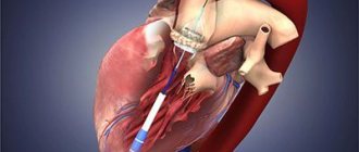
In this way, it will be possible to avoid the threat of developing concomitant diseases or a more complex stage of pathology development. If the disease continues to progress, experts recommend resorting to surgical treatment. To do this, prosthetic aortic valve or plastic surgery of its valves is performed. When performing prosthetics, a valve from an animal, a donor, or your own can be used.
Traditional methods are often used to treat the disease. But this can only be done after consultation with your doctor. This is necessary in order to avoid the risk of deterioration of the patient’s general condition.
A raw or vegetarian diet and acupuncture massage are quite widely used. However, you should not self-medicate. Only specialists can prescribe treatment that will give positive results.
If the aorta of the heart hardens, treatment can be prescribed after a full medical examination and a comprehensive diagnosis of the anomaly. In the case where the aorta is compacted, therapy includes:
- conservative methods;
- surgical intervention;
- preventive measures.
When the problem is caused by age-related changes in the body, the patient is most often prescribed drug therapy in conjunction with a pathology prevention program.
Medicines are aimed at:
- normalization of blood pressure;
- fight against atherosclerosis;
- normalization of metabolism;
- treatment of diseases that provoke the formation of compactions and thickenings.
Important! A patient with this diagnosis must be registered with a cardiologist and be checked from time to time for changes or complications. Surgery is also a fairly common way to eliminate the disease.
Based on the severity of the pathology, plastic surgery, prosthetics or organ transplantation may be prescribed
Surgery is also a fairly common way to eliminate the disease. Based on the severity of the pathology, plastic surgery, prosthetics or organ transplantation may be prescribed.
Prevention of seals in the aorta of the heart consists of observing several important points:
- adjusting the nutritional program - you don’t have to go on a strict diet, but you should stick to a healthy eating program (reduce salty, fatty, fried foods, less carbohydrates);
- giving up all bad habits (cigarettes, alcoholic drinks, caffeine);
- prevention of nervous disorders, stressful situations - if necessary, it is worth using psychotropic drugs;
- limiting heavy physical activity - sports are needed, but within reasonable limits and in accordance with one’s capabilities;
- spend more time outdoors;
- Constant monitoring by a specialist - routine medical examinations to monitor changes.
Means of preventing aortic thickening also include treatment with folk remedies. But any therapy must be agreed with the attending physician.
Since most traditional methods are not only ineffective, but can also significantly harm the patient’s health, doctors do not recommend their use. But in this case, drinking large amounts of freshly squeezed juices, fresh fruits and vegetables, and avoiding fatty foods and foods containing cholesterol can be considered quite acceptable.
Aortic stenosis
Treatment of aortic wall dissection
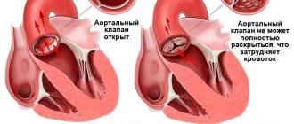
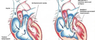
What is stenosis in medicine? Stenosis means narrowing of the lumen of a vessel. Aortic stenosis is a narrowing of the valve that separates the left ventricle of the heart from the aorta. There are minor, moderate and severe. This defect can affect the mitral and aortic valves.
With a minor valve defect, a person does not feel any pain or other warning symptoms, because the increased work of the left ventricle will be able to compensate for the poor functioning of the valve for some time. Then, when the compensatory capabilities of the left ventricle are gradually exhausted, weakness and poor health begin.
«>
The aorta is the main blood vessel. If the valve is malfunctioning, all vital organs will suffer from lack of blood supply.
The causes of heart valve stenosis are:
- Congenital valve defect: fibrous membrane, bicuspid valve, narrow ring.
- A scar formed by the connective tissue just below the valve.
- Infectious endocarditis. Bacteria that enter the heart tissue change the tissue. Due to the colony of bacteria, connective tissue grows on the tissue and on the valves.
- Deforming osteitis.
- Autoimmune problems: rheumatoid arthritis, lupus erythematosus. Due to these diseases, connective tissue grows in the place where the valve is attached. Growths form on which more calcium is deposited. Calcification occurs, which we will remember later.
- Atherosclerosis.
Unfortunately, in most cases, aortic stenosis is fatal if valve replacement is not performed in time.
Treatment
Treatment of the disease is aimed at eliminating its cause, if possible, and correcting hemodynamic disturbances.
If the thickening of the artery walls was caused by increased pressure, drug correction of blood pressure numbers is prescribed. In case of decompensation and development of heart failure, it is supported by the prescription of diuretics and cardiac glycosides. Atherosclerosis and blood cholesterol levels are regulated by prescribing antilipidemic drugs (statins, fibrates), as well as by normalizing nutrition and a healthy lifestyle.
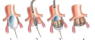
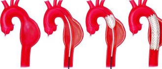
If the narrowing of the valve is significant, surgical correction of the condition is resorted to. In this case, treatment is aimed at restoring blood flow from the left ventricle of the heart to the aorta. A valvuloplasty operation is performed - the aortic valve expands, its valves are adjusted to a normal diameter. In case of severe aortic stenosis at any level, stenting is performed - surgical delivery of a special metal frame for further expansion of the vessel cavity.
Treatment options
How to treat aortic aneurysm (vasodilatation) and prognosis after myocardial infarction
When prescribing therapy, the specialist takes into account factors such as: the severity and duration of the disease, the presence of concomitant pathologies, and heredity.
Treatment is aimed specifically at eliminating the very cause of the aortic thickening.
To achieve this, measures are taken including:
- special food;
- motor mode;
- invasive and drug therapy;
- folk remedies.
In case of poor heredity, which is expressed in a predisposition to cardiovascular diseases, it is recommended to switch to a healthy lifestyle with an emphasis on proper nutrition.
Diet
For diseases of the vascular system (thrombosis, stenosis, atherosclerosis), the patient is prescribed dietary nutrition that helps restore the body.
From the diet you need to exclude fatty, spicy, fried foods, which cause the accumulation of cholesterol and clogging of blood vessels with plaques.
It is also contraindicated:
- sweet;
- flour dishes;
- fatty dairy products;
- fast food;
- coffee, black tea.
It is necessary to eat natural foods (vegetables, herbs, fruits, berries). Dietary meat (chicken, rabbit), fish, cereals, legumes, and nuts are allowed. Drinks are prepared based on berries or medicinal herbs. Green and white tea are beneficial.
Drug therapy and surgical interventions
Depending on the type of disease that led to hardening of the aorta, the doctor selects medications and prescribes a treatment regimen.
The following medications are prescribed:
- for atherosclerosis: Fenofibrate, Cholestyramine. Lovastatin, Cholestipol. They reduce cholesterol levels, improve the quality of hematopoiesis;
- for hypertension: adrenergic blockers - Bisoprolol, Atenolol; calcium channel blockers - Amlodipine, Nifedipine. They normalize high blood pressure;
- For sexually transmitted and infectious diseases, treatment with antibiotics and anti-inflammatory drugs is prescribed.
In cases of aortic obstruction or valve dysfunction, surgical intervention is indicated.
Transplantation of the valve leaflet, fibrous ring, and prosthetics of part of the vessel are used.
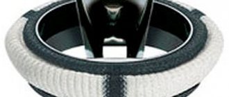
In severe cases of atherosclerosis, stenosis, blockage, a stenting operation is performed, which involves placing a stent inside the patient’s artery. It is a mesh tube that is inserted into the damaged vessel. Using a flexible catheter, it is brought to the site of narrowing. The stent balloon is then inflated using a special substance, after which the stent expands and expands the artery walls.
Herbal decoctions
One of them is infusions based on medicinal plants. To prepare drinks, herbs are used that help thin the blood and strengthen the walls of blood vessels.
These include:
- fennel;
- motherwort;
- sushi;
- rose hip;
- hawthorn;
- lovage
Herbs can be brewed individually or made into a mixture. Together they enhance the beneficial qualities of the drink. For 250 ml of boiling water, take a tablespoon of dried crushed herbs. The ingredients are poured with water and left to infuse for 20-30 minutes. After this, the composition is filtered. You need to take the infusion half a glass half an hour before meals two to three times a day. The course of treatment is 4-6 weeks.
Horseradish and honey
A mixture of horseradish and honey helps relieve blockage of veins. The dry root of the plant is ground into powder and mixed with a teaspoon of honey.
The composition is consumed an hour before meals 3-4 times a day.
Garlic tincture
Garlic infusion strengthens blood vessels well. Take a few cloves, peel and grind to a paste. Then pour a glass of boiling water.
Cover tightly with a lid and place in a dark, dry place. After a week, the composition must be mixed, strained and taken 20 ml three times a day.
Diagnostic examination
Considering the fact that the symptoms of thickened aortic walls do not appear immediately, the patient begins to notice the first signs when the disease has already progressed. In some cases, pathology is detected during a routine medical examination, using fluorography.
Ultrasound of the heart
To clarify the diagnosis, ultrasound of the heart muscle, circulatory system and contrast angiography of blood vessels are performed. For people who have a genetic predisposition to diseases of the cardiovascular system, heart diagnostics should be carried out once a year.
Compaction of the walls of the aorta and the aortic valve leaflets
Tricuspid valve insufficiency 2nd degree: what is it?
The aorta is the largest vessel in the human body, through which oxygenated blood is distributed to smaller arteries. It is directly connected to the left ventricle of the heart, and the flow of arterial blood into it is controlled by the operation of the muscular valve.
The aorta is the largest unpaired arterial vessel of the systemic circulation, coming from the heart and feeding all internal organs and systems, except the lungs. Throughout its entire length, the aorta has a uniform thickness and the same structure.
Aortic compaction can be detected using various diagnostic methods: radiographic, fluorographic or ultrasound examination. Having discovered such a defect, specialists must find the main causes of the pathology and prescribe the correct treatment.
Depending on the location of the lesion, the following forms of pathology are distinguished:
- Compaction of the aortic root,
- Consolidation of the aortic arch,
- Consolidation of the ascending aorta,
- Consolidation of the descending aorta.
Fibrosis of the aortic and mitral valve leaflets actually does not manifest itself in any way in the early stages of development. It is mostly discovered by chance during an annual examination.
Echocardioscopy (ultrasound of the heart) helps to see the enlarged valves. The doctor will assess the degree of pathological changes and prescribe the most effective course of treatment to stop the development of complications and improve the patient’s condition.
The presence of fibrosis of the aortic mitral valve requires its repair or replacement. When choosing a method of surgical intervention, the degree of functional insufficiency of the valve plays an important role.
The more affected it is, the more likely it is that it will need prosthetics. Some advanced clinics successfully use a minimally invasive method to restore the mitral valve, combining the possibility of catheterization and a minimal incision in the chest.
Correction of the defect is possible thanks to the use of the Metraclips method, which is also classified as a mini-invasive procedure. Its use has significantly expanded the scope of treatment of mitral valve defects.
Thanks to this technique, it became possible to operate on seriously ill patients for whom conventional surgery is contraindicated. Innovative methods have allowed a very large number of patients to regain health and return to a full lifestyle.
Infectious diseases should not be allowed to occur, as they can dramatically worsen your health. In winter, you should additionally take a complex of vitamins and minerals. Try to avoid stress.
Professional sports and heavy physical activity should be avoided, as this can even lead to death. If any disease appears, when prescribing medications for treatment, you should definitely notify the doctor about the presence of fibrosis of the mitral valve leaflets.
Mitral valve replacement is indicated for valve insufficiency, severe calcification or fibrosis of the leaflets. Surgical replacement of the mitral aortic valve is performed on a non-functioning heart using cardiopulmonary bypass and cardioplegia.
Mechanical valves are durable, but their use is associated with lifelong use of anticoagulants to prevent thrombosis. Tissue valves undergo biodegradation over time (calcification, leaflet rupture), but minimize the risks of thromboembolism and endocarditis of the prosthesis.
With limited changes in the valve, valve-preserving operations can be performed: suture valvuloplasty, annuloplasty, narrowing of the fibrous ring with a special rigid synthetic ring, restoration of subvalvular structures, as well as isolated replacement of valve leaflets with auto- or xenopericardium.
It should be noted that the results of surgical treatment of the mitral valve largely depend on the timely determination of indications for surgical treatment. Therefore, if you are scheduled for surgery, then in no case should you delay it.
Treatment and observation of a patient with fibrosis of the valve apparatus
Often on forums you can read the question of whether fibrosis can be treated using folk remedies. The answer is clear: there are no such recipes. This process is quite difficult in therapy even for modern medicine.
It is important to know that the prescription of medications is indicated only for the clinical picture of heart failure, in which the following are used:
- cardiac glycosides - Celanide, Digoxin, Strophanthin;
- diuretics - Trifas, Indap, Veroshpiron;
- if indicated, antihypertensive and antiarrhythmic drugs.
Medicines only minimize the symptoms caused by fibrosis without affecting the progression of the disease.
Radical treatment consists of the following methods:
valve prosthetics to replace the diseased structure with a mechanical or biological analogue. As a rule, a median sternotomy using a heart-lung machine is used;- mitral commissurotomy, closed or open, with the task of dissecting pathological connections between the valve leaflets;
- coronary artery bypass grafting;
- endovascular prosthetics. The essence of the method is to insert a catheter with an implant through the femoral vessels without general anesthesia. Indicated for patients with severe chronic diseases;
- valve transplantation (a relatively new technique).
Indications for surgical intervention in fibrosis:
- neglect of the process;
- wrinkling of valves, tendon threads;
- the presence of pronounced calcification.
After the operation, the patient should be under medical supervision of a cardiologist. The patient is indicated for annual examinations and treatment in a cardio- or cardio-rheumatological sanatorium.
The aorta is compacted: what does it mean, causes, symptoms, is treatment needed?
· You will need to read: 4 minutes
Compaction of the aorta of the heart: what is it and what to do about it?
From this article you will learn: what is aortic thickening, is it dangerous. Is it possible to get rid of the problem, and what needs to be done for this.
When the aorta thickens, the walls of the largest arterial vessel in the human body (aorta) thicken, increase in density, and decrease in elasticity. This condition is not a separate disease, but only one of the manifestations of various pathologies.
In itself, it does not bother a person at all and is detected accidentally during instrumental studies regarding various complaints. But if the compaction increases and intensifies, this threatens with dangerous consequences in the form of damage to the heart valves, circulatory disorders in the aorta, blockage, weakness and the threat of its rupture.
There is no need to specifically treat the thickening of the aortic walls. Only treatment of the underlying disease, which is manifested by this symptom, will either eliminate it or stop its progression (the resulting lumps no longer go away).
For medical help, you should contact a vascular surgeon or cardiac surgeon.
The essence of the problem: is it a symptom or a disease?
The most common manifestation of diseases affecting the aorta is a change in the normal structure of its walls. This means that the compaction of this vessel should not be regarded as a separate disease, but only as a symptom indicating the presence of a certain pathology.
If pathological changes in the aorta are limited only to thickening of its walls, this does not pose a great danger.
But this condition still has some prognostic significance. The aorta, as the central arterial vessel of the body, gives rise to arteries that bring blood to absolutely all organs and tissues except the lungs.
Therefore, even minimal pathological changes (including thickening of the aorta) over time can develop into severe and even life-threatening conditions:
- thrombosis - the formation of blood clots with blockage of the lumen;
- aneurysm - abnormal expansion with thinning of the walls;
- blocking the lumen of large arteries emanating from the aorta;
- dissection or rupture of the walls of the aorta, accompanied by lightning bleeding.
Comparative characteristics of the normal and compacted version of the vessel are given in the table:
| Soft-elastic | Dense, hard |
| Has uniform thickness throughout | Has areas of thickening |
| Pliable, capable of expansion and contraction | Rigid (unable to stretch) |
| Durable | Weakened |
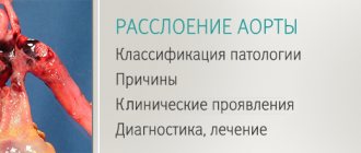
Eight causes of pathology
- Formation of cholesterol plaques in atherosclerosis.
- Natural age-related changes (the result of aging).
- Arterial hypertension (prolonged increase in pressure).
- An inflammatory process (aortoarteritis) caused by infections or autoimmune diseases (intestinal infections, viruses, systemic lupus, vasculitis, rheumatoid arthritis).
- Syphilis - the artery is affected in the late period of this disease (after several years or even more than 10).
- Tuberculosis - the aorta is always hardened secondary, against the background of pulmonary and extrapulmonary forms of complicated tuberculosis.
In more than 80%, the cause of aortic hardening is old age and atherosclerosis.
Risk factors that contribute to the development of pathology include:
- age over 50 years;
- smoking;
- increased blood cholesterol (abuse of fatty foods);
- diabetes;
- atherosclerosis of any arteries.
Are there any symptoms of pathology?
Changes in the walls of the aorta in the form of a compaction can be localized in any of its sections.
Regardless of location, aortic compaction itself does not cause any symptoms.
But if the compaction worsens, transforms into a more serious process, or affects the exit points of large arteries from the aorta, pathological symptoms may appear due to circulatory problems. They are briefly described in the table:
| In the root and ascending thoracic region | Chest pain, coronary heart disease and aortic valve damage |
| In the arch and descending thoracic region | Headaches, pain between the shoulder blades, signs of cerebrovascular accident |
| In the abdominal segment | Abdominal pain, poor circulation of the intestines, kidneys and other internal organs |
Sections of the aorta
Symptoms
Narrowing of the lumen between the left ventricle and the aorta leads to the following clinical picture:
- attacks of dizziness, even fainting, associated with minor physical activity;
- shortness of breath, attacks of suffocation;
- fast fatiguability,
- attacks of angina pectoris (pain behind the sternum of aching or burning nature).
Hardening of the aorta often causes angina attacks
For a long time, aortic stenosis is asymptomatic; severe symptoms appear when the opening narrows by more than 70%.
Symptoms of the disease are associated with the localization of the pathological process:
- when the lumen of the aortic root narrows, insufficient blood supply to the heart muscle occurs, this is manifested by the development of typical angina pectoris, including acute myocardial infarction;
- if the ascending part and arch of the aorta are compacted, neurological symptoms appear (headache, dizziness, weakness, fatigue, etc.);
- thickening of the descending aorta leads to malnutrition of the lower body and the development of intermittent claudication, acute abdominal pain, etc.
Specifics of treatment of pathology and prevention
There is no such thing as treatment for aortic thickening in medicine. Often, healing therapy involves eliminating the root cause that led to the precedent.
In a situation where a patient’s aortic walls have become enlarged due to atherosclerosis, medicine involves therapeutic measures aimed at lowering the cholesterol ratio in the blood. For this purpose, the patient may be prescribed drugs that thin the blood and anti-atherosclerotic agents. In parallel, the patient must follow a diet that includes enriching the menu with omega fatty acids and avoiding foods containing high concentrations of cholesterol.
In case of hypertension, as the primary source of pathology, the patient is prescribed drugs that normalize blood pressure, diuretics, and adrenergic blockers. If a patient’s vascular thickening progresses due to syphilis, treatment is provided by a venereologist, who will prescribe the appropriate medications, depending on the stage of the disease.
An increase in aortic parameters against the background of aging of the body requires supportive drug therapy and adherence to a healthy lifestyle.
When treating the disease, not only the primary source of development of aortic thickening is taken into account, but also its stage, namely the impact of the disease on the patient’s ability to live a full life. If the enlargement is significant and directly threatens the normal functionality of vital organs, surgical intervention may be provided, which is an operation to replace the vessel or its valves.
Often, in the complex therapy of pathology, its treatment with folk remedies is also used. Basically, alternative medicine is used as an auxiliary strengthening of vascular lines. The most popular and effective are the following folk recipes:
- Rowan bark decoction. Use one teaspoon three times a day, the duration of use should be at least three months to obtain noticeable results.
- Garlic infusion, with the addition of honey, helps strengthen blood vessels, cleanse them of cholesterol deposits, and increase the elasticity and firmness of the vascular epithelium. To prepare the potion, you need to peel and chop the garlic, add freshly squeezed lemon juice, and leave the potion in a dark place for a week, stirring once a day. It is recommended to take the drug three times a day, one spoon half an hour before meals.
- A herbal mixture of hawthorn, mint, hops, motherwort, sweet clover, rose hips, clover and oregano, collected in equal proportions, is taken as a freshly prepared infusion, half a glass, three times a day.
Garlic infusion with the addition of honey
Treatment with alternative medicine is permissible only after a thorough examination in medical institutions, consultation with a doctor and strictly in combination with drug therapy. It is not possible to eliminate pathology using exclusively folk methods.
Aortic thickening is a disease that, even after drug therapy and elimination of the source of pathology, does not self-destruct. Elimination of the disease provocateur only stops the evolution of the pathology and prevents its further progression. To prevent the ontogenesis of the disease and serious complications, the patient should continue to maintain a healthy lifestyle. The main preventive measures are:
- Refusal of harmful addictions.
- Maintaining proper nutrition.
- Vigorous physical activity.
- Complete rest.
- Regular walks in the fresh air.
- Maintaining the correct alternation of work and rest.
- Regular health monitoring and routine examinations.
Causes of compaction
Hypertension is the most common cause of the formation of aortic seals. An increase in blood pressure leads to the fact that the vascular wall loses its elasticity, becomes rigid and thickens with the formation of dense fibrous structures.
Various infectious and non-infectious diseases can lead to the formation of aortic seals.
”alt=””>
Abuse of foods containing large amounts of cholesterol and bad habits also actively contribute to the thickening and hardening of the walls of the aorta. As the body ages, the condition of the walls changes under the influence of age-related factors, and atherosclerotic plaques form, narrowing the lumen of the vessel.
The most common cause of aortic hardening is hypertension. Sometimes atherosclerosis also contributes to this. Aortic compaction - what is it? This is the process of deformation of the walls of blood vessels, when they lose their elasticity, become rigid and thicken. A number of other diseases besides those mentioned above can lead to this result.
With hypertension, due to loss of elasticity of the aortic walls, fibrous structures of increased density begin to form. They change the rigidity of the walls and transform their thickness. The result is a thickening of the aorta.
Bad habits and foods high in cholesterol contribute to thickening of the aorta. Vessels also narrow due to the aging of the body, and if compaction occurs in young people, then most likely it is inherited.
The onset of the formation of atherosclerotic plaques and thickening of the aortic root may be associated with the following predisposing factors:
- hereditary factor;
- obesity;
- gender (mostly male);
- existing thyroid disorders;
- smoking and alcohol abuse;
- impaired blood clotting function;
- disruption and changes in hormonal levels in women;
- hypertonic disease;
- diabetes;
- adynamia and or complete lack of physical activity;
- exposure to frequent emotional stress and depression;
- unhealthy diet with a predominance of fatty and fried foods.
If it is possible to eliminate any risk factors from life, it is necessary to do this as quickly as possible.
Atherosclerosis is a slow process of cholesterol deposition on the vessel wall, leading to the formation of plaque.
Thickening of the aortic walls can occur due to:
- Hypertension. In the body of people suffering from this disease, the blood vessels bear a huge load, since the blood flows in them under very high pressure. As the largest blood vessel, the aorta experiences tremendous stress every day. Holding it back, the walls eventually lose their former elasticity. Their inner surface is overgrown with fibrous structures, increasing the thickness and density of the walls. As a result, the thickened aorta becomes more rigid.
- Natural aging of the body, typical for people over 55 years of age. As a rule, most representatives of this age category experience a significant decrease in the elasticity of blood vessels.
- Atherosclerosis is a disease accompanied by the formation of cholesterol plaques, which provoke a narrowing of the lumen of blood vessels and lead to uneven compaction of the aortic walls.
- Ever had a venereal disease.
- Diseases of infectious and non-infectious etiology (scarlet fever, sepsis, brucellosis, rheumatism).
- Smoking addictions.
- Eating foods high in cholesterol.
- Alcohol abuse.
- Hereditary predisposition. It is this reason that provokes thickening of the aortic root of the heart muscle in children. In clinical practice, cases have been described in which the pathology began in a child, then paused for several decades in order to become more active with the onset of aging processes.
As a rule, this phenomenon is due to the age of the patient. Since over many years of continuous work the body (including the heart valves) of a person belonging to the older age category inevitably wears out, doctors consider this to be the norm.
Compaction of the mitral valve leaflets can occur due to defects in the development of the connective tissues that form them. The consequence of this pathological phenomenon may be stenosis (narrowing) or disruption of the obturator function of the damaged valve.
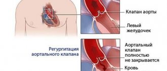
2.Why does mitral regurgitation occur?
The mitral valve is a kind of valve that separates the left atrium from the ventricle. Due to various factors that impair its functioning, the valve does not fit tightly enough to the walls of the ventricle. As a result, the two connecting plates - components of the valve - cannot effectively block the reverse flow of blood, placing additional stress on the heart to normalize the circulation of blood flow. This phenomenon does not pass without leaving a trace on the human body. The consequences of mitral valve regurgitation can include diseases such as arrhythmia, heart failure and endocarditis.
There are two main forms of mitral regurgitation: chronic and acute. Let us consider in more detail their differences and features of the course.
- Acute mitral regurgitation.
Acute regurgitation of the mitral valve occurs against the background of various cardiac pathologies: dysfunction or rupture of the papillary muscles, acute rheumatic fever, infective endocarditis, rupture of the mitral valve leaflets, dilatation of the left ventricle. Acute regurgitation develops instantly. Due to possible severe bleeding caused by rupture of cardiac tissue, it can be life-threatening. - Chronic mitral regurgitation.
The causes of chronic regurgitation of the mitral valve are also associated with heart diseases - mitral valve prolapse, expansion of its ring. The chronic form of regurgitation is much more common than the acute form. It is characterized by slow and gradual development, so the disease is most often diagnosed in older people.
Visit our Cardiology page
Hardening of the aortic walls: treatment methods
Thickening of the aortic walls is treated based on the patient’s symptoms, the degree of neglect of the pathology, as well as the cause that caused it.
Traditional drug therapy includes the following:
- Prescription of statin drugs (Lovastitin, Cholestyramine). This is practiced if the thickening of the aortic walls is caused by atherosclerotic syndrome or elevated cholesterol levels.
- Prescription of diuretics for the myocardium (Hypothiazide).
- If the thickening of the aortic root of the heart is caused by infectious diseases, the person is prescribed drugs based on penicillin - antibiotics.
A CT scan and chest x-ray will help determine whether a person needs surgical treatment or not. Using these studies, you can see the thickness of the compaction of the aortic root of the heart, as well as the exact localization of the deposition of cholesterol plaques.
During the period of therapy, the patient needs to be under medical supervision, take prescribed medications, follow a diet and avoid stress.
As an additional treatment with folk remedies, the following can be used:
- Herbal treatment (herbal medicine).
- Acupuncture.
- Physiotherapeutic treatment.
Traditional medicine also offers the following recipes that will help eliminate compaction of the aortic root of the heart and general compaction of the aortic walls:
- Chop the garlic and pour boiling water over it. Add lemon juice and leave for three days. Take the finished product one teaspoon twice a day an hour before meals. The duration of treatment is at least four months.
- Make a tincture of water and rowan. Boil for an hour. Strain and take a teaspoon once a day.
- Mix rose hips, hops, mint and oregano in equal quantities. Pour boiling water and leave for two hours. Strain and take a spoon an hour before meals.
Prevention of compaction of the aortic root of the heart consists of giving up bad habits, avoiding stress, moderate sports activities and a healthy lifestyle.
It is also important to eat a balanced diet and promptly treat serious illnesses. A cardiologist can recommend Orthomol Cardio vitamins
Compaction of the aorta is a pathology that can lead to a heart attack, aneurysm and many other dangerous pathologies, so do not underestimate this disease. It needs to be treated immediately after diagnosis.
The aorta is the largest unpaired arterial vessel of the systemic circulation, coming from the heart and feeding all internal organs and systems, except the lungs. Throughout its entire length, the aorta has a uniform thickness and the same structure. Under the influence of unfavorable exogenous and endogenous factors, its structure is disrupted: atherosclerotic plaques or fibrous growths appear on the walls, disrupting normal blood flow.
Aortic compaction can be detected using various diagnostic methods: radiographic, fluorographic or ultrasound examination. Having discovered such a defect, specialists must find the main causes of the pathology and prescribe the correct treatment. Otherwise, the disease can lead to serious consequences: dissection of the aortic walls and rupture of the blood vessel. These complications are accompanied by large blood loss and often result in the death of the patient.
Depending on the location of the lesion, the following forms of pathology are distinguished:
- Compaction of the aortic root,
- Consolidation of the aortic arch,
- Consolidation of the ascending aorta,
- Consolidation of the descending aorta.
The main etiological factors of aortic compaction:
- Atherosclerosis is the main cause of the pathology. According to its structure, the aorta belongs to the vessels of the muscular-elastic type. They are the ones most exposed to LDL. Lipids are deposited on the inner lining of the aorta and lead to the development of rough fibrous tissue. Plaques form, protruding into the lumen of the vessel and growing with scar tissue. Compaction of the aortic root is always accompanied by spasm of the coronary arteries, which leads to the development of hypoxia and myocardial ischemia.
serious atherosclerotic changes in the aorta with the formation of a life-threatening aneurysm
aortic valve seal
Factors contributing to thickening of the aorta: bad habits, foods high in cholesterol, hereditary predisposition, systematic overeating. Under the influence of these factors, the aorta automatically thickens and becomes highly sensitive to traumatic damage. The consequence of the pathology is sclerosis of the valve leaflets and the aortic ring. They lose their mobility, aortic stenosis and aortic regurgitation develop.