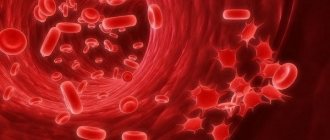
Human blood is a liquid substance consisting of plasma and formed elements, or blood cells, suspended in it, which make up approximately 40-45% of the total volume. They are small in size and can only be seen under a microscope.
There are several types of blood cells that perform specific functions. Some of them function only within the circulatory system, others go beyond its boundaries. What they have in common is that they are all formed in the bone marrow from stem cells, the process of their formation is continuous, and their lifespan is limited.
All blood cells are divided into red and white. The first are erythrocytes, which make up the majority of all cells, the second are leukocytes.
Platelets are also considered to be blood cells. These small blood platelets are not actually full-fledged cells. They are small fragments separated from large cells - megakaryocytes.
Red blood cells
Red blood cells are called red blood cells. This is the most numerous group of cells. They carry oxygen from the respiratory organs to the tissues and take part in the transport of carbon dioxide from the tissues to the lungs.
The place of formation of red blood cells is the red bone marrow. They live for 120 days and are destroyed in the spleen and liver.
They are formed from precursor cells - erythroblasts, which, before becoming an erythrocyte, go through different stages of development and divide several times. Thus, up to 64 red blood cells are formed from the erythroblast.
Red blood cells lack a nucleus and are shaped like a disk concave on both sides, the diameter of which is on average about 7-7.5 microns, and the thickness at the edges is 2.5 microns. This shape increases the ductility required for passage through small vessels and the surface area for gas diffusion. Old red blood cells lose their plasticity, which is why they linger in the small vessels of the spleen and are destroyed there.
Most red blood cells (up to 80%) have a biconcave spherical shape. The remaining 20% may have another: oval, cup-shaped, simple spherical, sickle-shaped, etc. Violation of the shape is associated with various diseases (anemia, deficiency of vitamin B12, folic acid, iron, etc.).
Most of the cytoplasm of the red blood cell is occupied by hemoglobin, consisting of protein and heme iron, which gives the blood its red color. The non-protein part consists of four heme molecules with an Fe atom in each. It is thanks to hemoglobin that the red blood cell is able to carry oxygen and remove carbon dioxide. In the lungs, an iron atom binds with an oxygen molecule, hemoglobin turns into oxyhemoglobin, which gives the blood a scarlet color. In tissues, hemoglobin gives up oxygen and adds carbon dioxide, turning into carbohemoglobin, as a result the blood becomes dark. In the lungs, carbon dioxide is separated from hemoglobin and removed by the lungs to the outside, and the incoming oxygen is again associated with iron.
In addition to hemoglobin, the cytoplasm of the erythrocyte contains various enzymes (phosphatase, cholinesterase, carbonic anhydrase, etc.).
The erythrocyte membrane has a fairly simple structure compared to the membranes of other cells. It is an elastic thin mesh, which ensures rapid gas exchange.
On the surface of red blood cells there are different types of antigens that determine the Rh factor and blood group. The Rh factor can be positive or negative depending on the presence or absence of the Rh antigen. The blood group depends on which antigens are on the membrane: 0, A, B (the first group is 00, the second is 0A, the third is 0B, the fourth is AB).
In the blood of a healthy person, there may be small amounts of immature red blood cells called reticulocytes. Their number increases with significant blood loss, when replacement of red cells is required and the bone marrow does not have time to produce them, so it releases immature ones, which are nevertheless capable of performing the functions of red blood cells in transporting oxygen.
What is a microscope
This is an optical device that produces images magnified several times.
Both visible and invisible objects are subject to research. Most often these are microorganisms, cells of living tissues, elements of the structure of substances and much more. Microscopes can be simple or complex. The first device consists of one plano-convex lens (for example, a regular magnifying glass), the complex one combines a combination of two simple lenses. Interesting!
Modern microscopes can provide magnification up to 2000x, at which the image quality will be excellent.
A compound microscope is used to examine blood, as it provides high magnification and has high resolution. The action is based on a two-step scheme:
- One objective (lens) is brought close to the object. It enlarges the image with the required resolution.
- The second lens (eyepiece) is placed directly close to the observer’s organ of vision.
Two lens systems are located at opposite ends of the device tube. The tripod is the main part of the tube.
Leukocytes
Leukocytes are white blood cells whose main task is to protect the body from internal and external enemies.
They are usually divided into granulocytes and agranulocytes. The first group is granular cells: neutrophils, basophils, eosinophils. The second group does not have granules in the cytoplasm; it includes lymphocytes and monocytes.
Neutrophils
This is the most numerous group of leukocytes - up to 70% of the total number of white cells. Neutrophils got their name due to the fact that their granules are stained with dyes with a neutral reaction. Its grain size is fine, the granules have a purple-brownish tint.
The main task of neutrophils is phagocytosis, which consists of capturing pathogenic microbes and tissue breakdown products and destroying them inside the cell with the help of lysosomal enzymes found in granules. These granulocytes fight mainly bacteria and fungi and to a lesser extent viruses. Pus consists of neutrophils and their remains. Lysosomal enzymes are released during the breakdown of neutrophils and soften nearby tissues, thus forming a purulent focus.
A neutrophil is a rounded nuclear cell, reaching a diameter of 10 microns. The core may have the shape of a rod or consist of several segments (from three to five) connected by cords. An increase in the number of segments (up to 8-12 or more) indicates pathology. Thus, neutrophils can be band or segmented. The first are young cells, the second are mature. Cells with a segmented nucleus make up up to 65% of all leukocytes, and band cells in the blood of a healthy person make up no more than 5%.
The cytoplasm contains about 250 types of granules containing substances through which the neutrophil performs its functions. These are protein molecules that affect metabolic processes (enzymes), regulatory molecules that control the work of neutrophils, substances that destroy bacteria and other harmful agents.
These granulocytes are formed in the bone marrow from neutrophilic myeloblasts. A mature cell stays in the brain for 5 days, then enters the blood and lives here for up to 10 hours. From the vascular bed, neutrophils enter the tissues, where they remain for two to three days, then they enter the liver and spleen, where they are destroyed.
Basophils
There are very few of these cells in the blood - no more than 1% of the total number of leukocytes. They have a round shape and a segmented or rod-shaped nucleus. Their diameter reaches 7-11 microns. Inside the cytoplasm there are dark purple granules of varying sizes. They got their name due to the fact that their granules are colored with dyes with an alkaline, or basic, reaction. Basophil granules contain enzymes and other substances involved in the development of inflammation.
Their main function is the release of histamine and heparin and participation in the formation of inflammatory and allergic reactions, including the immediate type (anaphylactic shock). In addition, they can reduce blood clotting.
They are formed in the bone marrow from basophilic myeloblasts. After maturation, they enter the blood, where they remain for about two days, then go into the tissues. What happens next is still unknown.
Eosinophils
These granulocytes make up approximately 2-5% of the total number of white cells. Their granules are stained with an acidic dye, eosin.
They have a rounded shape and a slightly colored core, consisting of segments of the same size (usually two, less often three). Eosinophils reach 10-11 microns in diameter. Their cytoplasm is painted pale blue and is almost invisible among the large number of large round granules of yellow-red color.
These cells are formed in the bone marrow, their precursors are eosinophilic myeloblasts. Their granules contain enzymes, proteins and phospholipids. A mature eosinophil lives in the bone marrow for several days, after entering the blood it remains in it for up to 8 hours, then moves to tissues that have contact with the external environment (mucous membranes).
The function of the eosinophil, like all leukocytes, is protective. This cell is capable of phagocytosis, although it is not their main responsibility. They capture pathogenic microbes mainly on the mucous membranes. The granules and nucleus of eosinophils contain toxic substances that damage the parasite membrane. Their main task is protection against parasitic infections. In addition, eosinophils take part in the formation of allergic reactions.
Lymphocytes
These are round cells with a large nucleus occupying most of the cytoplasm. Their diameter is 7 to 10 microns. The kernel can be round, oval or bean-shaped and has a rough structure. Consists of lumps of oxychromatin and basiromatin, resembling blocks. The core can be dark purple or light purple, sometimes it contains light inclusions in the form of nucleoli. The cytoplasm is colored light blue; around the nucleus it is lighter. In some lymphocytes, the cytoplasm has azurophilic granularity, which turns red when stained.
Two types of mature lymphocytes circulate in the blood:
- Narrow plasma. They have a rough dark purple nucleus and a narrow blue rim of cytoplasm.
- Wide-plasma. In this case, the kernel has a paler color and bean-shaped shape. The rim of the cytoplasm is quite wide, gray-blue in color, with rare ausurophilic granules.
From atypical lymphocytes in the blood you can find:
- Small cells with barely visible cytoplasm and a pyknotic nucleus.
- Cells with vacuoles in the cytoplasm or nucleus.
- Cells with lobed, kidney-shaped, jagged nuclei.
- Bare kernels.
Lymphocytes are formed in the bone marrow from lymphoblasts and undergo several stages of division during the process of maturation. Its complete maturation occurs in the thymus, lymph nodes and spleen. Lymphocytes are immune cells that mediate immune responses. There are T-lymphocytes (80% of the total) and B-lymphocytes (20%). The former matured in the thymus, the latter in the spleen and lymph nodes. B lymphocytes are larger in size than T lymphocytes. The lifespan of these leukocytes is up to 90 days. Blood for them is a transport medium through which they enter tissues where their help is required.
The actions of T-lymphocytes and B-lymphocytes are different, although both take part in the formation of immune reactions.
The former are engaged in the destruction of harmful agents, usually viruses, through phagocytosis. The immune reactions in which they participate are nonspecific resistance, since the actions of T lymphocytes are the same for all harmful agents.
Based on the actions they perform, T-lymphocytes are divided into three types:
- T-helpers. Their main task is to help B-lymphocytes, but in some cases they can act as killers.
- Killer T cells. Destroy harmful agents: foreign, cancerous and mutated cells, infectious agents.
- T-suppressors. Inhibit or block overly active reactions of B-lymphocytes.
B-lymphocytes act differently: against pathogens they produce antibodies - immunoglobulins. This happens as follows: in response to the actions of harmful agents, they interact with monocytes and T-lymphocytes and turn into plasma cells that produce antibodies that recognize the corresponding antigens and bind them. For each type of microbe, these proteins are specific and are capable of destroying only a certain type, therefore the resistance that these lymphocytes form is specific, and it is directed primarily against bacteria.
These cells provide the body's resistance to certain harmful microorganisms, which is commonly called immunity. That is, having encountered a harmful agent, B-lymphocytes create memory cells that form this resistance. The same thing - the formation of memory cells - is achieved by vaccinations against infectious diseases. In this case, a weak microbe is introduced so that the person can easily survive the disease, and as a result, memory cells are formed. They can remain for life or for a certain period, after which the vaccination must be repeated.
Monocytes
Monocytes are the largest of the leukocytes. Their number ranges from 2 to 9% of all white blood cells. Their diameter reaches 20 microns. The monocyte nucleus is large, occupies almost the entire cytoplasm, can be round, bean-shaped, mushroom-shaped, or butterfly-shaped. When stained it turns red-violet. The cytoplasm is smoky, bluish-smoky, less often blue. It usually has an azurophilic fine grain size. It may contain vacuoles (voids), pigment grains, and phagocytosed cells.
Monocytes are produced in the bone marrow from monoblasts. After maturation, they immediately appear in the blood and remain there for up to 4 days. Some of these leukocytes die, some move into the tissue, where they mature and turn into macrophages. These are the largest cells with a large round or oval nucleus, blue cytoplasm and a large number of vacuoles, which is why they appear foamy. The lifespan of macrophages is several months. They can be constantly in one place (resident cells) or move (wandering).
Monocytes form regulatory molecules and enzymes. They are able to form an inflammatory response, but can also inhibit it. In addition, they participate in the wound healing process, helping to speed it up, and promote the restoration of nerve fibers and bone tissue. Their main function is phagocytosis. Monocytes destroy harmful bacteria and inhibit the proliferation of viruses. They are able to carry out commands, but cannot distinguish between specific antigens.
Clinical blood analysis: from light microscope to hematological analyzers
A general clinical blood test is the most common diagnostic test that a doctor prescribes to a patient.
Over the past decades, the technology of this routine, but very informative research has made a colossal leap - it has become automatic. High-tech automatic hematology analyzers came to the aid of the laboratory diagnostics doctor, whose tool of work was an ordinary light microscope. In this post we will tell you what exactly happens inside the “smart machine” that sees right through our blood, and why you should trust it. We will consider the physics of processes using the example of the UniCel DxH800 hematology analyzer
global brand Beckman Coulter. It is on this equipment that studies ordered from the laboratory diagnostics service LAB4U.RU are performed. But to understand automated blood testing technology, we'll look at what the lab technicians saw under the microscope and how they interpreted that information.
Blood test parameters
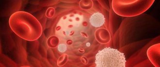
So, there are three types of cells in the blood:
- leukocytes that provide immune protection;
- platelets, responsible for blood clotting;
- red blood cells that transport oxygen and carbon dioxide.
These cells are found in the blood in very specific quantities. They are determined by the person’s age and state of health. Depending on the conditions in which the body is located, the bone marrow produces as many cells as the body requires. Therefore, knowing the number of a certain type of blood cells and their shape, size and other qualitative characteristics, you can confidently judge the condition and current needs of the body. It is these key parameters - the number of cells of each type, their appearance and qualitative characteristics
- that make up a general clinical blood test.
When conducting a general blood test, the number of red blood cells, platelets and leukocytes is counted. It’s more complicated with leukocytes: there are several types of them, and each type performs its own function. There are 5 different types of leukocytes:
- neutrophils, which mainly neutralize bacteria;
- eosinophils, neutralizing antigen-antibody immune complexes;
- basophils involved in allergic reactions;
- monocytes are the main macrophages and utilizers;
- lymphocytes providing general and local immunity.
In turn, neutrophils according to the degree of maturity are divided into:
- stab,
- segmented,
- myelocytes,
- metamyelocytes.
The percentage of each type of leukocyte in their total volume is called the leukocyte formula, which has important diagnostic value.
For example, the more pronounced the bacterial inflammatory process, the more neutrophils in the leukocyte formula. The presence of neutrophils of varying degrees of maturity indicates the severity of the bacterial infection. The more acute the process, the more band neutrophils in the blood. The appearance of metamyelocytes and myelocytes in the blood indicates an extremely severe bacterial infection. Viral diseases are characterized by an increase in lymphocytes, and in allergic reactions - an increase in eosinophils. In addition to quantitative indicators, cell morphology is extremely important. A change in their usual shape and size also indicates the presence of certain pathological processes in the body.
An important and most well-known indicator is the amount of hemoglobin in the blood - a complex protein that ensures the supply of oxygen to tissues and the removal of carbon dioxide. The concentration of hemoglobin in the blood is the main indicator in the diagnosis of anemia.
Another important parameter is the erythrocyte sedimentation rate (ESR). During inflammatory processes, red blood cells tend to stick together, forming small clots. Having greater mass, clumped red blood cells settle faster under the influence of gravity than single cells. The change in their sedimentation rate in mm/h is a simple indicator of inflammatory processes in the body.
How it was: scarifier, test tubes and microscope
Blood collection
Let's remember how they used to donate blood: a painful puncture of the pad with a scarifier, endless glass tubes into which precious drops of squeezed blood were collected.
How a laboratory assistant traced one piece of glass over another, where there was a drop of blood, scratching a number on the glass with a simple pencil. And endless test tubes with different liquids. Now it already seems like some kind of alchemy. Blood was taken specifically from the ring finger, for which there were quite serious reasons: the anatomy of this finger is such that its injury poses a minimal threat of sepsis in case of infection of the wound. Taking blood from a vein was considered much more dangerous. Therefore, venous blood analysis was not routine, but was prescribed as necessary, and mainly in hospitals.
It is worth noting that already at the sampling stage significant errors began. For example, different thicknesses of the skin give different depths of the injection; tissue fluid entered the test tube along with the blood - hence the change in blood concentration; in addition, with pressure on the finger, blood cells could be destroyed.
Remember the row of test tubes where the blood collected from your finger was placed? To count different cells, you actually needed different tubes. For erythrocytes - with saline solution, for leukocytes - with a solution of acetic acid, where the erythrocytes were dissolved, for the determination of hemoglobin - with a solution of hydrochloric acid. There was a separate capillary for determining ESR. And at the last stage, a smear was made on the glass for subsequent calculation of the leukocyte formula.
Blood test under a microscope
To count cells under a microscope in laboratory practice, a special optical device was used, proposed back in the 19th century by a Russian doctor, after whom this device was named - the Goryaev camera.
It made it possible to determine the number of cells in a given microvolume of liquid and was a thick glass slide with a rectangular recess (chamber). A microscopic mesh was applied to it. The top of Goryaev’s chamber was covered with a thin cover glass. This grid consisted of 225 large squares, 25 of which were divided into 16 small squares. Red blood cells were counted in small striated squares located diagonally in Goryaev’s chamber. Moreover, there was a certain rule for counting the cells that lie on the border of the square. The number of red blood cells per liter of blood was calculated using a formula based on the dilution of the blood and the number of squares in the grid. After mathematical abbreviations, it was enough to multiply the calculated number of cells in the chamber by 10 to the 12th power and enter it into the analysis form.
Leukocytes were counted here, but larger grid squares were used, since leukocytes are a thousand times larger than red blood cells. After counting leukocytes, their number was multiplied by 10 to the 9th power and entered into the form. An experienced laboratory technician took an average of 3-5 minutes to count cells.
Methods for counting platelets in the Goryaev chamber were very labor-intensive due to the small size of this type of cell. Their number had to be estimated only on the basis of a stained blood smear, and the process itself was also very labor-intensive. Therefore, as a rule, platelet counts were calculated only upon specific request from the physician.
Leukocyte formula
, that is, the percentage composition of each type of leukocytes in their total number could only be determined by a doctor - based on the results of studying blood smears on glasses.
Visually identifying the different types of leukocytes in the field of view by the shape of their nucleus, the doctor counted the cells of each type and their total number.
Having counted 100 in total, he received the required percentage of each type of cell. To simplify counting, special counters with separate keys for each type of cell were used. It is noteworthy that such an important parameter as hemoglobin was determined by the laboratory assistant visually (!) by the color of hemolyzed blood in a test tube with hydrochloric acid. The method was based on the conversion of hemoglobin into brown hydrochloric acid hematin, the color intensity of which is proportional to the hemoglobin content. The resulting solution of hematin hydrochloride was diluted with water to the color of the standard corresponding to the known concentration of hemoglobin. In general, the last century
How it became: vacuum containers and hematology analyzers
Let's start with the fact that the technology of blood sampling has now completely changed. Scarifiers and glass capillaries with test tubes were replaced by vacuum containers. The blood sampling systems now in use are low-traumatic, the process is completely unified, which has significantly reduced the percentage of errors at this stage. Vacuum tubes containing preservatives and anticoagulants allow blood to be stored and transported from the point of collection to the laboratory. It is thanks to the advent of new technology that it has become possible to take tests as conveniently as possible - at any time, anywhere.
At first glance, it seems impossible to automate such a complex process as identifying and counting blood cells. But, as usual, everything ingenious is simple. Automatic blood testing is based on fundamental physical laws. The technology for automatic cell counting was patented back in 1953 by Americans Joseph and Wallace Coulter. It is their name that is in the name of the global brand of hematological equipment Beckman&Coulter.
Cell counting
The aperture-impedance method (Coulter method or conductometric method) is based on counting the number and assessing the nature of impulses that occur when a cell passes through a small-diameter hole (aperture), on both sides of which two electrodes are located. As a cell passes through a channel filled with electrolyte, the resistance to electric current increases. Each passage of the cell is accompanied by the appearance of an electrical impulse. To find out what the cell concentration is, it is necessary to pass a certain volume of sample through the channel and count the number of pulses that appear. The only limitation is that the sample concentration must ensure that only one cell passes through the aperture at a time.
Automated hematology analysis technology has come a long way over the past 60 years. At first, these were simple cell counters that determined 8-10 parameters: red blood cell count (RBC), white blood cell count (WBC), hemoglobin (Hb) and several calculated ones. These were first class
.
Second class
analyzers have already determined up to 20 different blood parameters. They are significantly higher in the level of differentiation of leukocytes and are able to isolate populations of granulocytes (eosinophils + neutrophils + basophils), lymphocytes and an integral population of medium cells, which included monocytes, eosinophils, basophils and plasma cells. This differentiation of leukocytes has been successfully used in the examination of practically healthy people.
The most technologically advanced and innovative analyzers today are third-class
, which determine up to hundreds of different parameters, carry out detailed differentiation of cells, including by degree of maturity, analyze their morphology and signal the laboratory assistant about the detection of pathology.
Third-class machines, as a rule, are also equipped with automatic systems for preparing smears (including their coloring) and displaying images on the monitor screen. These advanced hematology systems include BeckmanCoulter equipment, such as the UniCel DxH 800 Cell Analysis System
.
Modern BeckmanCoulter devices use multiparameter flow cytometry based on patented VCS (Volume-Conductivity-Scatter) technology.
VCS technology involves assessing cell volume, its electrical conductivity and light scattering. The first parameter, cell volume, is measured using the Coulter principle based on an estimate of the resistance as a cell passes through an aperture at a constant current. The size and density of the cell nucleus, as well as its internal composition, are determined by measuring its electrical conductivity in high-frequency alternating current. Scattering of laser light at different angles allows one to obtain information about the structure of the cell surface, the granularity of the cytoplasm and the morphology of the cell nucleus.
The data obtained from three channels is combined and analyzed. As a result, the cells are distributed into clusters, including separation according to the degree of maturity of erythrocytes and leukocytes (neutrophils). Based on the obtained measurements of these three dimensions, many hematological parameters are determined - up to 30 for diagnostic purposes, more than 20 for research purposes and more than a hundred specific calculated parameters for highly specialized cytological studies. Data is visualized in 2D and 3D formats. A laboratory technician working with a BackmanCoulter hematology analyzer sees the analysis results on the monitor looking something like this:
And then he decides whether they need to be verified or not.
Needless to say, the information content and accuracy of modern automatic analysis is many times higher than manual analysis? The productivity of machines of this class is about a hundred samples per hour when analyzing thousands of cells in a sample. Let us remember that during microscopy of a smear, the doctor analyzed only 100 cells!
However, despite these impressive results, microscopy still remains the “gold standard” for diagnosis. In particular, when the apparatus detects pathological cell morphology, the sample is analyzed manually under a microscope. When examining patients with hematological diseases, microscopy of a stained blood smear is performed only manually by an experienced hematologist. This is exactly how, manually, in addition to automatic cell counting, the leukocyte formula is assessed in all children's blood tests according to orders made using the online laboratory service LAB4U.RU.
Instead of a resume
Technologies for automated hematological analysis continue to develop rapidly.
Essentially, they have already replaced microscopy in routine preventive tests, leaving it for particularly significant situations. We mean children's tests, tests of people with confirmed diseases, especially hematological ones. However, in the foreseeable future, in this area of laboratory diagnostics, doctors will receive devices capable of independently performing morphological analysis of cells using neural networks. By reducing the burden on doctors, they will at the same time increase the requirements for their qualifications, since only atypical and pathological cell conditions will remain in the human decision-making zone. The number of informative parameters of blood analysis, which has increased many times over, raises the requirements for professional qualifications and for the clinician, who needs to analyze combinations of values of a mass of parameters for diagnostic purposes. Expert systems come to the aid of doctors on this front, which, using analyzer data, provide recommendations for further examination of the patient and give a possible diagnosis. Such systems are already available on the laboratory market. But this is a topic for a separate article.
Platelets
These blood cells are small, anucleate plates and can be round or oval in shape. During activation, when they are near the damaged vessel wall, they form outgrowths, so they look like stars. Platelets contain microtubules, mitochondria, ribosomes, and specific granules containing substances necessary for blood clotting. These cells are equipped with a three-layer membrane.
Platelets are produced in the bone marrow, but in a completely different way than other cells. Blood plates are formed from the largest cells of the brain - megakaryocytes, which, in turn, were formed from megakaryoblasts. Megakaryocytes have a very large cytoplasm. After the cell matures, membranes appear in it, dividing it into fragments that begin to separate, and thus platelets appear. They leave the bone marrow into the blood, stay in it for 8-10 days, then die in the spleen, lungs, and liver.
Blood plates can have different sizes:
- the smallest are microforms, their diameter does not exceed 1.5 microns;
- normoforms reach 2-4 microns;
- macroforms – 5 microns;
- megaloforms – 6-10 microns.
Platelets perform a very important function - they participate in the formation of a blood clot, which closes the damage in the vessel, thereby preventing blood from leaking out. In addition, they maintain the integrity of the vessel wall and promote its rapid recovery after damage. When bleeding begins, platelets adhere to the edge of the injury until the hole is completely closed. The adhered plates begin to break down and release enzymes that affect the blood plasma. As a result, insoluble fibrin threads are formed, tightly covering the injury site.
Functions
The general functions of leukocytes are as follows:
- Protective – consists in the formation of specific and nonspecific immunity. The main mechanism is phagocytosis (capture of a pathogenic microorganism by a cell and deprivation of its life).
- Transport - lies in the ability of white cells to adsorb amino acids, enzymes and other substances found in plasma and transport them to the right places.
- Hemostatic - involved in blood clotting.
- Sanitary – the ability, with the help of enzymes contained in leukocytes, to dissolve tissues that have died due to injury.
- Synthetic – the ability of some proteins to synthesize bioactive substances (heparin, histamine and others).
Each type of leukocyte has its own functions, including specific ones.
Neutrophils
The main role is to protect the body from infectious agents. These cells capture bacteria into their cytoplasm and digest them. In addition, they can produce antimicrobial substances. When an infection enters the body, they rush to the site of penetration, accumulate there in large quantities, absorb microorganisms and die themselves, turning into pus.
Eosinophils
When infected with worms, these cells penetrate the intestines, are destroyed and release toxic substances that kill the helminths. In allergies, eosinophils remove excess histamine.
Basophils
These leukocytes take part in the formation of all allergic reactions. They are called first aid for bites of poisonous insects and snakes.
Lymphocytes
They constantly patrol the body in order to detect foreign microorganisms and out-of-control cells of their own body, which can mutate, then quickly divide and form tumors. Among them there are informants - macrophages, which constantly move throughout the body, collect suspicious objects and deliver them to lymphocytes. Lymphocytes are divided into three types:
- T-lymphocytes are responsible for cellular immunity, come into contact with harmful agents and destroy them;
- B lymphocytes identify foreign microorganisms and produce antibodies against them;
- NK cells. These are real killers that maintain normal cellular composition. Their function is to recognize defective and cancer cells and destroy them.