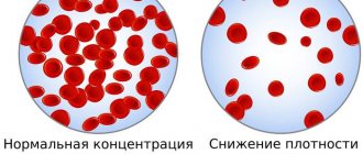
To make any accurate diagnosis, each of us undergoes a procedure such as donating blood for analysis. In most cases, a sample from a finger is enough, but sometimes you have to take biomaterial from a vein. Often, during research, doctors use such a definition as a shift in the leukocyte formula. Having heard such an expression somewhere, not everyone will be able to understand what they are actually talking about.
It is worth noting that each person’s blood composition is individual, and it can change due to various biological processes. The leukocyte formula tells about these changes. And it is precisely this that will be discussed further in the topic of this article.
What is the leukocyte formula?
There are several types of white blood cells in our blood (more on this in the next section) and each of them performs its own task. The leukocyte formula, or leukogram, is the percentage of all types of blood cells. It also allows you to determine the overall level of leukocytes, thereby identifying a possible shift in the leukocyte formula. This has nothing to do with mathematics. Thanks to this formula, you can assess the general state of a person’s health, as well as identify various possible deviations.
In some cases, it is possible not only to recognize the disease, but also to determine the extent of its progression and subsequent outcome. In most cases, an analysis to determine the leukocyte formula is prescribed with general studies during a routine medical examination, if leukemia is suspected, and also as a control preventive measure.
What to do in case of deviations
A shift of the formula to the left is not a disease in itself, and therefore does not require treatment. This is just a symptom that signals disturbances in the normal functioning of the body. Therefore, once you receive the analysis in your hands, you should not try to decipher it yourself; you should consult a doctor.
If he reveals violations, then treatment will be aimed at the cause that caused the neutrophilia. For minor lesions, medication treatment at home is sufficient; for more serious lesions, in a medical facility. In all cases, the doctor usually prescribes additional tests and refers you to specialists for consultation. After the final diagnosis is made, appropriate therapy is prescribed.
The leukogram is used very often in medical practice. This is an effective, safe method that allows you to make a preliminary diagnosis quite accurately and begin treatment in a timely manner.
Types of leukocytes
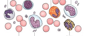
In the blood of the human body, as mentioned above, there is more than one type of leukocyte. These important cells, which fight infectious threats and respond to tissue damage, are produced in the bone marrow. There are five types of them:
- lymphocytes;
- neutrophils;
- monocytes;
- basophils;
- eosinophils.
In this case, monocytes, basophils and eosinophils are considered heavy, and lymphocytes and neutrophils are considered light. Each of these types of blood cells differs from each other not only in structure, but also performs its own function. When examining the issue related to the shift in the leukocyte formula, it is worth getting to know them better.
Lymphocytes - these cells belong to the group of agranulocytes and represent the foundation of our immune system. Their main task is to recognize and eliminate foreign antigens, including cancer cells. They also take part in the production of antibodies. In turn, they are divided into three types:
- B cells;
- T cells;
- NK cells.
Monocytes are cells belonging to the mononuclear leukocyte group. They are oval in shape and contain a large nucleus, which contains chromatin, a large amount of cytoplasm with many lysosomes. When mature, they have a diameter of 18-20 microns. Monocytes are responsible for removing decaying cells, as well as bacteria and other foreign bodies, from the body. In addition to neutralizing microorganisms, they participate in phagocytosis.
Neutrophils belong to the granulocytic group and are phagocytes in the classical sense. In many ways, it is for their reason that the leukocyte formula shifts to the right or left. They are divided into rod and segmented. In addition to being motile, cells are capable of chemotaxis and can capture bacteria. But at the same time, neutrophils absorb cells or particles of relatively small size. They take part in the production of certain bactericidal substances, thereby performing a pest control function.
Basophils - also belong to granulocytic leukocytes and have an S-shaped nucleus. They contain large quantities of substances such as:
- histamine;
- serotonin;
- leukotriene;
- prostaglandin.
The granules are born in the bone marrow and enter the bloodstream already mature. They are quite large in size, larger than neutrophils and eosinophils. When an inflammatory process occurs, basophils are responsible for transporting white cells to the site of the lesion. They also take an active part in allergic reactions.
Eosinophils, like neutrophils, are motile and participate in phagocytosis. They can absorb foreign bodies, but being microphages they are not able to fight large microorganisms. In addition, eosinophils are distinguished by their ability to absorb and bind histamine and some other mediators of allergy and inflammation. If necessary, they can release these substances in the same way as basophils do.
Main types of leukocytes and their functions
Leukocytes are immune cells; they perform a number of functions aimed at protecting the body from foreign agents and microorganisms. Depending on the presence of granules in the cytoplasm of leukocytes, they are divided into 2 types:
- Granulocytes - contain granules in the cytoplasm with various biological compounds, these include basophils, neutrophils, eosinophils.
- Agranulocytes - accordingly, do not contain granules in the cytoplasm; such cells include lymphocytes and monocytes.
Each type of these immune cells performs its main functions in the human body:
- Segmented neutrophils - protect the body from various foreign agents, primarily from pathogenic (disease-causing) bacteria and fungi. The defense mechanism is phagocytosis - the process of absorption and digestion of a foreign agent by a neutrophil. Young forms of these cells also circulate in the blood - rod and juvenile forms.
- Eosinophils - contain a large number of granules in the cytoplasm, which are filled with biologically active compounds. These cells are directly involved in the allergic reaction (hypersensitization reaction), which develops in response to allergens entering the body. At the same time, eosinophils release the contents of granules (histamine) into the blood, which leads to the development of allergy symptoms.
- Basophils - these cells also contain various mediators of the allergic reaction; they are more localized in the tissues of the area of the highest concentration of the allergen. When the contents of their granules are released, a local allergic reaction (redness, rash, itching) develops.
- Lymphocytes - these cells perform a large number of functions - the synthesis of antibodies (B-lymphocytes and plasma cells), the direct destruction of altered cells of their own tissues under the influence of viruses or a tumor process (killer T-lymphocytes). There are also helper T-lymphocytes, which regulate the functional activity of other immune cells, and memory B-lymphocytes (contain on their surface receptors for a foreign agent with which the human body has had contact at least once; when such an agent enters again, these cells begin to rapidly multiply and synthesize specific protective antibodies).
- Monocytes - they perform the function of phagocytosis; from the blood they rush into the tissues of the area of the inflammatory process, where they identify, absorb and digest foreign agents (fungi, bacteria, viruses).
Pus, which is formed as a result of various inflammatory processes, represents dead neutrophils and microorganisms.
Children's body
At a young age, especially for newborns, a more pronounced shift in the leukocyte formula of children is observed. And there is a simple explanation for this - the body of a child or a newly born baby is not yet fully formed and various biological processes are actively occurring in it.
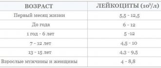
Moreover, unlike adults, the number of leukocytes in the blood, depending on the age of the child, is different. Throughout the entire childhood period, the child's leukocyte formula crosses twice. The first time this happens is after the baby is born. Since the mother’s body performed the main protective function for the fetus, the composition of the newborn’s blood is close to the normal level in adults.
When a baby is born, he immediately begins to get used to his surroundings, which is reflected in various processes occurring in his body. By the end of the first month of life, the level of lymphocytes increases significantly.
Being from the age of one to three, the child’s body is characterized by an unstable blood composition. That is, from time to time there is a shift in the leukocyte formula to the left in children or to the right. In this case, the concentration of lymphocytes and neutrophils can change throughout the day. Also, the reason for such a change may be certain conditions:
- hypothermia;
- a long walk in the sun;
- chronic diseases;
- changes at the gene level.
From 4 to 6 years, neutrophils take a leading position. However, in children over 6-7 years of age, the blood composition is identical to that of adults. During this entire period of hormonal changes, a 10-15% shift in the formula may be observed, which is normal.
The table below will show a more clear picture.
Changes in blood cell levels depending on the child's age
| Age | Name of blood cells | Norm, % |
| Newborns | lymphocytes | 20-35 |
| neutrophils | 65 | |
| monocytes | 3-5 | |
| basophils | 0-1 | |
| eosinophils | 1-2 | |
| First month of life | lymphocytes | 65-70 |
| neutrophils | 20-25 | |
| monocytes | 3-6 | |
| basophils | 1-2 | |
| eosinophils | 0,5-1 | |
| from 1 to 3 years | lymphocytes | 35-55 |
| neutrophils | 32-52 | |
| monocytes | 10-12 | |
| basophils | 0-1 | |
| eosinophils | 1-4 | |
| from 4 to 6 years | lymphocytes | 33-50 |
| neutrophils | 36-52 | |
| monocytes | 10-12 | |
| basophils | 0-1 | |
| eosinophils | 1-4 | |
| Over 6-7 years old | lymphocytes | 19-35 |
| neutrophils | 50-72 | |
| monocytes | 3-11 | |
| basophils | 0-1 | |
| eosinophils | 1-5 |
Thanks to such changes, the immunity of the child’s body is formed, while the child gets to know and understands the world around him.
Possible deviations from the norm in children
Each type of white blood cell is unique in nature due to its individual role in the body. Any deviations that the leukocyte formula undergoes, a shift to the left and to the right, indicate the presence of some disease.
An increased level of lymphocytes or lymphocytosis is observed in the event of a viral and bacterial infection (whooping cough, influenza, rubella, measles, tuberculosis). In addition to them, a high concentration of cells is indicated by the presence of bronchial asthma, an autoimmune disease (Crohn's or Lyme disease), as well as an innate tendency to allergies. Feeding a child predominantly carbohydrate foods in the first year of his life usually leads to an increase in the number of lymphocytes. Their significant deficiency (lymphocytopenia) indicates that the bone marrow is susceptible to pathology and can no longer reproduce blood cells in the required quantity.
A high content of neutrophils also has its own name - Neutrophilia or a shift in the leukocyte formula to the left. In some cases, this is explained by the body's natural defensive reaction to some threat. For example, extensive inflammation and systemic lupus erythematosus (SLE). If a hormonal imbalance occurs in the body, then neutropenia or a lack of neutrophils occurs. But besides this, it is affected by extensive intoxication of the body.
A high concentration of monocytes leads to monocytosis, which can occur as a result of a fungal or viral disease. Here the clinical picture can be judged by external signs:
- lymphadenopathy;
- inflammation of the nasopharynx and larynx with neoplasms;
- enlarged liver and characteristic pain in the right hypochondrium.
In addition, an altered leukocyte count with a shift to the left or right may be associated with a lack of these cells (monocytopenia). This often happens if the body does not receive enough B vitamins and folic acid. Iron deficiency anemia often occurs.
A large number of basophils is called basophilia. However, this phenomenon is very rare and develops in isolated cases. The cause may be a dangerous pathological change such as tuberculosis, damage to the lymph nodes, myeloid leukemia, or blood oncology.
A shift in the leukocyte formula may be indicated by a high level of eosinophils, which occurs for one of two possible reasons. The first is that when consuming dairy products, including lactose and gluten, an allergic reaction occurs. The second reason is related to the presence of parasitic worms, which have been ignored for a long time. It is worth noting that eosinophilia cannot be determined by external signs. But the process can proceed at a rapid pace and give rise to irreversible processes.
Preparing for the study
In order for the doctor to determine whether leukocytosis with a left shift is present, the patient needs to donate blood. The collection is made in the morning, from a finger or vein. It is recommended to drink some water to thin the blood. But you can’t eat in the morning; at least six hours must pass after dinner.
If possible, a week before the test you should stop taking any medications. In cases where this is not possible, the result must be interpreted taking into account the side effects of the drugs. Also, 2-4 days before visiting the laboratory, you should not eat spicy, fatty, salty foods, and give up nicotine and alcohol. The day before the collection, do not overwork, avoid psycho-emotional and physical stress.
In addition to these factors, the examination result may be affected by pregnancy and various physiotherapy procedures recently completed (X-ray, ultrasound). These circumstances should be reported to your doctor.
Indications for testing
Indications for donating blood to determine the leukocyte formula are the following cases:
- Mandatory examination by a doctor, which should be completed annually.
- If there are complications after the disease.
- If you experience severe fatigue.
As many experts note, such a blood test should not be underestimated. A shift in the leukocyte formula will make it possible to diagnose almost any acute or chronic disease, including oncology.
Only the study will give accurate answers if it is carried out in combination with other tests. Only in this case can an accurate diagnosis of the disease, as well as its development and outcome, be made.
Monocytes
Monocyte is translated from Greek as “single cell” or “single cell”. These are large cells without granules with a large non-segmented nucleus. Belongs to the class of phagocytes. The cytoplasm contains a large number of organelles - lysosomes, which are involved in the digestion of foreign proteins and microorganisms.
Normally, in peripheral blood they should be no more than 11 percent. In addition, most of them quickly move into tissues to perform their functions. An increase in the number of monocytes occurs in severe infectious processes, malignant tumors, systemic autoimmune connective tissue diseases, diseases of the hematopoietic system, and during the period of convalescence. In addition, an increase in monocytes is often observed after surgical interventions.
A decrease in the number of these cells is associated with long-term use of steroid drugs, sepsis, the development of aplastic anemia and hairy cell leukemia, infection with Salmonella typhoid, as well as physiological childbirth.
Analysis procedure
Before undergoing the blood donation procedure to determine the leukocyte formula, preparation is needed. It is simple, because all you need to do is not eat 3-4 hours before the test and avoid drinking alcohol. You also need to avoid overusing physical and emotional stress. Venous blood is taken for examination.
Moving directly to work, the laboratory assistant places the material on a special glass plate, which he places under a microscope. Next, the leukocyte formula of the blood is determined; a shift to the left or right is detected during the screening of several hundred blood cells so that the overall level of all leukocytes can be determined. The next step is to distribute the cells over the entire surface. In this case, the heavy granules are concentrated at the edges, and the light ones are placed in the center.
There are often two main methods used to count white blood cells:
- Schilling method - the smear is divided into 4 parts.
- Filipchenko's method - the smear is divided into three parts.
The transcript of the result will be ready after several days of examination, and its analysis will be carried out by the attending physician.
Decoding the results
Deciphering the leukocyte formula should only be carried out by an employee specially trained in this profile. But you can simply compare the results obtained with the norm. Often, when the leukocyte formula of blood is analyzed, shifts are determined during manual calculations. But some clinics have taken the modern route and use special equipment for this - an analyzer.
As a rule, it works in automatic mode, but in case of a sharp deviation from the norm, a specialist takes over the work. For comparison, a person will be able to examine 100-200 cells, while the apparatus is much larger – several thousand. But, despite the fact that modern equipment allows for more accurate calculations, errors inevitably occur. This may be influenced by several reasons: incorrect blood sampling, improperly prepared smear and other factors.
Technique
Preparing for a blood test is not difficult: the patient should simply refuse to eat four hours before the procedure, and the day before he should avoid emotional and physical stress.
The material for determining the formula is venous blood. Before this, the laboratory assistant must clamp the patient’s forearm with a special belt, and then insert a thin needle into the vein located in the bend of the elbow, through which the blood will flow into the test tube. This process cannot be called painless, but most often the pain is not too strong. As a result, a drop of blood is obtained and placed on a glass plate in order to determine the proportion of leukocytes and their number using a microscope. If the clinic has modern equipment, then the particles are counted by an analyzer - a special device, and human intervention is needed only in case of serious deviations from the norm or the presence of particles of an anomalous nature.
The time it takes to obtain results mainly depends on the institution where the study is carried out. Usually you have to wait no more than a few days. The attending physician should evaluate the obtained indicators. When deciphering, you can detect a shift to the left in the leukocyte formula.
Shift formula left
The term shift of the leukocyte formula to the left refers to a high concentration of band neutrophils, which indicates the occurrence of an inflammatory process. This can also happen due to:
- Infectious disease.
- Acid-base balance disorders.
- Comatose state.
- Physical overexertion.
Along with an increase in the concentration of neutrophils, a certain amount of metamyelocytes (not yet matured leukocytes) enters the blood.
In a healthy body they are found only in the red brain. However, due to the strong inflammatory response, most healthy neutrophils quickly die. In this case, the bone marrow has to send immature blood cells to the lesion.
Eosinophils
Eosinophils are a type of white blood cell, so named because they stain predominantly with acidic dyes. Their core consists of two segments connected by a constriction. These cells are able to move independently through blood vessels and tissues, and are prone to chemotaxis during inflammation or injury. They are also capable of absorbing and digesting foreign microorganisms and proteins.
But this is not the main role of eosinophils. On the surface of these cells there are receptors that attract class E immunoglobulins. This in itself is not scary, it is even useful, because the cytotoxic properties that appear in the eosinophil along with the addition of immunoglobulin make it possible to fight parasites. However, if there are many such “paired” cells, they can cause severe allergic reactions.
In adults, they should normally be no more than 5 percent; in children, this figure is slightly higher - up to 7 percent. A shift of the leukogram to the left (active inflammatory process) implies a decrease in the number of eosinophils, since the release of adrenal hormones leads to retention of cells in the bone marrow and inhibits their proliferation.
An increase in the absolute and relative number of eosinophils can be considered as evidence of the presence of allergic pathology, for example, bronchial asthma or urticaria. And also make the doctor think about a parasitic infection, the development of a tumor process in the hematopoietic organs, or an immunodeficiency state.
Shift formula right
The definition of a shift in the leukocyte formula to the right means a reduced content of band neutrophils. But at the same time the number of segmented cells increases. Often accompanied by chronic liver and kidney disease, including megablastic anemia. This can also be affected by blood transfusions.
The importance of the leukocyte formula is difficult to overestimate, since most changes occurring in the body lead to its shift. The concentration of some blood cells increases due to a decrease in the number of others.
Reasons for increased leukogram
Various infectious diseases and other pathological conditions cause an increase in the leukocyte count of the blood. By studying these numbers, the doctor can, together with the clinical manifestations, pinpoint the cause of this increase.
Lymphacytosis develops in various pathological conditions in the body:
- infection with mononucleosis, chickenpox, rubella, measles;
- chronic pathologies caused by pathogens of brucellosis, tuberculosis, syphilis;
- hyperthyroidism;
- development of lymphosarcoma, lymphocytic leukemia;
- plastic and hypoplastic anemia, folate deficiency;
- insufficient function of the adrenal cortex.
Neutrophilia will be present during the processes:
- acute blood loss;
- acute bacterial pathologies;
- tissue necrosis;
- acute intoxication;
- taking hormonal steroid drugs.
A high composition of eosinophils occurs:
- With scarlet fever.
- Infection with helminthiases.
- Infections in the recovery stage.
- For allergic reactions.
- Chronic skin diseases: eczema and psoriasis.
- With eosinophilic leukocytosis.
Elevated numbers of monocytes indicate the presence of:
- hemoblastoses;
- infectious diseases;
- autoimmune diseases;
Basophils increase:
- With erythremia.
- If chronic myeloid leukemia occurs.
- For allergic reactions.
Normal indicators
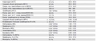
As is already known, any deviation from the norm implies the presence of significant changes in the body. Normal lymphocyte counts are 19-37% or 1.2-3x10⁹ pcs/l; neutrophils (specifically segmented ones) – 47-72% or 2-5.5x10⁹ pcs./l; band neutrophils – 1-6% or 0.04-0.3x10⁹ pcs./l; monocytes – 3-11% or 0.09-0.6x10⁹ pcs./l; basophils – 0-1% or 0-0.065x10⁹ pcs/l; and finally, the concentration of eosinophils is 0.5-5% or 0.02-0.3x10⁹ pcs./l.
Based on the data obtained from the results of the study, the doctor confirms or rejects the alleged diagnosis. And if a shift in the leukocyte formula has not occurred and everything is within normal limits, then there is no cause for concern.