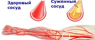
Dopplerography is a modern diagnostic method that makes it possible to visualize large vessels in real time. The method is based on the “Doppler effect” - the ability of an ultrasound beam to pass through motionless tissue. In most cases, Doppler is used to diagnose cardiovascular pathologies. Using this study, you can evaluate the speed and turbulence of blood flow, and also makes it possible to determine the condition of the walls of blood vessels. Based on the results of Doppler sonography, the doctor will be able to identify abnormalities, make a diagnosis and decide on treatment tactics.
Main advantages
Why is Doppler sonography popular among doctors and patients? This comes with a huge number of benefits. Here are the main ones:
- The procedure is completely safe for the body, since it is not associated with radiation exposure;
- Doppler ultrasound is a non-invasive diagnostic method; it does not cause pain or any discomfort;
- Doppler sonography can be performed on almost everyone - even newborns, pregnant and lactating women;
- This technique allows for observations in real time;
- The study allows us to identify pathological processes and deviations even at an early stage of development;
- The study can be repeated many times. This may be necessary to assess the dynamics of the disease during treatment;
- The procedure does not require special preparation.
Description of the method
This is a non-traumatic and absolutely safe way to obtain detailed information about the structure of changes in arterial walls. The method does not require much time or special preparatory measures. Can be used without being in a hospital.
CT calcium scoring is a fairly informative diagnostic method. Thanks to the test you can:
- detect deposits of calcium salts on the walls of blood vessels;
- check the aorta and trunk of the pulmonary artery;
- assess the condition of neighboring organs.
Preparation involves removing jewelry and metal products that are located in the chest area. They make diagnostics difficult and can cause errors during decoding. Additional studies (ultrasound, CT, MRI and laboratory tests) allow a more accurate diagnosis.
A CT scan for coronary calcium takes up to 15 minutes. The patient is only required to lie on his back, first raising his arms up. At the doctor's command, you need to hold your breath for a few seconds.
Indications
In what cases can a doctor prescribe a Doppler ultrasound of the heart?
- Pain in the heart area is the most common symptom when Doppler ultrasound is recommended. If the patient is bothered by discomfort and pain in the chest, Dopplerography of the heart vessels is a mandatory part of the examination.
- Heart murmurs discovered at an appointment with a therapist or cardiologist. This symptom may indicate a heart defect, so additional diagnostics are necessary.
- Patient's complaints about heart failure. These may be arrhythmias or extrasystoles, tachycardia or bradycardia.
- Complaints of frequent dizziness and shortness of breath. These symptoms may be associated with heart failure, therefore, to clarify the diagnosis, Doppler sonography is required in combination with other types of examination.
- Pathological changes on the ECG.
- Suspicion of a blood clot. Thanks to diagnostics, it is possible to identify its size and exact location.
- Suspicion of compaction in the pericardium.
Doppler ultrasound of the heart and blood vessels can also be prescribed for preventive purposes, for example, if there is a predisposition to heart disease. In addition, the study is necessary for patients who are planning to undergo heart surgery.
If your doctor advises you to undergo a Doppler ultrasound, you should not refuse this procedure. Pathologies in the early stages of development are much easier and faster to treat.
Who is the study for?
Ultrasound of the heart is prescribed for the following purposes:
- Diagnosis of diseases of the cardiovascular system in the presence of complaints.
- Preventive examinations.
- Monitoring the course of existing pathology - arrhythmias, myocardial dystrophy, pathology of the valve apparatus, angina pectoris, etc.
- Examination before surgery and invasive procedures.
- The presence of some non-cardiac diseases - varicose veins, systemic pathologies, pneumonia and other lung diseases, etc.
The following symptoms may indicate heart problems:
- Pain in the chest or upper abdomen. Often it radiates to the arm or shoulder blade.
- Blue skin.
- Dyspnea.
- Swelling, especially if there are no kidney problems.
- Weakness, dizziness, loss of consciousness.
- Feeling of heart sinking.
- High blood pressure.
Preparation
No special preparation is required for the study. However, the attending physician should warn the patient not to drink alcohol or energy drinks several hours before the examination. You should also refrain from smoking - this contributes to the narrowing of blood vessels, and therefore the results of the study after a smoke break may be distorted.
Be sure to tell your doctor if you are taking any medications. Some medications may need to be stopped several days before the test.
Ultrasound of the heart: when is it prescribed and what will it show?
To clarify a cardiac diagnosis, one of the most frequently prescribed studies is an ECG. However, often the doctor refers the patient to an ultrasound of the heart. What does this diagnosis show and is it worth doing it at all?
Today we are having an unusual conversation, as our consultants will be two doctors.
Olga Nikolaevna Vitkova, and her colleague, an ultrasound diagnostics doctor in pediatric practice, Inna Vyacheslavovna Popova, tell us about heart ultrasound and its capabilities.
— Olga Nikolaevna, Inna Vyacheslavovna, please tell us what ultrasound of the heart is and when is it prescribed?
This is a research method based on the use of ultrasound with certain characteristics for diagnostic purposes. Ultrasound is beyond the audibility of the human ear. During the examination, the ultrasound machine generates a stream of ultrasonic waves. The doctor directs them using a sensor to one or another part of the body. Ultrasound, having reached anatomical formations, is reflected from them and returns. These signals are recorded by an ultrasound machine, which, based on their characteristics, “constructs” an image of the organ being examined on the monitor screen.
Ultrasound examination allows us to judge the size of the heart, its layers, valves, and the characteristics of blood circulation.
It is prescribed for pain in the heart, high blood pressure, edema, shortness of breath, wheezing in the lungs; for dizziness, loss of consciousness, general weakness, prolonged increase in body temperature of unknown origin, patients with diabetes mellitus, and those with a history of myocardial infarction; people whose relatives suffered from certain diseases.
It's off scale! We are looking for the causes of high blood pressure. Read here
In children, ultrasound is performed on the direction of a cardiologist or pediatrician - most often if a heart defect is suspected. More than half of young patients come for examination precisely for this reason.
Ultrasound is prescribed for children with serious infections, in particular bacterial ones. Occasionally - if a heart tumor is suspected, it is extremely rare, but still a common diagnosis in children.
Preventive examinations are also performed for babies up to one year old.
— Are cardiac ultrasound and echocardiography the same study or are there differences between them? And if so, what is the difference?
Ultrasound of the heart and echocardiography are the same thing.
— Can ultrasound of the heart replace an ECG? What is the difference between these studies?
No, these studies are not interchangeable. It is correct to say that they complement each other. These methods characterize different aspects of the heart’s work. An ECG indicates the function of the conduction system of the heart and its possible disorders. With its help, you can indirectly judge the size of the cavities of the heart.
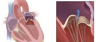
However, the ECG is not informative regarding the structural features of the organ - for example, the valve apparatus, the presence of defects, blood clots or inflammatory changes in the heart. In these cases, compared with ECG, ultrasound is the method of choice. It will allow you to directly evaluate the chambers of the heart, its layers, valves and their defects, aneurysms after a heart attack, the amount of blood ejected by the chambers of the heart, etc.
Specific example. The patient's ultrasound revealed tachycardia (rapid heartbeat), but we cannot say anything about its origin or type. This is the power of an ECG.
What causes tachycardia? Elena Viktorovna Novikova, a cardiologist at the Expert Kursk Clinic, says:
— What diseases can ultrasound of the heart diagnose?
Taking into account what has already been listed above, ultrasound also reveals signs of congestive heart failure (for example, fluid in the pericardial cavity), neoplasms and aneurysms of the heart, dilation of the heart chambers and blood clots in them, dissecting aortic aneurysm.
— In order to identify heart pathology, is it enough for a patient to undergo only an ultrasound scan or should the diagnosis be comprehensive?
No, ultrasound alone is definitely not enough. The examination must be comprehensive and must include a survey and examination of a doctor, an ECG and/or - as indicated - Holter monitoring (24-hour ECG), laboratory tests (depending on the intended diagnosis), ultrasound of the vessels of the head and neck, gastrointestinal tract, as indicated - coronary angiography (x-ray examination of the heart vessels using a contrast agent).
Why are x-rays dangerous? Yulia Aleksandrovna Rutskaya, head of the radiation diagnostics department at Clinic Expert Kursk, says:
— Is there an alternative to cardiac ultrasound?
It depends on what you mean by alternative. If the method is safe, non-invasive and accessible, then there is no alternative. If it refers to diagnostic capabilities, to a certain extent, transesophageal echocardiography, as well as spiral computed tomography with a contrast agent, can be an alternative. These methods can be used in cases where traditional echocardiography is difficult for some reason or does not answer some of the doctor’s questions.
— How safe is heart ultrasound and how often can this study be done?
Completely safe. It can be performed as often as required.
— How is heart ultrasound performed for adults and children?
There are no fundamental differences between adults and children. It is performed with the patient lying down, more often on the left side. Using a sensor, the doctor examines all heart formations and evaluates a number of functional parameters.
The peculiarity of children is that it is often difficult to convince them to lie on their left side, and therefore studies are often performed on their back. Another point is the location of the sensor. In childhood, research is often carried out through the so-called subcostal access (when the sensor is located in the epigastrium). In adults, this approach is rarely used due to their anatomical features.
— Is preparation necessary for an ultrasound examination of the heart?
There is no special preparation for adults.
It is important to calm children before the test. Due to their inherent mobility and restlessness, they may not lie as they should. Therefore, in this regard, parents play a big role, who must explain to the child what is happening and calm him down. Before the planned examination, you can play ultrasound with your child at home.
It is recommended that before the study the child is well-rested and well-fed (but preferably not immediately after eating, i.e. not with a full stomach). Or, if conditions permit, a small child can first be euthanized and only then examined.
Occasionally, examinations are performed under anesthesia - usually in a hospital setting and for certain indications.
— Can ultrasound examination of the heart be recommended as a preventive method?
Yes. In addition, in children under 1 year of age, echocardiography for preventive purposes is mandatory, not to mention examination after infectious diseases.
— In order to undergo a heart ultrasound at the Expert Clinic, do you need a doctor’s referral?
You can undergo the examination with or without a referral. The advantage of a referral is that the doctor can provide some additional information about the patient (for example, about a heart murmur detected during an examination). Algori makes more sense.
You can sign up for a cardiac ultrasound here
Please note: diagnostics are not available in all cities
For reference:
Vitkova Olga Nikolaevna
Graduate of the Faculty of Medicine of the Smolensk State Medical Academy in 2012.
From 2012 to 2014 she completed clinical residency in the specialty “Therapy”.
In 2014, she underwent professional retraining in the specialty “Cardiology”, in 2015 - in the specialty “Ultrasound Diagnostics”.
Since 2021, he has been working at Clinic Expert Smolensk LLC as an Ultrasound Doctor.
Popova Inna Vyacheslavovna
Graduate of the pediatric faculty of the Smolensk State Medical Academy in 2007.
In 2008 she completed an internship in the specialty “Pediatrics”
In 2009, she underwent professional retraining in the specialty “Functional Diagnostics”, and in 2010 - in the specialty “Ultrasound Diagnostics”.
Since 2021, he has been working at Clinic Expert Smolensk LLC as an Ultrasound Doctor.
How the research works
The study is carried out in a special room. The patient lies down on the couch and exposes the chest area. A special gel is applied to the skin for better glide of the sensor.
The operating principle of Doppler is similar to ultrasound - the ultrasound machine sends a wave aimed at the blood flowing in the vessels. Ultrasound, reflected from them, forms a certain pattern, which is displayed on the screen. The examination is carried out in various modes: first in black and white B-mode, which allows the specialist to see on the monitor a two-dimensional image of the vessels and their possible pathologies.
Then the study is carried out in the ultrasound mode. If color mapping is done, at this stage the doctor sees a color image of the blood flow on the monitor.
The doctor evaluates how the blood moves in the cavities of the arteries and veins, its consistency, and the degree of filling of the vessels; uniformity, blood flow speed, etc.
The duration of the diagnostic study is about 30 minutes. At the end of the procedure, you should wait about 15-20 minutes while the specialist prepares the examination results and prints them.
Carrying out echocardiography (ultrasound of the heart) at the “Family Doctor”
You can get an echocardiography (ultrasound of the heart) in Moscow at JSC “Family Doctor”. In the Family Doctor clinics, you can undergo an examination (including Doppler analysis) quickly and in comfortable conditions. Doppler echocardiography allows you to evaluate the speed and direction of blood movement in each of the chambers of the heart, which makes it possible to use the diagnostic capabilities of cardiac ultrasound to the fullest extent possible.
No special preparation is required for echocardiography (ultrasound of the heart). However, it is advisable that echocardiography be performed no earlier than three hours after the last meal, as a high position of the diaphragm may interfere with some aspects of the study. The procedure is performed in a lying position or on the left side. The average duration of the study is no more than half an hour.
Sign up for diagnostics Do not self-medicate. Contact our specialists who will correctly diagnose and prescribe treatment.