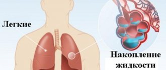
Aortic aneurysm
This is a pathology in which a thickening appears on the walls of the artery - something like a sac where blood accumulates. There are many reasons for the appearance of an aneurysm: atherosclerosis, trauma, inflammation, deterioration of vascular elasticity in old age, severe physical activity, hypoplasia, tumor, as well as congenital pathologies and even genetic predisposition.
Rice. Aortic aneurysm
Aortic rupture can occur instantly, or it can last for several months or even years while the vascular membranes dissect. In acute cases, as a rule, doctors simply do not have time to provide timely assistance. However, the disease in its chronic form can be caught by examination by doctors.
How does the disease manifest?
Symptoms and manifestations of aortic rupture also depend on the stage. If the internal lining of a vessel is damaged, the patient feels pain, drowsiness, weakness, and blood pressure increases. Detachment of the middle wall of the aorta can be accompanied by sharp and burning pain, and blood pressure drops. When an injury occurs to the outer wall of a vessel, shock occurs, the pressure drops sharply, and the temperature rises.
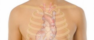
Damage to blood vessels can occur in the thoracic aorta or in the abdominal aorta - the symptoms may also differ. For example, when the abdominal aorta ruptures, a person feels shock, severe abdominal pain and weakness, vision may be blurred, and kidney failure is possible. When the thoracic aorta ruptures, severe pain is felt in the chest, shortness of breath and swelling of the neck are possible, the skin turns blue, and the veins swell.
Rice. Thoracic aortic aneurysm
How is this treated?
If suspicions arise, doctors refer the patient for angiography - this is an x-ray with contrast, which examines all parts of the aorta. However, if the inner lining of the blood vessels is damaged, this method may not produce results. In addition, the patient is given an ECG to make sure that the cause of the pain is not myocardial infarction. The most reliable method (and the most expensive) is computed tomography.
The only treatment option is prosthetic surgery using a stent graft. Unfortunately, it will not be possible to “glue” a ruptured vessel together with medication.
What is an aneurysm
This is what an arch aneurysm looks like
An aneurysm is a sac-like stretching of the aortic wall. In this case, the wall becomes thinner over time, and it grows in size. A distinction is made between thoracic aortic and abdominal aneurysms. In addition, it can form in other vessels, including the brain. After a heart attack, a cardiac aneurysm may develop.
An aneurysm can form at any age and can even be congenital. But more often the reasons for its development are atherosclerotic vascular lesions. Less commonly, injury may be the cause.
Syphilitic damage to the aorta also leads to the formation of a defect in its wall, but in our time this is rare.
Diagnostic methods
As a rule, pathology is discovered accidentally during various chest examinations: radiography or ultrasound. However, if there is a suspicion of an aortic aneurysm, the treating specialist recommends:
- x-ray examination,
- EchoCG,
- CT and MRI of the thoracic aorta.
The identified pathology requires constant monitoring, since the treatment tactics completely depend on the speed of its growth. The patient is monitored until the aneurysm significantly enlarges.
Why is an aneurysm dangerous?
As mentioned above, an aneurysm can exist for many years and not cause problems. But it's not that simple. In fact, it is an explosive with a ticking clock mechanism. Only the time is not shown. The wall can at any moment become thinner to such an extent that it cannot withstand the pressure and ruptures. In such a situation, unfortunately, no one will have time to provide medical assistance. Massive bleeding and cardiac arrest. In a situation with a ruptured aorta, it is difficult to save a person, even if at that moment he is on the operating table.
That is why it is so important to diagnose this pathology and eliminate the aneurysm in a timely manner.
Various forms of aneurysms
There is also such a thing as dissecting aortic aneurysm.
If the rupture is characteristic of the abdominal aorta, dissection is the prerogative of a thoracic aneurysm. The dissection can also lead to rupture or other complications. It all depends on which branches it will spread to.
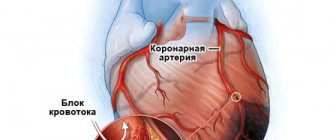
The essence of the dissection is the detachment of the inner lining of the vessel - the intima. The reasons for this are the tearing of the damaged intima and the influence of blood pressure driven under this tear. The dissection can extend either downward or upward, extending into the heart and damaging the valve and papillary muscles. If it spreads to the coronary vessels, it can lead to myocardial infarction.
The main difference between the rupture of a thoracic aortic and abdominal aneurysm is precisely the above. In the thoracic region, aortic rupture is preceded by dissection, which manifests itself with severe symptoms.
If the dissection is recognized early, there will be time for treatment and surgery to avoid rupture. An abdominal aortic aneurysm ruptures immediately and without warning.
How the disease develops
First, the wall of the vessel begins to delaminate, blood enters the gap between the layers, which contributes to even greater delamination of the tissue and rupture of the outer layers of the uveal membrane (there are three in total). The blood creates excess pressure and the outer layers stretch (aneurysm). If the integrity of the last layer is violated, an aortic rupture is diagnosed. A crack in a wall rarely lasts longer than 24 hours from the start of peeling (usually less than a few hours).
There is another scenario: the patient is treated himself. After an aneurysm (protrusion of a thin vascular wall), blood pressure flowing from the heart ruptures not the outer layer, but the intimal layer. The interlayer pressure then normalizes and the aneurysms heal spontaneously.
Types of abdominal aortic rupture
Rupture of an abdominal aortic aneurysm can occur in several scenarios:
- Into the abdominal cavity. It manifests itself as a sharp drop in pressure as a result of hemorrhagic and painful shock. Death occurs in a matter of minutes.
- Break through the peritoneum. Characterized by sharp back pain in the lumbar region. The pain is very intense and cannot be relieved with narcotic analgesics. The hematoma can compress the iliac arteries, leading to limb ischemia.
- It can break into the abdominal organs (stomach, small intestine).
- Characterized by hemorrhagic shock, vomiting blood and bloody stools.
Forecasts
Treatment of aortic rupture is exclusively surgical.
All options for the course of aortic rupture are deadly, even if the patient is in the hospital, since only a few minutes or hours remain for specialists to provide assistance. An even lower survival rate is observed among victims in whom damage to the integrity of this main artery occurs outside a medical institution.
The prognosis for aortic ruptures is always unfavorable. The probability of death from this vascular accident is 90%.
Signs of a dissecting aneurysm
Main symptoms:
- The pain is intense, localized behind the sternum, in the epigastrium, and can be between the shoulder blades and along the spine. When the dissection passes to the brachiocephalic vessels or to the vessels of the kidneys, it can spread to the head and neck, lower back and legs.
- The nature of the pain is one of the main distinguishing features. The pain is acute, sudden, sharp, and has a migrating nature.
- Vomiting, loss of consciousness, and neurological disorders are possible.
- Hemodynamic disturbances - there may be high or low blood pressure, rapid pulse.
- Signs of heart failure, shortness of breath, swelling.
- Symptoms are caused by the spread of dissection into branches and concomitant acute organ ischemia.
Schematic representation of the delamination
Symptoms may be similar to myocardial infarction, pleurisy, and mediastinal diseases.
Causes
The exact causes of thoracic aortic aneurysm are unknown, but factors that contribute to its development include:
- Marfan syndrome is a genetic disorder that affects connective tissue in the body. People born with Marfan syndrome are especially at risk of developing a thoracic aortic aneurysm. People with Marfan syndrome have weakened aortic walls, which makes them more susceptible to developing an aneurysm. People with this syndrome have specific physical characteristics: tall height, very long arms, chest deformities, and vision problems.
- Other connective tissue diseases. Ehlers-Danlos syndrome contributes to the development of thoracic aortic aneurysm. In Ehlers-Danlos syndrome, the skin, joints and connective tissue become very fragile and the skin stretches easily.
- Heart valve damage. Sometimes people with damage to the heart's aortic valve (the valve through which blood flows out of the heart) are at increased risk of developing a thoracic aortic aneurysm. This is especially true for people with bicuspid aortic valve, where the aortic valve has only two leaflets instead of three.
- Previous injuries to the aorta. If there has been previous damage to the aorta, such as a tear in the aortic wall (aortic dissection), the risk of developing a thoracic aortic aneurysm increases dramatically.
- Traumatic injury. Thoracic aortic aneurysms develop after injuries from falls or road traffic accidents.
Treatment
The resulting rupture leads to death. Therefore, the main treatment is to prevent aortic rupture.
If there is an aneurysm, how to behave:
- Avoid intense physical activity.
- Do not allow blood pressure to rise above 140/80.
- Avoid stress.
- Avoid injury.
- Be under the supervision of a doctor and follow all his recommendations.
- If possible, surgery to remove the aneurysm should be performed.
Surgical treatment of aneurysm
There are open operations in which the abdominal cavity is opened, the dilated part of the aorta is removed and a synthetic prosthesis is placed in its place. This operation is technically very complex, traumatic and lengthy. But the result is worth it.
A modern method of treating aneurysms is the installation of a special intravascular endoprosthesis. Through an incision in the thigh and through the femoral artery under X-ray control, the endoprosthesis is brought to the site of the aneurysm. Under the influence of air pumped into it through a catheter, it straightens and closes the aortic defect.
Here is such a complex stent for the spread of an aneurysm to the iliac arteries
Drug treatment
Treatment of patients with dissecting aneurysm and threat of rupture is carried out in the intensive care unit.
In case of dissection, narcotic analgesics (morphine, fentanyl) are prescribed for pain relief. The pain must be relieved. Drugs that reduce cardiac output (beta blockers) and drugs to lower and control blood pressure at a given level (Ebrantil, Enap) are also prescribed. An increase in blood pressure above 180/100 is unacceptable in this situation.
Aneurysm rupture is an extremely serious complication of vascular diseases with a high mortality rate. It is important to diagnose the pathology in time and contact experienced doctors for treatment and surgery.
Acute traumatic aortic injury: imaging and management
Acute traumatic injury of the aorta (ATI) is fatal in most cases - with such damage, about 80% of victims die before arriving at the hospital. Among the causes of death as a result of road accidents, traumatic brain injury is second only to traumatic brain injury in frequency. With improved imaging options and advances in surgical treatments and percutaneous interventions, mortality among patients with OTPA has decreased from 50% to 5%. OTPA occurs in approximately 2% of blunt chest trauma cases, approximately 70% of which occur in motor vehicle accident victims.
The exact pathogenesis of OTPA is not entirely clear, but the proposed mechanisms are a sharp increase in intravascular pressure (like a water hammer effect) and rupture of the aorta, sandwiched between the vertebral body and the sternum displaced towards the spine (Fig. 1). .
Figure 1 | Mechanisms of aortic injury in patients with blunt chest trauma
The method of choice for diagnosing OTPA is CT angiography, which replaces conventional angiography. It allows you to evaluate intimal disorders and indirect signs of aortic damage, such as periaortic hematoma. In emergency settings, performing ECG-gated CT to visualize the aorta is impractical due to lengthy setup times, the need for additional preparation, and prolonged patient breath holding. With the advent of new dual-source CT scanners with ultra-fast acquisition speeds, it has become possible to obtain virtually “still” images without the motion artifacts seen in the heart and aortic root with traditional CT scanning. With these images, radiological assessment of OTPA can be used to determine patient management and prognosis.
Next, we will describe current aspects of imaging, radiological classification, diagnosis and management of patients with OTPA, as well as recent advances that have a significant impact on the accuracy of imaging in patients with this injury.
Due to its diagnostic capabilities and availability of the method, CT angiography is the study of choice for diagnosing OTPA. The most commonly injured areas are the aortic isthmus, ascending aorta, and aortic hiatus, and arterial phase CT scans of the chest and upper abdomen should be obtained in patients with blunt trauma. Most often, CT angiography is performed as part of a general computed tomography when examining patients with polytrauma.
The advent of dual-source CT scanners and scanners with 64 or more slices has revolutionized the use of CT in cardiac and vascular imaging. These scanners have spatial resolutions ranging from 0.25 to 0.47 mm, with most new scanners providing a resolution of 0.33 mm. Also, such scanners have very fast gantry rotation, which leads to a higher speed of image acquisition - still images of even the ascending aorta can be obtained without ECG synchronization (Fig. 2). In addition, motion artifacts from the patient's breathing are minimal, which provides the necessary diagnostic image quality.
Figure 2
ECG-unsynchronized axial CT image at the level of the sinuses of Valsalva (the space between the semilunar valves and the aortic wall) obtained using a new dual-source CT scanner (A) MSCT on a 64-slice scanner in the same patient. Note the absence of ripple artifacts (indicated by arrows) in the new dual-source scanner, even without ECG synchronization.
The rationale for arterial phase imaging of the chest and upper abdomen is that most vascular injuries anatomically occur in these areas unless the patient has additional risk factors such as a pelvic fracture and therefore possible pelvic vascular injury. In addition, arterial phase imaging of the chest alone does not preclude the detection of any serious pulmonary pathologies.
A low-dose delayed phase scan of the abdomen and pelvis is performed if damage is confirmed during the portal venous phase of the study or if kidney or bladder damage is suspected. An initial noncontrast phase is useful in diagnosing intramural hematoma, but is not performed as part of the trauma protocol. The chest and abdominal CT protocol for patients with blunt trauma is shown in Table 1.
Table 1 | CT examination protocol for blunt trauma of the chest, abdomen and pelvis
In each case, sagittal and coronal 3D models are generated through images in the axial plane. Other 3D methods, including maximum intensity projection (MIP) and volume rendering (VR), are created on a separate workstation. Curved aortic reconstructions are created only in cases where stent placement is planned later during surgery. This 3D data modeling is always read in combination with raw data, which greatly assists in surgical planning.
MRI is almost never used to identify injuries in patients with blunt trauma. However, MR angiography is also used to diagnose trauma: this method is mainly used after placement of an endovascular stent or if renal failure precludes CT angiography. The MRI protocol for aortic imaging is highlighted in Table 2. If CT and MR angiography cannot be performed due to renal failure (eGFR < 30), patients are best treated with non-contrast CT for interval changes in aortic diameter.
Table 2 | Protocol of MR angiography of the aorta
Although CT angiography is the definitive modality in most cases, the initial evaluation of injury often begins with a chest x-ray. Data suspicious for OTPA with this research method include:
- mediastinal widening of 8 cm at the level of the aortic arch (or superior mediastinum, measuring approximately 25% of the width of the chest);
- uneven contour of the aortic arch;
- inability to determine the aortopulmonary window;
- darkening of the border of the lung with the aorta;
- deviation of the trachea or nasogastric tube to the right;
- omission of the left main bronchus;
- dilated left paratracheal stripe.
CT angiography has very high sensitivity (approximately 98%) and almost 100% specificity for diagnosis. Signs of OTPA on CT angiography can be divided into direct and indirect.
Indirect signs of PAAD include mediastinal or periaortic hematoma, retrocrural hematoma, or a small caliber of the aorta distal to the injury site. The location of the mediastinal hematoma is critical because if there is a clear layer of fatty tissue between the aorta and the hematoma, alternative sources of bleeding (from a vein, intercostal artery, or bone) should be considered (Figure 3). Periaortic hematoma without any obvious signs of aortic injury may be associated with occult intimal injury. Conventional angiography may not provide sufficient diagnostic imaging in patients with occult intimal injury, so additional use of intravascular ultrasound or transesophageal echocardiography is better. .
Figure 3
A 36-year-old man presented with a mediastinal hematoma and hemothorax. Sagittal (A) and coronal images (B, C) of CT angiography. A clearly defined layer of adipose tissue is identified between the descending aorta and the hematoma (indicated by arrows). This suggests that the aorta is not the source of the hematoma. The source of ongoing bleeding is the intercostal artery (arrow in image C)
Direct signs of OTPA include active contrast extravasation, rupture (or traumatic pseudoaneurysm), intramural thrombus, aortic displacement, and aortic contour abnormality (including sudden change in caliber, also known as pseudocoarctation). When there is a high risk of a mechanism of injury, as in a motor vehicle injury, evaluation of other associated conditions is also critical. Typically, PAH is accompanied by severe head injuries, damage to the lungs and heart, rupture of the diaphragm, intra-abdominal bleeding, and fractures of the pelvis and long bones.
The term minimal aortic injury (MAI) is an evolving concept that was introduced in 1999 by the Presley Center Aortic Trauma CT Grading System (Table 3) to describe injuries that are minor in nature.
Malhotra et al. were the first to describe this injury and defined it as a small (<1 cm) post-traumatic intimal valve with minimal or no periaortic hematoma. Most of these injuries cannot be diagnosed by other imaging modalities before routine use of CT angiography. MPA was initially found in approximately 10% of patients with OTPA, but more recent studies have shown that it occurs in approximately 25% to 35% of OTPA cases. The increase in the detection rate of MPA may be due to a number of factors, such as increased availability of CT angiography, improved CT technology, and improved vehicle safety. The descending aorta is the most common location of MPA, with the aortic isthmus being the second most common site. Although the generally accepted criterion for MPA is minimal or absent mediastinal hematoma, there is no standard finding that defines MPA. Variations in the definition of minimal or negligible aortic injury in various studies are given in Table 3.
Table 3 | Definition of the group of patients with minor/minimal aortic injuries in various studies
The use of different signs of minimal or minor aortic injury is misleading to radiologists and surgeons, as this may influence tactical decisions. Among the key features, most studies considered a “small” intimate flap, intraluminal thrombus, or intramural hematoma without any external contour abnormalities and with minimal or no periaortic hematoma as MPA (Figs. 4, 5). In most studies, an intimal flap was considered “small” if it measured less than 1 cm, as untreated intimal flaps or thrombi larger than 1 cm had a higher incidence of disease progression. .
Figure 4
Frontal (A) and sagittal (B) CT images of the chest of a 32-year-old man (jumped from the roof of a building). A tiny intraluminal thrombus (arrows) is identified in the aorta at the level of the isthmus, without external hematoma or irregular contour shape suggesting aortic damage.
Figure 5
Axial CT image of the chest in a 41-year-old man (fall from a motorcycle). A small intimal flap (indicated by a black arrow) is identified in the anterior descending thoracic aorta without external hematoma or deformation of the external contour, which corresponds to minimal damage to the aorta. We also see a fracture of the left rib (indicated by a white arrow) and bilateral accumulation of fluid in the pleural cavity.
As with the diagnosis of MPA, there is also no consensus regarding the management and monitoring of these injuries. Most studies suggest that they can be treated noninvasively by monitoring heart rate and blood pressure and anticoagulant or antiplatelet therapy. The choice of appropriate treatment strategy should be individualized, taking into account other concomitant diseases. The initial use of negative inotropes (most often beta-blockers) is suggested to achieve target blood pressure and heart rate values to reduce vascular shear stress—systolic blood pressure of 120 mmHg. Art. or an average blood pressure of 80 mm Hg. Art., as well as a heart rate less than 80 beats per minute. Second-line drugs are calcium channel blockers or arterial vasodilators. The Society of Vascular Surgery (SVS) guidelines suggest a “watchful waiting” approach with serial monitoring of the intimal condition.
Recent Eastern Association for the Surgery of Trauma (EAST) guidelines note that there is insufficient evidence to make recommendations for the conservative treatment of MPA, with more research needed. Even the SVS guidelines lack any specific recommendations on the “expected” course of the disease and follow-up protocol. Visual observation of MPA usually varies depending on the experience of each individual institution and its ongoing observations. Although a recent study from Harborview Medical Center by Heneghan et al. suggest that after conservative treatment of MPA there is no need for follow-up imaging, a previous study from the same institute in 2014, Gunn et al. suggested the use of CT angiography on day 3, 1 week, and 1 month after injury. In another study, Mosquera et al. recommended CT angiography at 1, 3, 6, and 12 months after injury, followed by annual cardiac and vascular MR scanning. Although there is no consensus regarding the timing of prophylactic testing, most investigators suggest that surveillance usually ends when the aorta returns to normal appearance. Almost all studies show that in most patients the disease does not progress, and if it does, it occurs within the first month after injury. This pattern of disease progression is also supported by experimental animal studies, which show that maximum intimal cellular activity and thickening occur 3–4 weeks after injury. If after this time there are signs of stabilization or improvement in the imaging appearance of the injury, further observation may not be necessary. Ultimately, there is a need for a prospective multicenter study using a “fixed” anatomical definition of “minor” aortic injury, in which a cohort of patients with MPA should be serially monitored with a predetermined CT frequency and duration of follow-up.
There are several different OTPA classification systems (Table 4). One of the most widely used OTPA assessments was proposed by Azizzadeh et al. (approved by the Society of Vascular Surgery) and divides injuries into 4 degrees: class 1 - intimal tears (Fig. 5), class 2 - intrathoracic hematoma or large intimal valve (Fig. 6), class 3 - pseudoaneurysm (Fig. 7, and 4 grade—free tear (Figure 9).Although this grading system provides a straightforward description of the injury, it is not a guide to treatment.
Table 4 | OTPA classifications
Figure 6
A 45-year-old man with blunt chest trauma. Axial (A), coronal (B) and sagittal (C) planes of CT angiography image. A PA is detected in the proximal descending aorta, causing an intramural hematoma in the area of the aortic arch, branches of the aortic arch (curved arrow in image C) and the descending thoracic aorta (white arrows) with thrombus (hollow arrows in images A and C).
Figure 7
A 63-year-old woman suffered as a result of a motorcycle injury. Axial (A) and sagittal (B) sections of CT scan of the chest. A small pseudoaneurysm (indicated by white arrows) is identified in the area of the aortic isthmus with a mediastinal hematoma (indicated by a hollow arrow).
Figure 8
Male, 33 years old, OTPA. Axial (A) and coronal (B) images of CT angiography with ECG synchronization. A pseudoaneurysm of the ascending thoracic aorta is visualized from the area above the sinotubular junction. The dimensions of the pseudoaneurysm are 1.4 x 0.7 cm. There are no signs of active contrast extravasation. An open arrow indicates slight bleeding from mediastinal fat.
Figure 9
A 70-year-old woman with a traumatic aortic rupture. Axial (A) and coronal (B) CT images. There are signs of complete rupture of the anterior wall of the descending thoracic aorta (indicated by an arrow), causing extravasation of contrast material (indicated by an asterisk). Note the mediastinal hematoma and bilateral fluid collections in the pleural cavities.
There are many treatment options for a patient with OTPA, such as traditional open surgery, endovascular stenting, and the choice of conservative management for some groups of patients. Thus, evaluation of CT angiography of the OTPA is highly valuable for treatment planning. In addition to information about the severity and type of injury, CT findings should include a description of the aortic arch anatomy, signs of vertebral artery dominance, any significant pre-existing atherosclerotic disease or stenosis, previous postoperative changes (such as coronary artery bypass graft), length of aortic injury, greater aortic diameter and below the injury, as well as the proximity of the left subclavian artery (in case of injury to the aortic isthmus). This information helps the doctor choose treatment options and type of surgery. For example, surgical repair of the aorta typically involves a left posterolateral thoracotomy, often requiring the patient to be placed on a heart-lung machine. Imaging helps the surgeon determine the exact area of injury and provides information about other injuries that can be treated using the same approach.
A few additional important radiological findings to consider when planning endovascular stenting are the presence of any focal lesion or thrombus, information about the proximal and distal stent site (preferred length is 2 cm), the presence of calcification in the stent site, aortic tortuosity and diameter of access vessels.
There are differing opinions regarding the best management option for patients with this type of injury. Studies have shown that the increased use of endovascular stents among patients requiring surgical treatment has led to a decrease in the incidence of early complications and shorter hospital stays. Recent EAST guidelines recommend the use of endovascular grafting in patients who have no contraindications to the procedure.
The guidelines also suggest that in hemodynamically stable patients, definitive treatment with OTPA should be delayed until other acute life-threatening injuries have resolved. Early treatment with OTPA may be associated with a higher incidence of paraplegia and renal failure. But if the patient is hemodynamically unstable and there is direct evidence of aortic injury on CT, urgent treatment of the aortic injury is necessary. The proposed management algorithm for OTPA in patients with blunt trauma, based on the recommendations of the EAST Practice Work Group, is shown in Table 5.
Table 5 | Tactics for OTPA, based on the recommendations of the Eastern Association of Surgeons and Traumatologists (EAST practice work group)
Analysis of a national inpatient sample by Ultee et al. showed that treatment using endovascular methods (TEVAR - thoracic endovascular aortic repair) increased from 6.5% of surgical volume in 2005 to 87% in 2011. Overall mortality from aortic trauma has also decreased, due to the use of TEVAR, expanded indications for use, improved surgeon skills, and increased technical capabilities. Aortic injury, relatively common in younger age groups, presents many unique challenges related to its management and outcome. The average aortic size in this population is much smaller than in aneurysmal disease, and therefore early experience with endovascular aortic repair (EVAR) encountered problems in determining graft size. The arch geometry in younger patients is also more acute, which is associated with poor compatibility of the endograft with the aortic arch. A 0–18% rate of infolding, migration, stent collapse, and even acute aortic occlusion requiring reintervention has been reported, with most complications occurring early after surgery. Aortic remodeling in these patients may also differ in that the aorta distal to the left subclavian artery dilates at a faster rate than the proximal segments. In addition to technical advances, the use of procedural modifications, such as limiting excess graft size to 10% to accommodate subsequent remodeling, has been proposed. The influence of these factors on long-term results requires further study.
With hybrid operating rooms becoming more common, parallel advances in imaging such as cone beam CT, fusion imaging, and intravessel ultrasound (IVUS) are also facilitating operative and interventional procedures by minimizing radiation exposure and use of contrast agents. This, along with better image quality, is critical to the technical success of complex endovascular aortic procedures. Better planning software and fusion visualization are helpful for accurate graft deployment and this can be further confirmed by IVUS. The use of IVUS in aortic procedures improves understanding of complex anatomy and has been shown to reduce the amount of contrast used and overall operative time.
The patient with blunt trauma requires a multidisciplinary approach to treatment. Because OTPA is a life-threatening condition with almost always unpredictable outcomes, multidisciplinary teams are needed to rapidly assess, triage, and manage patients suspected of having OTPA (based on physical examination, external or CT findings, or known prior history). This group includes specialists in cardiovascular imaging, interventional radiology, cardiac surgery, vascular and endovascular surgery, cardiology, and anesthesiology and resuscitation. The team should provide expert judgment for all acute situations and discuss various options for approaches to the treatment of OTPA, including the use of endovascular techniques, minimally invasive and open surgery. In the researchers' experience, it is the interdisciplinary team approach to the management of patients with OTPA that is critical for appropriate treatment.
Sources:
- Fox N, Schwartz D, Salazar JH, et al. Evaluation and management of blunt traumatic aortic injury: a practice management guideline from the Eastern Association for the Surgery of Trauma. J Trauma Acute Care Surg. 2015;78(1):136–46.
- Neschis DG, Scalea TM, Flinn WR, Griffith BP. Blunt aortic injury. N Engl J Med. 2008;359(16):1708–16.
- Heneghan RE, Aarabi S, Quiroga E, Gunn ML, Singh N, Starnes BW. Call for a new classification system and treatment strategy in blunt aortic injury. J Vasc Surg. 2016;64(1):171–6.
- Ungar TC, Wolf SJ, Haukoos JS, Dyer DS, Moore EE. Derivation of a clinical decision rule to exclude thoracic aortic imaging in patients with blunt chest trauma after motor vehicle collisions. J Trauma. 2006;61(5):1150–5.
- Steenburg SD, Ravenel JG, Ikonomidis JS, Schonholz C, Reeves S. Acute traumatic aortic injury: imaging evaluation and management. Radiology. 2008;248(3):748–62.
- Nagpal P, Khandelwal A, Saboo SS, Bathla G, Steigner ML, Rybicki FJ. Modern imaging techniques: applications in the management of acute aortic pathologies. Postgrad Med J 1078;2015(91):449–62.
- Seimens Healthineers USA
- Starnes BW, Lundgren RS, Gunn M, et al. A new classification scheme for treating blunt aortic injury. J Vasc Surg. 2012;55(1): 47–54.123
- Lamarche Y, Berger FH, Nicolaou S, et al. Vancouver simplified grading system with computed tomographic angiography for blunt aortic injury. J Thorac Cardiovasc Surg 2012;144(2):347–54.
- Rogalla P, Kloeters C, Hein PA. CT technology overview: 64-slice and beyond. Radiol Clin North Am. 2009;47(1):1–11.
- Voitle E, Hofmann W, Cejna M. Aortic emergencies-diagnosis and treatment: a pictorial review. Insights Imaging. 2015;6(1):17–32.
- Nagpal P, Saboo SS, Khandelwal A, Duran-Mendicuti MA, Abbara S, Steigner ML. Traumatic right atrial pseudoaneurysm. Cardiovasc Diagnosis Ther. 2015;5(2):141–4.
- Soto JA, Anderson SW. Multidetector CT of blunt abdominal trauma. Radiology. 2012;265(3):678–93.
- Fishman EK, Ney DR, Heath DG, Corl FM, Horton KM, Johnson PT. Volume rendering versus maximum intensity projection in CT angiography: what works best, when, and why. Radiographics. 2006;26(3):905–22.
- Sammer M, Wang E, Blackmore CC, Burdick TR, Hollingworth W. Indeterminate CT angiography in blunt thoracic trauma: is CT angiography enough? AJR Am J Roentgenol. 2007;189(3):603–8.
- Fabian TC, Richardson JD, Croce MA, et al. Prospective study of blunt aortic injury: Multicenter Trial of the American Association for the Surgery of Trauma. J Trauma.1997;42(3):374–80 (discussion 80–83).
- Kram HB, Appel PL, Wohlmuth DA, Shoemaker WC. Diagnosis of traumatic thoracic aortic rupture: a 10-year retrospective analysis. Ann Thorac Surg. 1989;47(2):282–6.
- Gavant ML. Helical CT grading of traumatic aortic injuries. Impact on clinical guidelines for medical and surgical management. Radiol Clin North Am. 1999;37(3):553–74.
- Malhotra AK, Fabian TC, Croce MA, Weiman DS, Gavant ML, Pate JW. Minimal aortic injury: a lesion associated with advanced diagnostic techniques. J Trauma.2001;51(6):1042–8.
- Forman MJ, Mirvis SE, Hollander DS. Blunt thoracic aortic injuries: CT characterization and treatment outcomes of minor injury. Eur Radiol. 2013;23(11):2988–95.
- Gunn ML, Lehnert BE, Lungren RS, et al. Minimal aortic injury of the thoracic aorta: imaging appearances and outcome. Emerg Radiol. 2014;21(3):227–33.
- Paul JS, Neideen T, Tutton S, et al. Minimal aortic injury after blunt trauma: selective nonoperative management is safe. J Trauma. 2011;71(6):1519–23.
- de Mestral C, Dueck A, Sharma SS, et al. Evolution of the incidence, management, and mortality of blunt thoracic aortic injury: a population-based analysis. J Am Coll Surg. 2013;216(6):1110–5.
- Osgood MJ, Heck JM, Rellinger EJ, et al. Natural history of grade I–II blunt traumatic aortic injury. J Vasc Surg. 2014;59(2):334–41.
- Kidane B, Abramowitz D, Harris JR, DeRose G, Forbes TL. Natural history of minimal aortic injury following blunt thoracic aortic trauma. Can J Surg Journal canadien de chirurgie.2012;55(6):377–81.
- Mosquera VX, Marini M, Lopez-Perez JM, et al. Role of conservative management in traumatic aortic injury: comparison of long-term results of conservative, surgical, and endovascular treatment. J Thorac Cardiovasc Surg. 2011;142(3):614–21.
- Erbel R, Aboyans V, Boileau C, et al. 2014 ESC Guidelines on the diagnosis and treatment of aortic diseases: document covering acute and chronic aortic diseases of the thoracic and abdominal aorta of the adult. The Task Force for the Diagnosis and Treatment of Aortic Diseases of the European Society of Cardiology (ESC). Eur Heart J 2014;35(41):2873–926.
- Lee WA, Matsumura JS, Mitchell RS, et al. Endovascular repair of traumatic thoracic aortic injury: clinical practice guidelines of the Society for Vascular Surgery. J Vasc Surg. 2011;53(1):187–92.
- Neville RF, Padberg FT Jr, DeFouw D, Hernandez J, Duran W, Hobson RW 2nd. The arterial wall response to intimal injury in an experimental model. Ann Vasc Surg 1992;6(1):50–4.
- Azizzadeh A, Keyhani K, Miller CC 3rd, Coogan SM, Safi HJ, Estrera AL. Blunt traumatic aortic injury: initial experience with endovascular repair. J Vasc Surg. 2009;49(6):1403–8.
- Forcillo J, Philie M, Ojanguren A, et al. Outcomes of traumatic aortic injury in a primary open surgical approach paradigm. Trauma Mon. 2015;20(2):e18198.
- Fanelli F, Dake MD. Standard of practice for the endovascular treatment of thoracic aortic aneurysms and type B dissections. Cardiovasc Intervent Radiol. 2009;32(5):849–60.
- Ultee KH, Soden PA, Chien V, et al. National trends in utilization and outcome of thoracic endovascular aortic repair for traumatic thoracic aortic injuries. J Vasc Surg.2016;63(5):1232–9.
- Forbes TL, Harris JR, Lawlor DK, Derose G. Aortic dilatation after endovascular repair of blunt traumatic thoracic aortic injuries. J Vasc Surg. 2010;52(1):45–8.
- Farber MA, Mendes RR. Endovascular repair of blunt thoracic aortic injury: techniques and tips. J Vasc Surg. 2009;50(3):683–6. 36. Kolbel T, Dias N, Resch T, Holst J, Sonesson B, Malina M. In situ bending of thoracic stent grafts: clinical application of a novel technique to improve conformance to the aortic arch.
- J Vasc Surg. 2009;49(6):1613–6.
- Neschis DG, Moainie S, Flinn WR, Scalea TM, Bartlett ST, Griffith BP. Endograft repair of traumatic aortic injury—a technology in evolution: a single institution's experience. Ann Surg. 2009;250(3):377–82.
- Stangenberg L, Shuja F, Carelsen B, Elenbaas T, Wyers MC, Schermerhorn ML. A novel tool for three-dimensional roadmap- ping reduces radiation exposure and contrast agent dose in complex endovascular interventions. J Vasc Surg. 2015;62(2):448–55.
- Song TK, Donayre CE, Kopchok GE, White RA. Intravascular ultrasound use in the treatment of thoracoabdominal dissections, aneurysms, and transections. Semin Vasc Surg. 2006;19(3):145–9.
- Guo BL, Shi ZY, Guo DQ, et al. Effect of intravascular ultrasound-assisted thoracic endovascular aortic repair for ''complicated'' type B aortic dissection. Chin Med J (Engl). 2015;128(17):2322–9.
- Dijkstra ML, Eagleton MJ, Greenberg RK, Mastracci T, Hernandez A. Intraoperative C-arm cone-beam computed tomography in fenestrated/branched aortic endografting. J Vasc Surg. 2011;53(3):583–90.
- Bashore TM, Bates ER, Berger PB, et al. American College of Cardiology/Society for Cardiac Angiography and Interventions Clinical Expert Consensus Document on cardiac catheterization laboratory standards. A report of the American College of Cardiology Task Force on Clinical Expert Consensus Documents. J Am Coll Cardiol. 2001;37(8):2170–214.
- Alberta HB, Secor JL, Smits TC, et al. Comparison of thoracic aortic diameter changes after endograft placement in patients with traumatic and aneurysmal disease. J Vasc Surg. 2014;59(5):1241–6.
- Watson J, Slaiby J, Garcia Toca M, Marcaccio EJ Jr, Chong TT. A 14-year experience with blunt thoracic aortic injury. J Vasc Surg. 2013;58(2):380–5.
- Fearn S, Lawrence-Brown MM, Semmens JB, Hartley D. Follow-up after endovascular aortic aneurysm repair: the plain radiograph has an essential role in surveillance. J Endovasc Ther. 2003;10(5):894–901.
- Riesenman PJ, Brooks JD, Farber MA. Acute blunt traumatic injury to the descending thoracic aorta. J Vasc Surg. 2012;56(5):1274–80. 47. Estrera AL, Miller CC 3rd, Guajardo-Salinas G, et al. Update on blunt thoracic aortic injury: fifteen-year single-institution experience. J Thorac Cardiovasc Surg. 2013;145(3 Suppl):S154–8. 48. Rabin J, DuBose J, Sliker CW, O'Connor JV, Scalea TM, Griffith BP. Parameters for successful nonoperative management of traumatic aortic injury. J Thorac Cardiovasc Surg. 2014;147(1):143–9.
Risk factors
Risk factors for developing thoracic aortic aneurysm include:
- Age. Pathologies most often occur in people aged 60 years and older.
- Tobacco use. The longer you smoke or chew tobacco, the greater the risk.
- High blood pressure: Hypertension damages blood vessels in the body, increasing the risk of developing an aneurysm.
- Plaque deposition in the arteries (aortic atherosclerosis). Atherosclerosis damages the mucous membrane of the blood vessel, increasing the risk of developing aneurysmal dilatation.
- Male gender. Men are more likely than women to develop aortic aneurysms. However, women with aneurysms are at higher risk of rupture than men.
- Race. Aortic aneurysms are more common in white people than in other races.
- Family history. People with a family history of aortic aneurysm are at increased risk of developing one. In people with a family history of aneurysms, they develop at a younger age and are more likely to rupture.
Replacing parts
Conservative treatment is used only in case of serious contraindications to surgical intervention (old age, presence of concomitant systemic diseases, etc.). The most commonly used surgical methods.
Implantation of a synthetic prosthesis. The artificial vessel has good biocompatibility and can exist in the body for an unlimited time. Carrying out this operation requires the surgeon to have the highest professionalism and mastery of technique. It is most difficult to operate on thoracic and thoraco-abdominal aneurysms, since these rather lengthy and complex surgical procedures are carried out under conditions of artificial circulation. Abdominal surgery is performed for all types of aneurysm, but a less traumatic method of treatment can be used on the abdominal aorta.
Stenting method. By puncture of the femoral artery, a metal stent covered with a special fabric is inserted into the aorta. Opening in the aorta, it strengthens the walls of the vessel and protects it from rupture. After surgery, the patient must maintain normal blood pressure at all times and avoid excessive strain.
Surgical interventions
Surgical treatment is recommended for aneurysm sizes of 5.6 cm or more. If you have Marfan syndrome, other connective tissue diseases, or a family history of an aortic aneurysm, your doctor may recommend surgery for even small aneurysms.
Depending on your condition and the location of the thoracic aortic aneurysm, your doctor will recommend:
- Open chest surgery. An operation to open the chest to treat a thoracic aortic aneurysm involves removing the damaged section of the aorta and replacing it with a synthetic tube (prosthesis), which is sewn in place of the removed section. This procedure requires opening the abdomen or chest, takes more than 4 hours and takes several months to fully recover.
- Endovascular intervention. Through a small incision in the groin area, a special stent graft is inserted into the area of the aortic expansion. After which the stent-graft is opened, its position is assessed and secured. The aortic stenting operation is performed in an angiographic operating room, lasts 1 – 1.5 hours and the recovery period takes approximately 1.5-2 weeks.
- Other heart surgeries. If you have a medical condition that contributes to the formation of an aneurysm, such as damage to the heart valves, your doctor will recommend additional surgeries to repair or replace damaged valves to prevent the aneurysm from getting worse.
Preparing for a doctor's appointment
If you suspect you have a thoracic aortic aneurysm or are concerned about your risk of developing an aneurysm due to your family history, make an appointment with your doctor at a vascular clinic. If an aneurysm is detected early, treatment may be easier and more effective.
Because most thoracic aortic aneurysms are discovered during a routine medical examination or during an examination for another reason, there is no need to make special preparations for your visit to the doctor. If you are being evaluated for an aortic aneurysm, your doctor will likely ask if anyone in your family has had an aortic aneurysm, so be prepared to answer this question. Be prepared to discuss your diet, physical activity and bad habits. If you are not following a diet or exercise routine, be prepared to talk to your doctor about problems you may encounter when starting treatment. Be sure to tell your doctor if you smoke or have quit smoking.
Search and don't give up
A dissecting aneurysm (longitudinal rupture of the aorta) is practically asymptomatic. At the time of rupture of the vessel, the patient experiences severe “tearing” pain in the chest, which is often mistaken for symptoms of another cardiovascular disease, such as myocardial infarction. If prompt surgical measures are not taken, death after aortic rupture can occur within 2 hours.
In order not to lead to such a situation, you need to undergo examination on time. An ultrasound examination can detect an aneurysm even in cases where there is no dangerous dilation of the vessel yet, but it is about to appear. X-ray is very informative for an abdominal aneurysm, since it is clearly visible due to the deposition of calcium on the walls of the vessel. More detailed studies - computer and magnetic resonance imaging - are carried out after the ultrasound method. They allow you to determine the strategy for further treatment. It is believed that if the diameter of the aneurysm is less than 5 cm, the risk of rupture of the vessel is low. With larger sizes the risk increases.