The constant movement of blood through a closed cardiovascular system, which ensures gas exchange in tissues and lungs, is called blood circulation. In addition to saturating organs with oxygen, as well as cleansing them of carbon dioxide, blood circulation is responsible for delivering all necessary substances to the cells.
Everyone knows that blood can be venous and arterial. In this article you will learn through which vessels darker blood moves, and you will find out what is included in this biological fluid.
This system includes blood vessels that penetrate all tissues of the body and the heart. The process of blood circulation begins in the tissues, where metabolic processes occur through the capillary walls.
The blood, which has given up all the useful substances, flows first to the right half of the heart, and then into the pulmonary circulation. There, it, enriched with useful substances, moves to the left, and then spreads in a large circle.
The heart is the main organ in this system . It is endowed with four chambers - two atria and two ventricles. The atria are separated by the interatrial septum, and the ventricles by the interventricular septum. The weight of a human “motor” is from 250-330 grams.
The color of the blood in the veins and the color of the blood moving through the arteries are slightly different. You will learn a little later about which vessels the darker blood moves through and why it differs in shade.
An artery is a vessel that carries biological fluid saturated with useful substances from the “motor” to the organs. The answer to a fairly frequently asked question: “What vessels carry venous blood?” simple Venous blood is transported exclusively by the pulmonary artery.
The arterial wall consists of several layers, these include:
- outer connective tissue membrane;
- medium (consisting of smooth muscles and elastic hairs);
- internal (consisting of connective tissue and endothelium).
Arteries divide into small vessels called arterioles. As for capillaries, they are the smallest vessels.
The vessel that carries carbon dioxide-enriched blood from tissues to the heart is called a vein. The exception in this case is the pulmonary vein, since it carries arterial blood.
Dr. W. Harvey first wrote about blood circulation back in 1628. The circulation of biological fluid occurs through the pulmonary and systemic circulation.
The movement of biological fluid in a large circle begins from the left ventricle , due to increased pressure, the blood spreads throughout the body, nourishes all organs with beneficial substances and takes away harmful ones. Next, the transformation of arterial blood into venous blood is noted. The last stage is the return of blood to the right atrium.
As for the small circle, it begins from the right ventricle . First, the blood gives off carbon dioxide, receives oxygen, and then moves to the left atrium. Next, through the right ventricle, the flow of biological fluid into the systemic circle is noted.
The question of which vessels carry darker blood is quite common. Blood is red in color; it differs only in shades due to the amount of hemoglobin and oxygen enrichment.
Surely many remember from biology lessons that arterial blood has a scarlet tint, and venous blood has a dark red or burgundy tint. Veins located near the skin also appear red when blood circulates through them.
In addition, venous blood differs not only in color, but also in function. Now, knowing through which vessels darker blood moves, you know that its color is due to its enrichment in carbon dioxide. The blood in the veins has a burgundy tint.
It contains little oxygen, but at the same time it is rich in metabolic products. It is more viscous. This is due to an increase in the diameter of red blood cells due to the influx of carbon dioxide into them. In addition, the temperature of the venous blood is higher, and the pH is lower.
It circulates through the veins very slowly (due to the presence of valves in the veins that slow down the speed of its movement). There are many more veins in the human body compared to arteries.
Causes of arterial bleeding
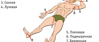
The main arteries of the human body: 1 - carotid, 2 - subclavian, 3 - axillary, 4 - femoral, 5 - brachial
Arterial bleeding occurs due to:
- traumatic injuries: thermal or mechanical;
- the presence of vascular diseases and tumors;
- existing blood clotting diseases, as well as liver diseases;
- general diseases, including: diabetes, infectious diseases, lack of vitamins and others;
- the presence of organ damage due to exposure to other diseases.
How to provide first aid for venous bleeding? 12800
Bruise after tests: why
Usually, only a small dot at the puncture site can “tell” about the blood being taken. However, in some cases, the “consequences” of the procedure are difficult to miss. What could be the reason for the formation of a hematoma after taking blood? And who is more at risk of “getting” such a bruise than others?
Ignoring recommendations after taking
The leading positions in the “formation” of bruises after tests are occupied by straightening the arm too early and removing the pressure bandage.
The fact is that the absence of a pressure factor before the formation of a full-fledged clot in the vessel leads to “leakage” of blood under the skin and the formation of a bruise.
Therefore, according to generally accepted recommendations, the bandage should not be removed for 0.5-1 hour after taking it, as well as lifting heavy objects for several hours.
Thinness and fragility of blood vessels
It may seem surprising, but this situation is not at all uncommon. And the point here is not so much in the genetic features of the structure (although this, of course, matters), but in the disruption of the synthesis of collagen fibers and the permeability of the vascular wall.
In the first case, the cause may be age-related changes, estrogen deficiency, in the second - a lack of vitamins C and P (rutin).
By the way, this vitamin deficiency is usually accompanied by bleeding gums, frequent nosebleeds and the formation of bruises even with minor exposure. And people who have such “symptoms” are more likely than others to “get” a hematoma after taking blood.
The presence of vascular pathology, as a rule, remains unnoticeable for a long time and is often a consequence of existing diseases. People with autoimmune diseases and metabolic diseases (diabetes mellitus, gout and others) are at increased risk for “bruising”.
Bleeding disorders
This cause of hematoma obviously requires attention and treatment.
Unpopular, but true - most coagulation factors (as well as anticoagulants) are formed in the liver. Impaired liver function can lead to insufficient synthesis of coagulation factors.
The size of the hematoma, at the same time, often reaches large sizes, and bleeding from the wound does not stop for a long time. This situation threatens the development of internal bleeding, and the level of greatest importance here is:
- INR
- prothrombin,
- APTT,
- fibrinogen,
- antithrombin III.
You can also comprehensively assess blood coagulation using the “Coagulogram. Expanded”, which allows, in addition to the risk of bleeding, to assess the risk of thrombosis.
A slight “bruising” can result from a decrease in platelets.
These cells are not associated with liver health, but are sensitive to many drugs (for example, “gastric” ranitidine, or the anti-epileptic carbamazepine), as well as various infections (Epstein-Barr virus, hepatitis C and others).
A threat of bleeding is considered to be a decrease below 50 x 109, and a “routine” general blood test can check platelet levels.
Thus, the appearance of a large bruise after taking blood is not at all a normal option and requires attention to exclude serious diseases if this situation recurs.
Source
What is arterial bleeding, how to determine it?
Arterial bleeding is a disorder in which arterial blood leaks out. This phenomenon is associated with various types of traumatic factors.
Arteries are vessels that carry blood from the heart to the internal organs. Their walls are thick and strong, the blood flowing through them is saturated with oxygen and moves under high pressure. The color of arterial blood is bright scarlet, so in most cases bleeding can be determined by this sign alone. In addition, there are a number of symptoms - specific signs of arterial bleeding.
Characteristic signs of arterial bleeding
Scarlet arterial blood pours out of the wound under high pressure. Doctors who arrive to help often record the following clinical picture: the patient quickly loses blood, which is released from the wound under pressure in a pulsating stream in time with the heart contractions. Measures to stop bleeding must be taken immediately.
In case of severe blood loss, the patient's condition deteriorates sharply. Doctors note the following symptoms:
- Stopping arterial bleeding - signs and first aid
- paleness of the skin;
- confusion;
- fainting;
- The pulse is practically not palpable.
Indirect signs of arterial bleeding
External arterial bleeding is easy to diagnose, unlike internal bleeding.
With this type of disorder, doctors pay attention to indirect signs of arterial bleeding, which include:
- weakness;
- disturbance of consciousness;
- severe nausea;
- uncontrollable vomiting;
- dizziness;
- pallor of the skin.
To accurately determine the location of bleeding and provide medical care, it is necessary to determine the location of the damaged arterial vessel. The nature and algorithm for providing first emergency aid will directly depend on this. This plays an important role in stopping bleeding.
Venous and arterial bleeding - differences
Venous and arterial bleeding have many similarities. Their main difference is the type of damaged vessel. As noted, arteries deliver blood to the body’s organs from the heart, veins deliver blood in the opposite direction: from tissues and organs to the heart. Venous blood flows through them. It has a dark red color due to the presence of high concentrations of carbon dioxide in it. With arterial bleeding, the blood is scarlet in color.
Other differences between venous and arterial bleeding include:
- Pressure in the vessel:
the pressure in the artery is higher, so when it is injured, blood flows out in a stream, a “fountain”. - Rate of blood loss:
When an artery is damaged, blood flows out faster and is more difficult to stop. - Volume of blood loss:
with venous bleeding and timely provision of medical care, the patient rarely loses more than 500 ml of blood, with arterial bleeding - about 1 liter or more.
Study Information
Total bilirubin is one of the pigments (orange-yellow) of blood and other biological fluids (bile, urine, etc.).
It is a breakdown product of hemoglobin. Hemoglobin is the main part of erythrocytes (red blood cells). Its function is to deliver oxygen to tissues from the respiratory organs and reverse transport of carbon dioxide. As a result of the breakdown of indirect (unbound) bilirubin is formed , which is then released into the circulating blood. Liver cells "capture" indirect (unbound) bilirubin from the blood, bind to another metabolic component (glucuronic acid) and convert it into direct , or conjugated, bilirubin . The attached glucuronic acid gives bilirubin the ability to dissolve in liquid, which allows it to dissolve in bile, after which, in its composition, it is first excreted into the intestine and then removed from there along with feces.
Together, both fractions make up the total bilirubin. Based on the concentration of total bilirubin and the percentage of each fraction, one can judge the state of human health. The blood serum of a healthy person should contain from 3 to 19 mmol/l of total bilirubin, and the ratio of free to bound bilirubin should be 3:1.
Bilirubin test
The reason for checking the concentration and fractional composition of bilirubin in the blood can be different indications, including:
- diagnosis of jaundice;
- liver diseases;
- cholestasis;
- hemolytic anemia.
Ways to stop arterial bleeding
The integrity of the artery can be completely restored only in stationary conditions by suturing the vessel. Until the patient is hospitalized, methods of temporarily stopping arterial bleeding are actively used. Depending on the situation and conditions, various techniques can be used to save the victim’s life.
Among the effective techniques:
- Pressure of a large artery.
This kind of first aid for injuries that occur can completely stop blood loss. It is used for small lesions, when it is possible to quickly hospitalize. - Application of a tourniquet.
A tight rubber bandage helps to completely stop blood circulation in the artery, resulting in stopping bleeding. In case of arterial bleeding, a tourniquet is applied above the wound site. - Pressure bandage.
A temporary method of stopping bleeding, effective for minor arterial injuries, used in the prehospital stage.
- Types of bleeding and first aid for them. This is important for everyone to know! IN
What kind of blood is in the pulmonary vein?
The pulmonary arteries ensure the flow of oxygenated blood into the aorta and its further circulation throughout the systemic circulation. The pulmonary vein returns some of the oxygenated blood to the heart to nourish the heart muscle. It is called a vein because it supplies blood to the heart.
- Arterial and venous blood: how do they differ in humans?
Types of bleeding
Experts distinguish two groups of bleeding: according to the type of vascular damage and according to external signs.
The first group includes the following bleeding:
- Arterial. The most dangerous type of bleeding because a large amount of blood can be lost in a short time.
- Venous bleeding. It is characterized by a lower rate of hemorrhage. Less dangerous than the previous type, however, if the vessels of the neck are damaged, death may occur due to the possible absorption of air.
- Capillary bleeding. It can often be observed with minor injuries such as abrasions, cuts and scrapes. It is characterized by slight bleeding, which does not pose any threat to life.
- Mixed bleeding. This type is characterized by the presence of signs of both arterial, venous and capillary hemorrhage. For example, mixed bleeding can be observed when a limb is torn off. It is very dangerous because there is arterial bleeding.
Based on external signs, bleeding is divided into the following:
- External. With it, as a rule, skin damage of varying degrees is observed.
- Internal. May be due to blunt trauma to various parts of the body, such as the chest and abdomen. In such situations, damage to human internal organs occurs. The main signs that indicate internal bleeding are weakness, thirst, dizziness, loss of consciousness, nausea, sometimes vomiting, skin changes, and low blood pressure.
Discharge color and disease
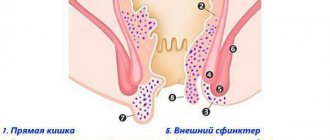
- bright red, scarlet blood from the anus on toilet paper or underwear, drops at the end of a bowel movement (stool) - hemorrhoids or anal fissure;
- red color of blood during anal bleeding - cancerous tumor, intestinal polyp;
- dark blood clots - tumors of the distal colon, diverticulosis;
- cherry-colored blood from the anus - pathology of the colon;
- black, tarry stools - diseases of the stomach, duodenum and small intestine.
IMPORTANT!
Bleeding is a serious symptom, after the appearance of which you cannot postpone a visit to the doctor. Unfortunately, blood from the anus can cause diseases such as a tumor of the rectum or colon. And in the worst case, this tumor may turn out to be malignant. Blood can also occur as a result of injury to a polyp - a benign tumor. Long-term polyposis can be a sign of colon cancer.
Pressure of arteries
In order not to get confused in case of bleeding and quickly find a suitable place to press the vessel, you should remember the following points of the body.
- Inguinal fold – in case of leakage from the thigh vessel.
- Popliteal region - with bleeding from the artery of the leg.
- The axillary region, the inner side of the biceps muscle - for the damaged arm vessel.
- The area on the neck (inner edge of the sternoclavicular muscle) - when flowing from the carotid artery.
- Supraclavicular surface - to stop bleeding from the subclavian vessel.
Menstrual cycle and vaginal bleeding - what's the difference?
The content of the article
The amount of blood during menstruation, its consistency and color vary individually, as does the length and frequency of the cycle. The color of menstruation can be very different - bright red, brown, dark and even black. On average, each adult woman loses 20-60 ml of blood during one menstrual cycle, which is from 4 to 12 teaspoons. The normal color of discharge is bright red.
Some women experience scanty bleeding, while others may have profuse bleeding, causing discomfort and the need for additional protection in the form of various hygiene products. Sometimes even clots are observed in the blood.
Clotting during menstruation is usually natural, especially when blood loss is significant. From time to time, small fragments resembling scales appear in menstrual blood.
Any change in color should be differentiated primarily from possible bleeding, which may be caused by diseases of the uterus, ovaries and vagina, as well as abnormal discharge. Strange periods may turn out to be bleeding. Any strange symptoms should be a warning sign and prompt the woman to visit a gynecologist.
Bleeding can last up to eight days, but the average duration is five days. In the first two days, the discharge is the most abundant. A typical symptom of menstruation is menstrual pain in the lower abdomen. Not all women experience pain in the ovaries, and usually this symptom also precedes the onset of menstruation.
Treatment
All diseases in which bloody stools are observed require medical attention. If this symptom is detected, you must contact a proctologist or surgeon and undergo an examination. Based on the results obtained, the doctor will prescribe treatment.
Surgical
If necessary, doctors at the Yauza Clinical Hospital perform endoscopic, laparoscopic or abdominal operations to remove tumors, ligate (ligate) bleeding vessels, and laser coagulate them.
Conservative
Conservative treatment at the Yauza Clinical Hospital may include:
If blood appears in the stool due to hemorrhoids or fissures, then conservative treatment methods are often sufficient. Drug therapy is aimed at eliminating pain and spasm of the sphincter. Medicines that normalize stool are also prescribed. The need for enzymatic support arises only in case of lactose deficiency and allergy to cow's milk proteins.
References
- Poluektov, M.G., Buzunov, R.V., Verbitsky, E.V. and others. Draft clinical guidelines for the diagnosis and treatment of chronic insomnia in adults, Consilium Medicum. Neurology and Rheumatology, 2016. - V. 2. - pp. 41–51.
- Bhattacharya, S., Patel, K., Dehari, D. et al. Melatonin and its ubiquitous anticancer effects. Mol Cell Biochem., 2021. - Vol. 462(1-2). — P. 133-155.
- Cipolla-Neto, J., do Amaral, F. Melatonin as a Hormone: New Physiological and Clinical Insights, Endocrine Reviews, 2021. - Vol. 39(6). — P. 990-1028.