A heart attack is not a diagnosis, but a variant of the course of one of the diseases of the cardiovascular system with temporary manifestations. The paroxysmal nature of the disease is always dangerous, since the symptoms appear suddenly, do not depend on the time and location of the patient, and significantly complicate the provision of medical care.
Severe attacks can occur in a passerby on the street, in a summer cottage, or in a store. The life of the victim depends on how familiar those around him are with the rules of first aid. Because, in any case, such manifestations serve as a signal of serious trouble in the condition of the heart.
The clinical picture is not always typical. Sometimes manifestations of osteochondrosis of the thoracic spine, muscle pain due to intercostal neuralgia, diaphragmatic hernia, gastric ulcer, acute pancreatitis or cholecystitis are masked as heart attacks.
Definition:
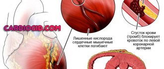
A heart attack is a condition that poses a serious threat to human life and health. A heart attack, also called a myocardial infarction, can be a fatal event. According to statistics, heart attacks are the most common cause of death and disability among the population in developed countries. A heart attack occurs when a blood clot blocks blood flow in a coronary artery (the vessel that supplies blood to the heart muscle). Interrupted blood flow to the heart can lead to damage and death of the heart muscle.
What is a “silent heart attack”?
In people over 75 years of age and with diabetes, a “silent heart attack” may occur, which is not accompanied by pain at all.
Experts estimate that approximately one fifth of such heart attacks are not diagnosed. Damage to the heart muscle in survivors of a “silent heart attack” progresses because it is not detected and treated. Abdominal fat destroys the heart
The maximum permissible waist circumference for women is 94 centimeters, for men – 102 centimeters. Exceeding these values increases the risk of cardiovascular disease. Find out more about the dangers of visceral (intra-abdominal) fat and how to get rid of it.
Causes
A heart attack is an acute condition that is accompanied by a sudden restriction of blood supply to the heart muscle. This happens with a disease associated with insufficient blood flow in the coronary vessels - coronary heart disease. The root cause is neoplasms and infections that lead to changes in the vascular walls. In most cases, heart attacks are caused by atherosclerosis of the heart vessels. This chronic disease affects large-caliber vessels: cholesterol deposits accumulate on their walls, which over time form dense plaques. They impede blood flow, causing oxygen deficiency in organ tissues. In addition, separated fragments of atheromatous plaque cause occlusion or stenosis. Atherosclerosis leads to the development of threatening conditions: acute coronary syndrome, heart attacks, strokes. Read more about the causes of a heart attack in the following publication.
How to recognize a heart attack?
People put off seeing a doctor because they confuse the symptoms of a heart attack with non-serious illnesses (such as digestive problems). They try to tolerate their symptoms and end up getting medical help too late. Treatment of myocardial infarction has changed dramatically in recent years. It is important to recognize the symptoms immediately and see a doctor if you suspect you are having a myocardial infarction.
Seek help immediately if pain in the heart and chest (angina attack) lasts more than 10 minutes and does not go away either at rest or after taking nitroglycerin. Pain (or obvious discomfort) may move to the shoulder, shoulder blades, neck or jaw. In such a situation, the patient is taken to a specialized cardiology clinic, where there is everything necessary to conduct a full examination and provide emergency care. Emergency cardiology specializes in such conditions.
When to think about a heart attack
It’s good when adults know that the threat of death from myocardial infarction, unfortunately, is not decreasing. The possibility of a heart attack is indicated by an increase in angina attacks. But a sudden development of the disease is also possible if a blood clot has blocked a large vessel and a large mass of heart muscle is immediately excluded from the operating mode.
A person who has had angina pectoris will not confuse it with any other attack
Main features:
- pain is localized behind the sternum;
- the intensity is much greater than during a normal attack, patients describe them as “dagger-like, burning”;
- radiates to the left scapula, shoulder joint (as with angina pectoris);
- paleness and blueness appear on the lips;
- sticky cold sweat on forehead;
- last more than half an hour;
- their intensity does not depend on physical activity;
- the pain cannot be relieved with a nitroglycerin tablet.
Atypical manifestations are possible: fainting, nausea and vomiting, abdominal pain.
What to do if you see a person with a developing myocardial infarction?
If you encounter an unconscious person due to a suspected myocardial infarction, call an ambulance. If you are trained in first aid, begin cardiopulmonary resuscitation (CPR). This will help deliver oxygen to the body and brain.
Begin CPR with chest compressions. Press down on the chest about 5 cm with each compression at a rate of about 100 compressions per minute. If you are trained in CPR, check the person's airway and give breaths every 30 compressions. If you are not trained in CPR, continue to perform chest compressions only.
Complications
Complications of myocardial infarction are associated with myocardial damage. This damage leads to the following conditions:
- Heart rhythm disturbances (arrhythmias). Types of arrhythmia are bradycardia and tachycardia. As a result of damage to the heart muscle due to a heart attack, electrical instability of the myocardium develops. It manifests itself as an abnormal heart rhythm and can be serious, even fatal. In this case, the patient is advised to install a pacemaker.
- Cardiogenic shock. Another deadly condition that accompanies myocardial infarction. In severe heart attacks, the heart loses its ability to pump blood—cardiogenic shock occurs.
- Myocardial rupture. An area of the myocardium weakened by the infarction may rupture, creating a hole in part of the heart. This complication is fatal.
- Heart failure. The amount of damaged heart muscle as a result of a heart attack can be so great that the remaining part of the myocardium is not able to adequately perform its job of delivering blood to the organs and tissues of the body. Reduced blood flow to organs and tissues leads to shortness of breath, increased fatigue and swelling of the legs. Heart failure is temporary and resolves after some of the damaged myocardium (stunned myocardium) has recovered, usually within a few days or weeks. There is also a chronic condition if a large area of the myocardium is irreversibly damaged.
- Heart aneurysms. Necrotic areas of the heart muscle are replaced by connective tissue. As a result, post-infarction cardiosclerosis develops, which leads to protrusion of the heart wall - an aneurysm.
- Problems with the heart valves. If parts of the heart valve apparatus are damaged as a result of a myocardial infarction, this leads to serious or even life-threatening valvular insufficiency.
What to do
A heart attack requires emergency medical attention. If someone has a heart attack, they should immediately call 112 for emergency help before doing anything else. Act quickly - it could help save someone's life.
First aid for myocardial infarction
Myocardial infarction is a type of ischemic heart disease characterized by irreversible damage to the heart muscle as a result of deterioration of blood flow through the coronary arteries
Characteristic signs (symptoms) of a heart attack (myocardial infarction):
- • sudden (paroxysmal) pressing, squeezing, burning, aching pain in the chest (behind the sternum) lasting more than 5 minutes;
- • similar pains are often observed in the left shoulder (forearm), left shoulder blade, left half of the neck and lower jaw, both shoulders, both arms, the lower part of the sternum along with the upper abdomen;
- • lack of air, shortness of breath, severe weakness, cold sweat, nausea often occur together and sometimes follow or precede discomfort/pain in the chest;
- • it is not uncommon for these manifestations of the disease to develop against the background of physical or psycho-emotional stress, but more often with some interval after them.
Uncharacteristic signs that are often confused with a heart attack:
- • stabbing, cutting, pulsating, boring, constant aching pain for many hours and not changing its intensity in the heart area or in a specific clearly defined area of the chest
Algorithm of actions in case of a heart attack
If you or someone else suddenly has the above characteristic signs of a heart attack, even with weak or moderate intensity, which last more than 5 minutes, do not hesitate, immediately call an ambulance team. Do not wait more than 10 minutes - in such a situation it is life-threatening.
If you have symptoms of a heart attack and there is no way to call an ambulance, then ask someone to take you to the hospital - this is the only right decision. Never drive yourself unless you have no other choice.
In the most optimal scenario, if a heart attack occurs, you must act according to the following algorithm:
- • Immediately after an attack occurs, sit down (preferably in a chair with armrests) or lie in bed with the head of the bed raised, take 0.25 g of acetylsalicylic acid (aspirin) (chew the tablet, swallow) and 0.5 mg of nitroglycerin (spray one inhalation dose into the cavity mouth while holding your breath, place one tablet/capsule under the tongue, first bite the capsule, do not swallow); free your neck and provide fresh air (open the vents or windows).
- • If after 5-7 min. After taking acetylsalicylic acid (aspirin) and nitroglycerin, the pain persists, it is imperative to call an ambulance team and take nitroglycerin a second time.
- • If pain persists 10 minutes after taking the second dose of nitroglycerin, it is necessary to take nitroglycerin a third time.
- • Give the patient a sedative (motherwort or valerian). There should be silence in the room, not allowing the sick person to become nervous.
- • If after the first or subsequent doses of nitroglycerin there is severe weakness, sweating, shortness of breath, you need to lie down, raise your legs (on a bolster, etc.), drink 1 glass of water and then, as with a severe headache, do not take nitroglycerin.
- If a person with a heart attack is unconscious and there is someone nearby who knows how to provide first aid in this situation, then you need to do it immediately!
Tests and diagnostics
If you have had or are suspected of having a heart attack, your diagnosis is most often made by emergency physicians. You will be asked to describe your symptoms and your blood pressure, pulse and temperature will be taken. You will be connected to a monitor and will immediately begin to diagnose your myocardial infarction.
Healthcare professionals will listen to your heart and lungs with a stethoscope. You will be asked about your medical history and your family history of heart disease. A subsequent cardiac examination will help determine what is causing the pain.
Examinations include:
- Electrocardiogram (ECG). This is the first test that is performed to diagnose myocardial infarction. It is carried out while you are being asked about the symptoms of the disease. This test records the electrical activity of the heart using electrodes attached to the skin. Electrical impulses are recorded as a waveform and recorded on a monitor or paper. Since myocardium damaged by a heart attack does not conduct electrical impulses in the normal way, an ECG will show that a myocardial infarction has occurred or is progressing.
- Blood analysis. If myocardial infarction develops, the content of cardiac enzymes in the blood increases. Emergency room doctors will take blood samples from you to check for these enzymes.
Additional Research
If you have a heart attack, your cardiologist will begin immediate treatment efforts. Additional research:
- X-ray examination of the chest organs. A chest x-ray will help the doctor evaluate the size of your heart and the vascular pattern of your lungs and the presence of fluid in the pleural cavity.
- Echocardiography. This test is based on the emission and recording of reflected sound waves that create an image of the heart on a monitor. Echocardiography helps visualize the part of the myocardium that is damaged as a result of a heart attack and is not able to normally push blood through the vessels.
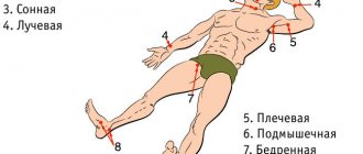
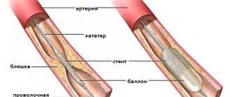
- Coronary angiography (catheterization of the coronary arteries). This type of coronary exam will show narrowing or blockage in the arteries that supply blood to the heart (coronary arteries). A liquid X-ray contrast agent fills the arteries supplying blood to the heart through a long, thin catheter. It is at the mouth of the coronary arteries through the femoral artery (in the groin area) or the radial artery (in the wrist area). When the arteries are filled with contrast, they become visible under X-ray guidance, revealing narrowing of the lumen and its complete blockage. In addition, since the artery is accessible, the doctor can treat coronary artery disease by performing coronary angioplasty and coronary artery stenting. During angioplasty, thin balloons are used that are delivered to the site of narrowing of the artery and inflated, expanding the lumen. In most cases, a mesh tube called a stent is then placed in this area of the artery. It keeps the artery open and prevents re-stenosis of the vessel lumen in the future.
- Stress test with load. Several days or weeks after your heart attack, you may be given a stress test. A stress test diagnoses how your heart and blood vessels respond to stress. You will walk on a treadmill or pedal a bicycle ergometer while an ECG or ECHO is recorded. If the patient is not ready for exercise, he is prescribed medications that stimulate the heart. A stress test will help your doctor determine the best treatment option. If your doctor wants to see an image of your heart during exercise, he will order a myocardial scintigraphy, a test similar to an exercise stress test that uses a special radiopharmaceutical and special imaging techniques.
- Coronary computed tomography (CT) or magnetic resonance imaging (MRI). This test is used to diagnose heart disease, including damage due to myocardial infarction. During a CT scan, you will lie on a table inside a tunnel-like machine. An X-ray machine rotates in this tunnel, recording and creating a series of images of your heart and chest organs. During this test, a radiopaque contrast agent is sometimes injected to visualize the coronary arteries. During an MRI, the patient lies on a table inside a similar tube-like machine that creates a magnetic field. The magnetic field arranges the atoms of cells in a certain way, and the device registers radio waves as a result of this rearrangement, depending on the type of tissue. These signals are processed and create an image of the heart.
Classification and symptoms of angina
Depending on the symptomatic picture, there are:
- a stable type of disease that occurs in an unchanged form and requires the most serious treatment;
- unstable angina - the most dangerous, of a primary nature or each time making itself felt with new symptoms;
- angina pectoris is an invariable sign of increased physical activity;
- angina at rest has no clear cause and can manifest itself even during sleep, accompanied by a feeling of panic, suffocation, and a set of autonomic disorders.
Characteristic signs allow you to distinguish angina pectoris from other diseases of the heart muscle:
- pressing burning pain;
- recoil under the left shoulder blade, in the neck or arm;
- noticeable fluctuations in pulse and blood pressure.
Any signs of angina pectoris should be a reason to contact a specialized doctor to clarify the diagnosis and prescribe appropriate treatment.
Are you experiencing symptoms of angina?
Only a doctor can accurately diagnose the disease. Don't delay your consultation - call
Medications
- Aspirin. You will receive aspirin from emergency personnel or upon arrival at a medical facility. Aspirin reduces blood clotting and helps maintain blood flow through a narrowed vessel.
- Other antiplatelet drugs. The emergency room doctor may give you other drugs that work similar to aspirin and prevent blood clots. These are clopidogrel (Plavix), ticagrelor (Brilinta) and other drugs - platelet aggregation inhibitors.
- Thrombolytics. These drugs, also called “clot killers,” are widely used in the treatment of cardiac ischemia. They help dissolve blood clots that block blood flow in the coronary arteries. The earlier treatment with thrombolytics is started during myocardial infarction, the greater the chance of survival and the smaller the area of damaged myocardium.
- Other drugs that thin the blood. These include heparin, which makes the blood less “viscous” and less capable of dangerous blood clots. Heparin is given intravenously or subcutaneously in the first few days after myocardial infarction.
- Medicines that relieve pain. If you experience severe chest pain, you will be prescribed painkillers such as morphine. This will reduce the unpleasant symptoms of vascular diseases.
- Beta blockers. These drugs help relax the heart muscle, slow the heart rate, and lower blood pressure, making it easier for the heart to pump. Beta blockers limit damage to the heart and prevent future heart attacks.
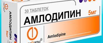
- Nitroglycerine. The drug is used to treat angina pectoris. It temporarily widens the arteries, improving blood flow to and from the heart.
- Cholesterol-lowering drugs. These are statins, niacin, fibrates and bile acid sequestrants. They help lower levels of unwanted blood cholesterol and are useful soon after a heart attack to improve survival.
Action tactics
If you suspect a heart attack, you should act immediately. First of all, you need to provide rest to the patient, stop performing any exercise, take a half-sitting or half-lying position, which will make breathing easier. You also need:
open a window or provide ventilation to the room;- free your neck, throat and chest from tight clothing;
- breathe steadily, taking deep, calm breaths in and out;
- if chest pain occurs, take a nitroglycerin tablet, placing it under the tongue (not recommended for pressure below 110/70 mmHg;
- if the pain lasts for more than 7–10 minutes, or if it intensifies or worsens, call someone for help, call an ambulance;
- for arrhythmia (rapid, uneven pulse and heartbeat), washing with cold water, a compress on the face or forehead can help;
- in case of dizziness and a state close to loss of consciousness, it is necessary to raise the position of the legs, which will ensure better blood flow to the upper body;
- if vomiting or nausea occurs, do not lie down to avoid aspiration (choking) of vomit;
- While waiting for a doctor, you should not take other medications or attempt self-medication.
Emergency care algorithm
In case of a prolonged attack of angina with chest pain or atypical symptoms lasting more than 5–15 minutes, medical intervention is necessary, otherwise there is a high risk of developing the most acute stage of a heart attack.
In the event of a heart attack, an emergency medical technician or a hospital emergency department doctor urgently provides the following measures:
If the pain syndrome persists, the patient is given one dose of Nitroglycerin, in a tablet, capsule or aerosol under the tongue. The drug is contraindicated in case of a sharp drop in blood pressure, hypotension or collapse.- Another necessary remedy is Acetylsalicylic acid (Aspirin Cardio or an analogue) in a dosage of 325 - 300 mg.
- At the same time, an electrocardiogram is taken to identify signs of myocardial ischemia (insufficient blood supply) to the heart area, confirming or refuting the diagnosis of unstable angina or heart attack. And an oxygen mask is put on to prevent hypoxia.
- In case of a heart attack with severe chest pain, the patient can be given an intravenous painkiller (Analgin 2.0 - 4.0 50% solution or Promedol, Morphine in a dosage of 1.0 - 2.0 ml 1%).
- For high blood pressure, medications to lower blood pressure are indicated: Propranolol 10-30 mg per day, or Metoprolol 50 mg.
In a hospital setting, the issue of further treatment of the patient is decided. If a heart attack is confirmed and an attack is detected in the first hour, the specialist may prescribe diagnostic coronary angiography or stenting at the Cardiology Center for Vascular Surgery. These minimally invasive surgical techniques are carried out with the aim of identifying a clear area of blockage of a heart vessel, expanding it with the help of a stent to restore patency, blood flow and nutrition of the heart muscle.
In the absence of signs of a heart attack and confirmation of unstable angina, inpatient treatment is required for a period of 10–14 days and subsequent prophylaxis to prevent complications.
Surgical methods
In addition to drug therapy, the following procedures are offered for effective treatment of myocardial infarction:
- Coronary angioplasty and stenting;
An emergency angioplasty procedure aims to open a blocked artery, allowing blood to flow freely to the myocardium. The doctor will insert a special long, thin tube (catheter) into the blocked coronary artery through an artery in the leg in the groin area or in the arm in the wrist area. Through this catheter, a special guide and balloon are brought to the affected area of the artery.
The balloon is guided through a guidewire to the affected area of the artery and inflated, facilitating the opening of the vessel. After this, a metal stent is implanted into this section of the artery - it will maintain the artery in a straightened state for a long time. It is better to implant a stent with a drug coating, which prevents restenosis - repeated narrowing of the vessel in this area. Read more about the benefits of balloon angioplasty here.
- Coronary bypass surgery;
In difficult cases, cardiac surgeons perform emergency coronary bypass surgery during a myocardial infarction. Coronary bypass surgery involves suturing a vein or artery to a coronary artery in a place after blockage or narrowing of the latter, thus restoring blood flow through the vessel.
Lifestyle changes and home rehabilitation
Your lifestyle affects your heart health. By following the recommendations for the prevention of coronary heart disease, you can not only prevent the development of myocardial infarction, but also improve your condition after it:
- Stop smoking. If you smoke, do one simple thing that will help your heart - stop smoking. Quitting smoking on your own can be difficult, so ask your doctor for a treatment plan. He will help you cope with a bad habit.
- Avoid secondhand smoke. Being around people who smoke is a potential trigger for heart attack because many of the chemicals in tobacco smoke damage arteries.
- Control your blood cholesterol levels. Get your cholesterol blood tested regularly and monitor it with your doctor. If your bad cholesterol levels are elevated, your doctor will prescribe dietary changes. He will prescribe medications that lower cholesterol and prevent vascular disease. How often you should test your cholesterol depends on how high your cholesterol levels are.
- Visit your doctor regularly and undergo preventive examinations. Some of the main risk factors for heart attack—high cholesterol, high blood pressure, and diabetes—do not appear in the early stages. Your doctor will perform tests to help identify these conditions. If a problem is detected, you and your doctor will begin treatment for early stages of cardiovascular disease. This will protect against the development of myocardial infarction in the future.
- Monitor your blood pressure levels.
- Exercise regularly. Regular exercise helps the heart muscle recover after a myocardial infarction. Exercise is an important part of a medical rehabilitation program after a heart attack. Physical activity helps prevent the onset of symptoms of coronary artery disease and the development of myocardial infarction. This is achieved by maintaining a healthy weight and controlling blood glucose levels, cholesterol levels and blood pressure. Walking for 30 minutes every day, five days a week, will improve your health.
- Maintain your weight within a healthy range. Excess weight increases stress on the heart and is associated with high cholesterol, high blood pressure and diabetes. Losing weight will reduce your risk of heart disease.
- Manage your stress. To reduce your risk of heart attack, reduce the number of stressful situations in your daily activities. Rethink your lifestyle at work and find a healthy way to minimize stressful situations in your life.
- Eat healthy. High levels of saturated animal fat and cholesterol in the diet lead to narrowing of the arteries of the heart. If you have had a myocardial infarction, limit your intake of fat, cholesterol, and salt. A diet with high salt levels increases blood pressure levels. Ask your doctor or nutritionist for advice on foods recommended for preventing atherosclerosis. Prepare healthy meals with your family members. Fish is one of the components of a healthy diet. It contains omega-3 unsaturated fatty acids, which help improve cholesterol levels and prevent blood clots. Eat enough vegetables and fruits. Fruits and vegetables contain antioxidants, components that help prevent damage to the wall of the coronary arteries.
- If you drink alcohol, consume it in moderation. Don't start drinking alcohol if you haven't done so before. Moderate alcohol consumption helps increase the level of high-density cholesterol, “good” cholesterol, and has a protective function in the development of myocardial infarction. Men are advised to drink no more than two drinks a day, and women no more than one. Excessive alcohol consumption increases blood pressure and triglyceride levels, increasing the risk of heart attack. One serving of alcohol is equivalent to 350 ml of beer, 120 ml of wine or 45 ml of spirits.
Angina pectoris
Atherosclerosis
Diabetes
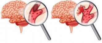
Stroke
24819 May 20
IMPORTANT!
The information in this section cannot be used for self-diagnosis and self-treatment.
In case of pain or other exacerbation of the disease, diagnostic tests should be prescribed only by the attending physician. To make a diagnosis and properly prescribe treatment, you should contact your doctor. Angina pectoris: causes, symptoms, diagnosis and treatment methods.
Definition
Angina is a clinical syndrome manifested by a feeling of tightness or squeezing pressing pain in the chest, which is most often localized behind the sternum and can radiate (“give”) to the left arm, neck, lower jaw, and to the epigastric region (epigastric region).
Pain occurs during physical activity or exposure to other factors that increase the heart's need for oxygen, and lasts from 1 to 15 minutes. It goes away with rest (when you stop exercising) or 1-3 minutes after taking nitroglycerin.
Translated from ancient Greek, angina means “narrow (weak, cramped) heart.”
Causes of angina
The development of angina is based on three mechanisms:
- atherosclerotic lesions of the coronary arteries;
- transient vascular thrombus formation;
- decreased coronary blood flow due to spasm or increased tone of the coronary arteries.
Angina pectoris makes itself felt during physical activity or stressful situations when there is a narrowing of the lumen of the coronary artery by 50-70%.
The severity of angina depends on the degree of coronary artery stenosis, its location and extent, the number and number of affected arteries. An atherosclerotic plaque can block a vessel completely or partially. With an increase in blood pressure, the inner layer of the coronary arteries (endothelium), damaged by the atherosclerotic process, can be easily damaged, blood penetrates into the plaque, the blood clotting process is activated and a blood clot is formed, which can partially or completely block the vessel.
The formation of a blood clot, especially against the background of a vessel spasm, can lead to its complete or partial blockage.
When myocardial tissue is damaged, pain mediators (serotonin, histamine, bradykinin, etc.) are released, which affect pain receptors.
There are modifiable (those that can be influenced) and non-modifiable factors that can provoke the development of angina. Modifiable diseases include dyslipoproteinemia (violation of the normal ratio of blood lipids), arterial hypertension, diabetes mellitus, smoking, low physical activity, obesity, and stress. Non-modifiable factors - male gender, age, family history of cardiovascular diseases (myocardial infarction or ischemic stroke in close relatives - up to 65 years (for women) and up to 55 years (for men)).
Classification of the disease
The most widespread classification of angina pectoris, developed
on the basis of WHO expert recommendations
:
- Stable angina pectoris (indicating functional class from I to IV).
- Class I – the patient tolerates physical activity well, angina attacks occur only during high-intensity exercise; Class II - slight limitation of usual physical activity, angina attacks occur when walking on level ground for a distance of more than 500 m, when climbing more than one floor; Class III – pronounced limitation of normal physical activity, attacks occur when walking at a normal pace on level ground for a distance of 100-150 m, or when climbing one floor; Class IV - angina occurs with little physical activity, walking on level ground at a distance of less than 100 m.
- Unstable angina:
- new-onset angina (less than 1 month from the onset of attacks);
- progressive angina (increasing attacks in frequency, duration, intensity with expanding localization and irradiation);
- early post-infarction (within 2 weeks after acute myocardial infarction) or post-operative angina.
- Spontaneous (vasospastic, variant, Prinzmetal) angina.
In practice, doctors also use the clinical classification of stable angina
:
- stable exertional angina (indicating the functional class);
- vasospastic angina;
- microvascular angina.
Symptoms of angina
Pain from angina can sometimes be perceived not as true pain, but as a feeling of discomfort, a feeling of heaviness, squeezing, tightness, distension, burning or lack of air.
Most often, the pain is localized behind the sternum or along the left edge of the sternum. It can radiate to the neck, lower jaw, teeth, interscapular space, and less often to the elbow or wrist joints, mastoid processes.
Pain with angina pectoris usually lasts from 1 to 15 minutes.
Pain occurs at the peak of physical or emotional stress. Stressful situations, due to increased activity of the sympathoadrenal system, lead to an increase in heart rate, increased blood pressure and myocardial contractility, which means the myocardial need for oxygen increases. After taking nitroglycerin or stopping the exercise, the pain stops, and the pain attack stops faster in a sitting or standing position.
As angina progresses, a moment comes when attacks occur even with minimal exertion, and then even under conditions of physical rest.
Angina at rest joins angina pectoris and is combined with it. In such cases, attacks occur during times of increased oxygen consumption by the heart muscle, for example, during REM sleep, when the heart begins to beat faster.
In some patients, an attack of angina may occur in a horizontal position due to increased blood flow to the heart.
Vasospastic angina develops independently of physical and emotional stress, is caused by spasm of the coronary arteries, and usually occurs at a younger age than exertional angina due to atherosclerosis of the coronary arteries. In patients with vasospastic angina, many typical risk factors for atherosclerosis cannot be identified. The disease may be accompanied by threatening disturbances in heart rhythm, leading to the development of myocardial infarction and/or sudden death.
A feature of vasospastic angina is very severe attacks, usually localized in a typical location. They occur at night or early in the morning, and also when exposed to cold on exposed areas of the body.
Microvascular angina is characterized by attacks that occur some time after physical activity, during emotional stress and at rest; they are poorly controlled by nitroglycerin. The cause of microvascular angina is considered to be dysfunction of small coronary arteries (100-200 μm in diameter) in the prearteriolar segment of the coronary bed. In more than 70% of cases, microvascular angina coexists with classical angina in patients with atherosclerotic stenoses.
Diagnosis of angina pectoris
The diagnosis of angina is established on the basis of a combination of complaints (pain characteristic of angina) and information received from the patient about the course of the disease.
For all patients with suspected coronary heart disease, the following questions are asked:
- current or past smoking;
- the presence of cardiovascular diseases and/or deaths from cardiovascular diseases in the patient’s immediate relatives (father, mother, siblings);
- previous cases of seeking medical help and the presence of previously registered electrocardiograms, studies and conclusions;
- the presence of concomitant diseases in order to assess additional risks;
- currently taken medications.
To clarify the diagnosis and existing complications, data from laboratory and instrumental examination methods are used.
All patients with suspected angina are recommended to have a lipid profile tested to detect dyslipoproteinemia:
- triglycerides (Triglycerides);
Patient support
Myocardial infarction is an unpleasant experience. Even if your doctor says everything is fine, you may still feel anxious. How will this affect your life? Will you be able to return to work or resume activities that bring you satisfaction? Will this happen again? Fear is one of the many experiences you and your loved ones will face. Other experiences after a heart attack include:
- Anger. You get angry and wonder, “Why did I have a heart attack and why now?” Feeling resentful after a myocardial infarction is a normal human reaction.
- Guilt. Family members feel guilty about your illness or take responsibility for actions that they believe caused the heart attack.
- Depression is common after myocardial infarction. You will feel that you are no longer able to do things that were previously acceptable, that you are no longer the same person you were before. Medical rehabilitation programs are effective in preventing and treating depression after myocardial infarction. It is important to tell your doctor about the signs and symptoms of depression.
These experiences arise most often and discussing them openly at a consultation with a cardiologist, with family members or a close friend will help to cope with them more effectively. You must take care of your physical and mental rehabilitation. Exercise and group activities with people who have also had heart attacks can help you cope with negative experiences and feelings.
What to do if you suspect a heart attack?
Call an ambulance immediately and try to calm down: you don’t need extra stress. Find a comfortable position and slowly chew one 300 mg aspirin tablet. (if there are no contraindications). Aspirin helps minimize the risk of developing a dangerous blood clot. It reduces the “stickiness” of platelets, minimizing the likelihood of them sticking together and blocking blood vessels.
Important! Don't forget to leave the door open so doctors can get to you at any time, or better yet, call a neighbor, friend or relative so you don't have to be alone. Try not to eat or drink anything until the doctors arrive.