October 15, 2012
What is blood made of? We talk about its main components - plasma, red blood cells, leukocytes and platelets.
Blood is a type of connective tissue, a distant relative of bones, cartilage and even fat. It circulates throughout the body with the help of the heart and blood vessels and has two main functions: transportation
nutrients and oxygen to cells and
protection
against infectious agents, including bacteria and viruses. What is blood made of? Let's talk about its main components - plasma, red blood cells, white blood cells and platelets.
Blood composition
Blood consists of 4 main components:
- red blood cells - red blood cells that transport oxygen from the lungs to the human organs;
- white blood cells - leukocytes responsible for fighting infections that attack the body;
- blood platelets - platelets that ensure blood clotting, thereby protecting the body from fatal blood loss due to injuries and cuts.
All these cells are suspended in blood plasma, which not only serves as a transport medium for blood cells, moving them throughout the human body, but also contains proteins and salts necessary for the body.
Hemoglobin
This is a complex protein that includes a pigment group. One third of the red blood cell consists of hemoglobin, which makes the cell red.
Hemoglobin consists of a protein - globin, and a non-protein pigment - heme, containing ferrous ion. Each hemoglobin molecule includes four hemes, which account for 4% of the total mass of the molecule, while globin accounts for 96% of the mass. The main role in the activity of hemoglobin belongs to the iron ion. To transport oxygen, heme reversibly binds to the O₂ molecule. Ferrous oxide is what gives blood its red color.
Red blood cells - erythrocytes
Red blood cells perform one of the important functions of blood. A drop of blood contains millions of red blood cells, which constantly circulate through the blood vessels, delivering oxygen to the organs and removing carbon dioxide formed during cellular respiration.
Red blood cells are called red blood cells because they contain the protein hemoglobin, which is bright red in color. It is hemoglobin that carries oxygen and carbon dioxide. As blood passes through the lungs, oxygen molecules attach to hemoglobin, which delivers it to every cell in our body. Freed from oxygen, hemoglobin attaches carbon dioxide molecules to itself. In the lungs, carbon dioxide is released and removed from the body through breathing.
The average lifespan of a red blood cell is 120 days. The bone marrow constantly produces blood cells, replenishing their natural loss.
Red pigments are helpers and protectors of photosystems of higher plants
Orange, yellow and red flamingos, vegetables, fruits and other foods high in carotenoids: pumpkin, carrots, oranges, persimmons. However, there are also a lot of orange carotenoids in the green parts of plants, leaves, and we will see this in the fall. (Photo by Luke SW, Shutterstock)
The article contains information on studies of some functions of carotenoids in higher plants. The results of laboratory experiments to study the physical and chemical properties of these pigments associated with the photosystems of chloroplasts of higher plants are presented. The participation of carotenoids in the adaptation of plants under natural growing conditions is considered.
By typing the word “carotenoids” into a search engine, you will see sets of fruits, vegetables and other tasty and bright plant products, the yellow-red color of which is provided by certain plant pigments - carotenoids (title illustration). What are the functions of these pigments in these products, we will not learn from the article under discussion *, since it is devoted to the role of carotenoids in green leaves. It is known that in the energy stations of plants - chloroplasts - the conversion of light energy into the energy of chemical bonds occurs, the latter is used by organisms to build their bodies and other life activities. The green color of leaves is provided by the main pigment of photosynthesis - chlorophyll, however, chloroplasts also contain other pigments - carotenoids, the presence of which we can observe with the naked eye in the leaves in the fall, when green chlorophyll is destroyed.
Carotenoids are hydrocarbon compounds containing a carbon “tail” with an alternating double bond (isoprene derivative, the so-called chromophore) (Fig. 1). It is the chromophore, justifying its name, that determines the absorption spectrum, i.e. pigment color ranges from yellow to red. The more conjugated double bonds in the “tail,” the more intensely colored the pigment. The authors write that to date, “600 structurally different carotenoids are known, the role of which continues to be actively studied (Karnaukhov, 1988),” although I ( KP
) this was somewhat confusing, because a recent work by Yabuzaki J. 2017 talks about 1117 natural carotenoids.
Nevertheless, there are two classes of carotenoids: carotenes
and their derivatives containing oxygen in the formula -
xanthophylls
(see Fig. 1). Enzymatic (i.e., provided by a special enzyme) oxidation of certain carotenes with atmospheric oxygen leads to the formation of three forms of oxygen-containing groups in the corresponding xanthophylls: hydroxy groups (–OH), or oxo groups (=O), or epoxy groups (–O–) between the carbon atoms of the ionone rings
Rice. 1. An example of the structural formulas of carotene and xanthophyll (zeaxanthin). (Image from studme.org/328680/geografiya/fotosinteticheskie_pigmenty)
The article examines ideas about the role of carotenoids in the work of the photosynthetic apparatus of chloroplasts: the participation of carotenoids in organizing the structure of photosystems; participation of carotenoids in the absorption of light energy; photoprotective function; violaxanthin cycle of xanthophyll transformation.
The photosystem of higher plants is a protein-pigment complex built into the thylakoid membrane of the chloroplast (Fig. 2). Carotenes take part in the functioning of the reaction center (RC) of the photosystem, and xanthophylls are located in the structures of light-harvesting complexes (LHC). The SKK proteins, encoded by the nuclear genome, have several specific binding sites for different xanthophylls (lutein, violaxanthin and neoxanthin). The weakest binding of SCK proteins is observed with violaxanthin, which is apparently determined by the participation of the latter in the violaxanthin cycle (VC) (see below). Xanthophylls influence the physicochemical characteristics of thylakoid membranes: penetrating the membrane bilayer as rigid rod-like molecules, they ensure the structural stability of the membrane, and also influence the temperature phase transitions of membranes, molecular dynamics, permeability and polarity gradient. Xanthophylls built into the membranes are additional pigments - they, like antennas, collect and transmit light energy of “their” range to the chlorophylls of the RC photosystem. Interestingly, xanthophylls can also receive energy from “energetically excited” chlorophyll in the case when the RC is closed and cannot use this energy for its intended purpose. The xanthophylls dissipate the resulting “extra” energy in the form of thermal radiation. A similar mechanism of interaction of carotenoids with reactive oxygen species “quenches” their “extra” energy. Thus, xanthophylls perform photoprotective and antioxidant functions.
Rice. 2. Scheme of the arrangement of light complexes in the thylakoid of a chloroplast. (Image from biocpm.ru/fotosintez-svetovaya-faza)
A noticeable mechanism for protecting the photosystem from the destructive effects of excess energy and reactive oxygen species is the cycles of interconversion of xanthophylls. One of these cycles, violaxanthin, is discussed in the article under discussion. In general terms, the work of VC is the interconversion reactions of violaxanthin and zeaxanthin catalyzed by various enzymes (essentially antagonists). With excess energy, the acidity of the lumen environment increases, which activates the enzyme (deepoxidase) located in the membrane on the lumen side. During the de-epoxidation reaction, violaxanthin loses a pair of oxygen molecules and turns into zeaxanthin. This transformation is accompanied by the release of thermal energy, which is dissipated. The reverse reaction of energy storage (collection) occurs during epoxidation (the addition of oxygen molecules to zeaxanthin) and is provided by epoxidase. This enzyme is localized on the stromal side of the thylakoid membrane and is activated by increasing pH (i.e., low light).
Domestic scientists obtained interesting results when studying environmental adaptations of plants. The authors write that “in the works carried out on the species of flora of the Arctic territories (Spitsbergen Island, Wrangel Island) and the taiga zone of the Republic. Komi, it has been shown that differences in the content and ratio of chlorophylls and carotenoids are determined by the life form, latitudinal range and ecological-coenotic conditions (Dymova et al., 2014; Markovskaya, Shmakova, 2017).” For example, the highest content of carotenoids was found in high-mountain plants of the Pamirs, and in desert plants - Kara-Kum and Gobi, which is associated with a high degree of insolation. Ephemeral plants under conditions of low temperature and high light exhibit b o
greater VC activity than plants growing in the summer season. It has also been shown that light-loving plants are distinguished by high (up to 80%) activity of de-epoxidation reactions (i.e., the photosystem actively gets rid of excess energy), while shade-loving plants are low. Moreover, there is evidence that the composition of plant carotenoids can change in different seasons of the year. In the winter season, for example, in conifers, the proportion of carotenoids that are resistant to low temperatures increases, which allows the plant to protect photosystems under conditions of low photosynthesis intensity but high light intensity.
* At the moment, more than 20 functions of carotenoids in living organisms can be identified. For a general idea of the biological role of carotenoids, you can consult the database (https://carotenoiddb.jp carotenoiddb.jp) on the biological functions, structure of carotenoids and relevant publications.
Read about the role and properties of other plant pigments on the pages of the Journal of General Biology: Synopsis “Sensitive plants also turn red” for the article ZOB, vol. 77, no. 6. Synopsis “Testing a fluorescent method for determining the reactivity of the photosynthetic apparatus” for the article ZOB, v. 80, no. 3.
Popular summary from the authors of the article
Maslova T.G., Markovskaya E.F.
Life on Earth depends on photosynthesis - the conversion of light energy into the energy of chemical bonds, the synthesis of organic matter and the formation of oxygen, in which chlorophylls and carotenoids participate. More than 800 types of carotenoids are known, the role of which continues to be actively studied. Together, all prokaryotic and eukaryotic organisms synthesize more than 110 million tons of yellow pigments annually. They are physiologically important for the structure and function of cells of photosynthetic organisms and are a biotechnologically valuable product for humans. Carotenoids are a variety of 40-carbon cyclic isoprenoid pigments that are orange, yellow, or red in color and are present in varying concentrations in all photosynthetic organisms. The problem of their function in the life of photosynthetic plants has constantly attracted the attention of researchers. Russian professor D.I. Sapozhnikov and the American scientist, Nobel Prize laureate M. Calvin, simultaneously in 1951, published works on the possible connection of carotenoids (as a redox system) with the oxygen component of photosynthesis. It turned out that only organisms that release oxygen during photosynthesis have carotenoids whose molecules contain epoxy oxygen.
If K.A. Timiryazev glorified the cosmic role of plants throughout the world in the study of chlorophyll, then an increased interest in the role of carotenoids in the life of plants among numerous researchers arose thanks to the work of D.I. Sapozhnikova and employees. In 1957, he discovered the “violaxanthin cycle” and put forward the first bold hypothesis about the participation of this cyclic process in the release of oxygen and in the photolysis of water. This discovery marked the beginning of the development of a new direction in plant biology - the study of the role of epoxyxanthophylls in photosynthesis.
Cyanobacteria, the first photosynthetics, began to actively release molecular oxygen during the photolysis of water, which turned out to be not only a byproduct of this reaction, but a strong oxidizing agent and a powerful environment former. Its accumulation led to the death of most anaerobes and the formation of an oxygen atmosphere. The released oxygen became a source of reactive oxygen species that could oxidize most organic molecules. Already at the early stages of evolution, a system of antioxidants appeared to protect against oxidation, among which the leading role is given to carotenoids and, to a greater extent, pigments of the violaxanthin cycle.
It is currently believed that plant carotenoids perform four main functions associated with the process of photosynthesis: photochemical, structural, light-harvesting, and photoprotective. The multifunctional activity of carotenoids is based on their structural features. Carotenoids are part of the terpenoid group. Based on their chemical structure, they are divided into carotenes (hydrocarbons) and xanthophylls (oxygen-containing) carotenoids. The central unit of carotenoids is a chain of conjugated double bonds that form a chromophore. The chromophore group with a certain number of conjugated double bonds in the carotenoid molecule determines its absorption spectrum and color. These pigments absorb light in the region of 280–550 nm, which is weakly absorbed by chlorophylls, and this additional energy from the sun makes a significant contribution to the cosmic role of plants on planet Earth. The dimensions of the poly chain of carotenoids are such that they can be integrated into the transverse structure of the membrane and, by changing their orientation, ensure the structural integrity of the membrane under different environmental conditions. The ability of carotenoids to deactivate reactive oxygen species is associated with their photoprotective function. The pigments of the violaxanthin cycle (violaxanthin, antheraxanthin and zeaxanthin), as well as lutein and neoxanthin, play a major role in almost all functions.
Multifaceted studies on the functions of carotenoids, which were led by D.I. for 30 years. Sapozhnikov in the pigment research group at the Botanical Institute of the Russian Academy of Sciences, received wide fame and great research resonance in all countries of the world. However, there remain some problems associated with the operation of the violaxanthin cycle of carotenoid interconversions, which have not been clearly resolved. This work is devoted to both modern problems of the functions of carotenoids and unresolved issues of the functioning of the violaxanthin cycle in higher vascular plants.
White blood cells - leukocytes
Leukocytes perform protective functions; as soon as an infection enters the body, white blood cells - leukocytes - come into play. Leukocytes are constantly on guard. Some white blood cells (lymphocytes) produce protective antibodies - proteins that neutralize or destroy viruses and pathogenic bacteria.
The life cycle of leukocytes is relatively short - from several days to several weeks. One cube of blood from a healthy person contains from 4 to 8 thousand leukocytes. If the body is fighting an infection, this number may increase. A persistently too high or too low number of white blood cells in the blood may indicate the presence of serious illnesses.
Development mechanism
Normally, stool is brown in color, which can vary from light to dark shades depending on dietary habits. This color is due to the presence of bilirubin metabolic products - stercobilin and mesobilifuscin. A change in the color of stool occurs when dyes are ingested in food or medications, or when there are pathological impurities (blood, bile pigments). With diarrhea, feces become golden yellow because their accelerated passage prevents the conversion of direct bilirubin to stercobilin.
Colorless or gray stools are characteristic of the occurrence of mechanical obstructions in the paths of bile outflow into the intestines (cholelithiasis, space-occupying formations of the pancreas, duodenum), as a result of which there are no coloring substances in the stool, which are normally formed from conjugated bilirubin. The stool takes on a grayish tint and a clayey consistency when barium sulfate contrast is taken orally, due to chemical reactions in the stomach and intestines.
Feces may turn greenish, which is often normal and is caused by eating a lot of fresh green vegetables. These products are rich in the pigment chlorophyll, which is not destroyed by digestive enzymes and provides a characteristic coloring. A change in the color of feces to green also occurs with pathological diarrhea, when biliverdin, a precursor of bilirubin, is massively released along with bile, which does not have time to go through all the stages of chemical transformations in the intestines.
Black coloring of feces appears when taking bismuth salts, which under the influence of saliva form insoluble complexes with black sulfur. Dark coloring also occurs when iron salts enter the feces. Tarry, black stools (melena) develop when there is bleeding from the stomach and upper small intestine. The dark color is due to the conversion of hemoglobin into hematin hydrochloride under the influence of enzymes and intestinal flora. When bleeding from the colon and rectum, hemoglobin is not destroyed, so the stool is red.
Platelets
It is very difficult for a person to tolerate massive blood loss. However, our body has a mechanism that protects it from blood loss, and platelets play a major role in this mechanism.
Platelets are colorless, irregularly shaped bodies that circulate in the blood. They have the ability to form clots (thrombi) that stop bleeding.
If bleeding begins, platelets gather at the wound and try to block the bleeding. Calcium, vitamin K and the protein fibrinogen help platelets form a clot to close a bleeding vessel. As the curd dries, it hardens, forming the well-known “crust”.
Classification
There are physiological changes in the coloring of feces, associated with certain dietary habits or the use of a number of medications, and pathological, caused by various inflammatory and destructive processes in the gastrointestinal tract and biliary system. To make a preliminary syndromological diagnosis, a classification is used taking into account the nature of stool coloring:
- The feces are grayish-white in color
. Grayish, “clayey” feces are often passed for several days after oral administration of barium sulfate contrast agent. Pale coloration of stool can be associated with impaired flow of bile into the duodenum due to biliary pathology, hepatitis, and pancreatic neoplasia. - The stool is yellow
. Yellow coloration is usually observed when food digestion is impaired due to enzyme deficiency. This color is characteristic of pancreatic diseases, malabsorption syndrome, and celiac disease. A golden-yellow color often indicates an excess of unchanged bilirubin. - Green stool
. A change in color appears when there is a large amount of lettuce leaves and green vegetables in the diet; the greenish-black color is associated with taking iron supplements. The color changes to green with diarrhea of various origins and severe dysbiosis, when the rate of transit of feces through the intestines is disrupted. - The stool is red
. The appearance of a brick-red color is normally associated with excess consumption of tomatoes, red berries and vegetables. A change in the color of stool to bright red usually occurs with heavy bleeding from the lower gastrointestinal tract, which occurs with anal fissures, hemorrhoids, and ulceration of neoplasms. - Black feces
. Stools become black when taking preparations of activated carbon and bismuth, blueberries and black currants. Black, tarry feces (melena) are a dangerous symptom that indicates profuse bleeding from the stomach and upper small intestine.
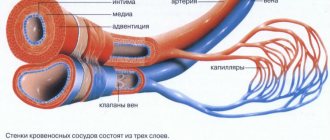
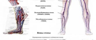
What are veins: their features and differences from arteries
Veins belong to the cardiovascular system of the human body and are involved in blood circulation. They all form the venous system.
Veins are blood vessels through which blood, which has given up oxygen and is filled with carbon dioxide along with cell breakdown products, flows from all organs back to the heart.
Veins are of two types: deep and superficial. The latter are located very close to the skin, so they can be seen. In addition, the visibility of veins depends on the color of a person’s skin - the lighter it is, the better they are visible. The amount of fatty tissue in the body also plays an important role; in obese people, veins are less noticeable.
Blood moves through these vessels due to muscle contraction [Verified source] - the muscles press on the veins, thus helping to push blood from the organs to the heart. Venous valves are located inside the veins to prevent rising blood from flowing down.
A portion of blood entering the vein is pushed upward after a muscle contraction, the valve in a certain section of the vein opens, it enters there and then it closes. When the valves are functioning normally, the downward flow of blood in the veins is impossible.
The pressure in these vessels is less than in the arteries, it is 15-20 mm. Hg Art. The arteries are located deep in the body and cannot be seen, but can only be felt, for example, the radial, carotid, and measure blood pressure.
The color of blood in arteries and veins is also different. So, in the former it is bright red due to oxygen saturation, and in the latter it is dark red with a bluish tint [Verified source] due to a small amount of oxygen and a high content of carbon dioxide. That's why blood is red.
However, there is one exception - the pulmonary veins. They transport oxygenated blood from the lungs to the left atrium, which is why it is bright red.
Brown color of blood during menstruation
If red-brown discharge occurs in the first and last days of menstruation, this is normal. The blood mixes with vaginal secretions and comes out slowly, having time to oxidize. Brown drops can be noticed for several days after menstruation. If brown discharge is present throughout menstruation or occurs on other days of the cycle, this may indicate:
- hormonal imbalance;
- diseases of the endocrine system;
- metabolic failure;
- weak immunity;
- recovery after childbirth or abortion;
- stress.
Scarlet blood during menstruation
The onset of menstruation may be accompanied by bright red discharge. This indicates an increased rate of discharge. The shade may not change during menstruation or become darker towards the end. The reason for this manifestation may be:
- infection. Infectious diseases provoke discharge between menstruation. They may have a scarlet color as they are caused by bleeding. In this case, you should urgently seek advice from a gynecologist;
- pregnancy. Scarlet smears during pregnancy (as well as smears of other colors) pose a serious health threat. This may indicate a threat of miscarriage. Require immediate medical attention;
- fibroids, polyps. Non-cancerous formations in the uterus provoke scarlet discharge both during menstruation and during other periods. Characteristic symptoms are pain and a feeling of pressure.