A healthy cow's heart pumps several thousand tons of blood per day. It is a hollow organ with a cone shape. It is located between the 3rd and 6th rib of the thoracic cavity. The main task of the cardiac system is to ensure the continuous movement of blood through the vessels. This organ has four chambers and contains lymph nodes and vessels. Heart sounds and murmurs are also distinguished, which make it possible to diagnose it.
How does the heart work in animals?
Why is it so important?
The heart is a vital organ with a complex structure. The most important muscle in the entire living organism. Very often the entire circulatory system (or, in other words, the cardiovascular system) is compared to a plumbing system, and the heart in it acts as a pump, only it pumps blood instead of water. Blood gives oxygen and nutrients to all tissues and organs, and takes away carbon dioxide and metabolic products.
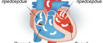
In you and me, and in our cats and dogs, like all mammals, the heart is almost equally tripled. Outwardly, it looks completely different from what you imagine. The organ looks more like an inverted cone. It is a hollow organ consisting of chambers and muscular walls. There are only 4 chambers. By analogy, the heart can be compared to a two-story house, the first floor represents the ventricles, right and left, the second - the right and left atrium. The right and left halves of the heart do not communicate in healthy animals. Thanks to this, the blood is clearly divided into arterial, oxygen-rich, and venous, oxygen-poor. The right half of the heart is venous; it collects oxygen-poor blood from the entire body, and then it is sent through the vessels to the lungs, where it is enriched with oxygen. Then from the lungs, through the pulmonary veins (they are called veins, but they carry arterial, that is, oxygen-rich blood), the blood enters the left atrium, then into the left ventricle and then spreads throughout the body through the aorta.
The atria, ventricles and main vessels are separated by valves. They are designed like doors that open only in one direction. They consist of valves.
The division of blood into systemic and pulmonary circulation (arterial and venous blood) is a fundamental feature of the structure of the mammalian heart.
The heart itself is supplied with blood by special vessels, which are called coronary vessels, because they form a network like a “crown”. They arise from the aorta at its very beginning.
What ensures constant contractions of the heart muscle?
The heartbeat, which is its rhythm, is provided by electrical impulses that are formed by the heart muscle itself. These impulses are formed in the right atrium, and then go through the interventricular septum and further through all the ventricles, causing them to contract.
Despite the fact that the heart of animals and humans is very similar, the diseases still differ . For example, animals almost never have myocardial infarctions, since there is no coronary heart disease or atherosclerotic plaques. Their coronary vessels are structured differently and fat metabolism occurs differently. Therefore, you should never self-medicate animals.
In order to suspect heart disease, it is necessary to detect various clinical manifestations, which you can read about in our article - WHEN CAN YOU SUSPECT HEART DISEASE IN AN ANIMAL?
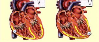
Myocardiofibrosis
This disease is the result of dystrophic or degenerative changes in the heart muscles, and is characterized by poor circulation. The clinical picture is manifested by the following symptoms:
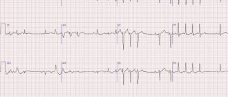
- Auscultation reveals muted tones;
- arrhythmia or tachycardia develops;
- the pulse is faintly audible;
- swelling occurs;
- breathing quickens.
This pathological process can develop over a long period of time, and can last for several months or even a year. A sick cow should be moved to a warm room and given balanced feed in small portions. Additionally, the doctor prescribes medications to improve blood circulation and prevent further development of the pathology.
Structure of the animal heart
The heart (core, cardia) is a hollow, cone-shaped muscular organ. It has a base (basis cordis), which faces caudodorsally, and an apex of the heart (apex cordis), which faces cranioventrally. All mammals have 4 chambers. 2 atria and two ventricles. The atria (atrium) are located at the base of the heart and occupy a small volume.
Externally, the atria are separated from the ventricles by the coronary groove. The atria have blind protrusions - ears that increase the volume of the atria. The inside of the ears have pectineal muscles, which, when the atria contract, contribute to a more complete ejection of blood. The ventricles (ventriculum) occupy the entire remaining area of the heart. From the inside, the heart is divided by a muscular septum into right and left halves, which do not communicate with each other. Communication occurs between the right atrium and right ventricle and the left atrium and left ventricle.
The atrium communicates with the ventricles through the atrioventricular (atrioventricular) openings.
According to the patterns of the course and branching of blood vessels, those vessels that carry blood from the heart are arteries. Those vessels that bring blood to the heart are veins. Regardless of what kind of blood circulates in these vessels. The aorta emerges from the left ventricle, which almost immediately at the base is divided into the brachiocephalic trunk and the thoracic aorta. The brachiocephalic trunk provides blood supply to the anterior part of the body.
The thoracic aorta passes in the chest cavity ventrally from the spinal column, then through the diaphragm and is called the abdominal aorta, which then in the region of the first sacral vertebrae passes into the middle sacral artery, then into the caudal artery. The pulmonary artery emerges from the right ventricle through a common trunk, which then divides into two trunks going to the right and left lungs. The pulmonary veins (2-7, 5-7) flow into the left atrium, and the cranial and caudal vena cava flow into the right atrium.
Valvular apparatus of the heart.
It is represented by leaflet valves that close the atrioventricular openings, with a bicuspid valve (mitral valve) on the left side and a tricuspid valve on the right.
The leaflet valves are attached to the pectineal muscles by tendinous strings. Semilunar (pouch) valves are located at the exit of the aorta and at the exit of the pulmonary artery. Valves allow blood to flow in one direction. The heart works strictly rhythmically. First, there is a simultaneous contraction of the atria, the leaflet valves open and blood from the atria enters the ventricles. The atria relax. Flap valves close. There is a simultaneous contraction of the ventricles.
The semilunar valves open and blood flows from the left ventricle into the aorta and from the right into the pulmonary artery. The ventricles relax, and a general pause occurs. The process is repeated.
Externally, the left ventricle is larger and thicker, the right ventricle is thinner.
The apex of the heart always belongs to the left ventricle.
The membranes of the heart.
Pericarditis
The development of pericarditis occurs against the background of previous diseases of infectious etiology. In some cases, injury to the pericardium causes inflammation. The main provoking factors for the development of the disease include a poor diet, which causes metabolic disorders. The main signs of pericarditis include:
- general weakness;
- periodic hyperthermia;
- loss of appetite;
- lack of chewing gum;
- deterioration in quantitative indicators of products;
- pulse increases to 120 beats;
- swelling appears in the neck, chest or abdomen;
- breathing becomes rapid.
A sick cow becomes restless, moans, and takes a body position in which the chest is higher than the hip area. There is weakness of heart beats, and murmurs are clearly audible on auscultation. If traumatic pericarditis develops, treatment will not be effective. In this case, the cow must be culled. If non-traumatic pericarditis develops, the animal needs to be kept at rest and fed light food.
The doctor prescribes antibacterial drugs, cold compresses and applications to the heart area. The doctor also prescribes medications intended to restore cardiac activity: camphor, glucose, caffeine, calcium chloride.
To prevent the development of this disease, it is recommended to promptly treat diseases that can cause its development.
Structure of the animal heart
The heart consists of 3 layers: epicardium (outer) - thin film, myocardium (middle) - muscular, endocardium - inner membrane.
Circulation circles.
The systemic or systemic circulation begins in the left ventricle with the aorta and ends in the right atrium with the vena cava.
The small circle or pulmonary begins in the right ventricle with the pulmonary artery and ends in the left atrium with the pulmonary veins.
Arterial blood circulates in the left half of the heart, and venous blood circulates in the right. Blood in the heart does not mix.
Features of fetal blood circulation.
1. There is a connection between the fetus and the mother’s body through the placenta.
2. There is a foramen ovale between the atria
3. Mixed blood circulates in the fetal body (through the systemic circulation)
4. There is a Botalov (arterial thoracic) duct that connects the pulmonary artery and thoracic aorta
5. The pulmonary circulation does not fulfill its functional purpose
The fibrous skeleton of the heart is represented by fibrous rings that surround the atrioventricular openings and are located at the exit of the aorta and pulmonary artery.
The structure of the pericardium (pericardium)
The neuromuscular conduction system of the heart is represented by the sinus node (sino-arterial) and the atrioventricular node.
The sinus node is located between the right cardiac appendage and the cranial vena cava. The atrioventricular node lies in the interventricular septum. The Hiss bundle is separated from it, which passes through the interventricular septum with a common leg and is divided into right and left legs running along the walls of the ventricles.
In the ventricular myocardium, the pedicles branch into Purkenje fibers. They are differentiated muscle fibers capable of independently conducting excitation. The sinus and atrioventricular nodes represent the sympathetic nervous system. Next to the nodes are the ganglia of the same name. The fibers of the vagus nerve end in these ganglia.
The ganglia are connected to each other by nerve fibers that form the intramural parasympathetic plexus.
Blood supply to the heart.
From the aorta, even before it branches into the brachiocephalic trunk and thoracic aorta, the right and left coronary arteries depart. Venous blood from the heart collects in the large middle and small cardiac veins, which drain into the right atrium.
Topography of the heart.
The heart is located in the chest cavity between the lungs in front of the diaphragm, shifted to the left.
The base of the heart is located in the middle of the first rib, the apex of the heart in the 5-6 intercostal space closer to the sternum. In carnivores, the base is located in the area of 1-3 ribs, the apex in the area of 6-7 intercostal space. In carnivores, the heart is located almost horizontally.
Vascular and nervous apparatus
The connection of the vessels takes place using anastomosis. Both vessels of the same type and vessels of different types communicate. There is the following classification of anastomoses:
- arterial;
- venous;
- arterial-venous.
Through anastomoses, longitudinal capillaries, networks, and collectors are formed. The heart also contains the autonomic nervous system. With the help of sympathetic nerve fibers, cardiac function is stimulated. And thanks to parasympathetic nerve fibers, cardiac activity slows down. Autonomic nerve fibers are in close communication with the neuromuscular system of the cardiac apparatus.
Animal Heart (Part 1)
The mammalian circulatory system is the highest form of blood circulation.
Like birds, it is characterized by a four-chambered heart and two circles - large and small.
This form promotes accelerated metabolism compared to other groups of vertebrates: in fact, we have “two hearts” installed in different parts of the vascular system.
The blood in both halves of the heart does not mix.
“Main” circulation
The large circle originates in the left ventricle.
The only arch of the aorta coming from it is the left one, and not the right one, like in birds. Branches from it carry blood throughout the body, saturating organs and tissues with oxygen and other necessary substances.
structure of the circulatory system of mammals photo
From them it takes in carbon dioxide and metabolic products.
Venous blood, saturated with carbon dioxide, is directed through the veins to the right atrium. Two vena cavae flow into it, the first of which carries blood from the head and forelimbs, and the second from the back of the body.
Composition of mammalian blood
The blood of mammals consists of liquid plasma, which contains a full set of so-called formed elements:
- Red blood cells are carriers of the iron-containing substance hemoglobin, they carry oxygen;
- Platelets are bodies responsible for blood clotting and serotonin metabolism;
- Leukocytes are white bodies responsible for immunity.
Red blood cells and platelets of mammals, unlike other groups of animals, do not contain nuclei.
Platelets are actually “blood platelets”; the absence of nuclei in red blood cells is explained by the need to accommodate a larger amount of hemoglobin.
Also, red blood cells do not have mitochondria, so they synthesize ATP without the use of oxygen, making them the most effective carriers of it.
Dropsy of the cardiac sac
The development of this disease is characterized by the accumulation of fluid in the sac, which is located near the heart. Often, the development of dropsy can be triggered by other diseases or insufficiency of chronic blood microcirculation. The main features include:
- weakness;
- deterioration in milk yield;
- the appearance of swelling in the space between the jaws;
- sudden jumps in blood pressure.
Therapeutic measures involve getting rid of the underlying disease. For the cow, you need to provide the most balanced nutrition possible and give a large amount of liquid.
Important! To reduce the amount of accumulated fluid, the doctor prescribes a course of cardiac, diuretic, diaphoretic and iodine medications.
Lymphatic system
The lymphatic system is closely connected with the circulatory system and is an intermediary between it and tissues in the exchange of nutrients.
It consists of blood plasma and lymphocytes.
It is noteworthy that mammals do not have “lymphatic hearts,” unlike reptiles and amphibians, which is the name given to areas of lymphatic vessels that can contract: lymph in mammals, which lead a much more active lifestyle, moves due to contraction of the skeletal muscles.
Mammals also have lymph nodes that cleanse the lymph from harmful microorganisms.