Hungry Heart
The heart, like any other organ, is supplied by blood vessels called coronary arteries. Narrowing of the coronary arteries leads to insufficient nutrition of the heart muscle, to oxygen “starvation,” which doctors call “ischemia.” Ischemia can occur when the heart requires more nutrients, such as during exercise. In this case, the patient experiences an attack of chest discomfort or angina. If blood flow through the coronary artery is suddenly blocked by a thrombus, then acute oxygen deficiency occurs and necrosis of part of the heart muscle, the so-called myocardial infarction, occurs.
Classification of coronary heart disease
Depending on the symptoms, the following main forms of the disease are distinguished:
Coronary death . Symptoms develop rapidly: loss of consciousness, pupils are dilated and do not respond to light. No pulse, no breathing.
Post-infarction cardiosclerosis . Among the characteristic signs: heart rhythm disturbances, manifestations of acute (attack of suffocation - “cardiac asthma”, pulmonary edema) and chronic heart failure (swelling of the legs, shortness of breath). The patient complains of a feeling of lack of air, shortness of breath, and swelling of his legs and feet.
Acute coronary syndrome. New-onset angina, progressive angina, myocardial infarction, etc.
Myocardial infarction . Often severe pressing and burning pain behind the sternum, radiating to the jaw, left shoulder blade and arm. Lasts up to half an hour or more, does not go away when taking nitroglycerin under the tongue. The patient also develops cold sweats, blood pressure decreases, weakness, vomiting and fear of death may appear.
Angina pectoris . A person complains of chest pain - squeezing, squeezing, burning behind the sternum during physical activity and sometimes at rest. Possible symptoms of angina include pain in the neck, left shoulder blade, lower jaw or left arm. The pain is usually short-lived.
Angina pectoris is one of the most striking manifestations of coronary heart disease. Self-treatment of angina pectoris with folk remedies is unacceptable! Only a doctor, based on his professional experience and diagnostic techniques, can draw conclusions about a person’s condition and the necessary treatment measures!
1 Ultrasound of the heart for angina pectoris
2 Ultrasound of the heart in “MedicCity”
3 Blood tests for ischemic heart disease
If angina pectoris occurs for the first time, if angina attacks begin to occur more often, last longer and manifest themselves more strongly, we are talking about acute coronary syndrome and a high risk of developing myocardial infarction. Such patients should be urgently hospitalized by ambulance to a hospital, where coronary angiography will be performed on an emergency basis and blood flow in the arteries of the heart will be restored, which will avoid the occurrence of myocardial infarction and, as a consequence, disability.
Silent myocardial ischemia
IHD may not be accompanied by pain. This ischemia is called silent ischemia.
The manifestation of the disease in the case of silent myocardial ischemia is often myocardial infarction or sudden coronary death. Therefore, it is very important to be regularly examined by a cardiologist, especially for people at risk (diabetics, hypertension, smokers, obese people, the elderly, etc.).
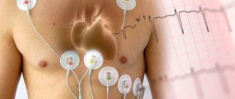
Such hidden ischemia can be detected using some instrumental techniques, for example, ECG with exercise (Veloergometry, treadmill). It is during a stress test on the electrocardiogram that changes specific to coronary heart disease are especially pronounced.
Two faces of a terrible disease
Often in the specialized literature two different terms are used - “angina pectoris” and “myocardial infarction”. Angina pectoris and myocardial infarction are two manifestations of one disease - coronary heart disease (CHD). At the same time, many patients who have had a heart attack are well aware of the symptoms of angina pectoris and, conversely, patients suffering from angina pectoris have ever suffered a myocardial infarction. The danger of myocardial infarction is that it often strikes suddenly - in about half of the cases, myocardial infarction occurs without any obvious harbingers of impending disaster.
First aid for emergency diseases of the cardiovascular system
assistance for emergency diseases of the cardiovascular system
May 27, 2021
There are situations when first aid must be provided in the first minutes and hours after the onset of a life-threatening condition and regardless of whether a medical professional is nearby.
Cardiovascular diseases are the leading cause of death among adults in almost all countries of the world. The peculiarity of emergency conditions provoked by these diseases is that they arise suddenly, are difficult, and the life of the injured person depends on the speed of providing first aid. The most common among them are: angina pectoris, myocardial infarction, hypertensive crisis and stroke.
Angina and myocardial infarction
Signs:
- The pain is pressing, squeezing, aching, a feeling of tightness or heaviness in the chest. The pain is retrosternal and can radiate to the shoulder, arm, neck, lower jaw or back.
- Labored breathing.
- Fast, slow, or irregular pulse.
- Pale or bluish skin.
- Increased sweating.
- Nausea or vomiting, often reminiscent of indigestion.
If severe pain and discomfort in the chest does not go away within 10 minutes after taking nitroglycerin, or such pain occurs for the first time in your life, immediately call an ambulance and begin first aid for a heart attack.
First aid:
- The patient should stop all physical activity.
- Help the patient get into a comfortable position (often a sitting position).
- Loosen your tie and waist belt.
- Help the patient take nitroglycerin under the tongue.
- After 5 minutes, if the pain has not gone away, the patient should take a second nitroglycerin tablet.
- Try to calm and reassure the patient. This helps the patient overcome anxiety and alleviates the pain somewhat.
Heart failure
The condition of cardiac arrest is critical because the brain and other vital organs remain viable for only a few minutes without oxygenated blood!
Signs of cardiac arrest are lack of consciousness, lack of breathing, and lack of pulse in the patient.
If there are signs of cardiac arrest, it is necessary to carry out cardiopulmonary resuscitation - this is artificial ventilation of the lungs with simultaneous rhythmic pressure of medium force on the lower third of the sternum, alternating 2 breaths through the patient’s mouth with simultaneous pinching of his nose and 30 pressures on the sternum. Without resuscitation procedures, brain death occurs within 5-6 minutes.
Hypertensive crisis
This is an emergency condition in which blood pressure rises rapidly and leads to significant and irreversible organ damage within a few hours.
Signs of a crisis:
- Headache.
- Dizziness.
- Facial redness.
- Increased heart rate, rhythm disturbance.
- Difficulty breathing.
First aid in a crisis:
- Make the patient sit down.
- Remind the patient to take quick-acting medication if they forget to take it.
- Call an ambulance".
- Do not allow aspirin to be taken.
Stroke
This is an acute disorder of cerebral circulation, leading to the death of brain cells as a result of hemorrhage from a ruptured blood vessel (hemorrhagic stroke) or lack of blood supply as a result of the formation of a blood clot in a brain vessel (ischemic stroke).
Signs of a stroke:
- Sudden weakness.
- Numbness of the face, arms, legs, usually on one side.
- Difficulty speaking or understanding speech.
- Sudden severe headache.
- Difficulty walking due to dizziness, loss of balance or coordination.
- Sudden visual disturbances.
- Uneven pupil dilation.
- Unconscious state.
First aid for stroke:
- Call an ambulance.
- Place the patient in the recovery position with the paralyzed side up to ensure evacuation of the oral contents. If saliva and vomit are present, remove them from the patient's mouth.
BU "Surgut City Clinical Hospital"
Link to website https://cmphmao.ru/node/174439
News December 17, 2021 The results of the social video competition “Live freely - choose health!” were summed up in Surgut.
December 17, 2021 Specialists from the Nyagan Children's Clinic took part in the School of Foster Parents
December 17, 2021 The Center for Occupational Pathology summed up the work of the information and diagnostic platform of the AIDS Center
News archive
When the clock counts...
Angioplasty and stenting of arteries
- Cost: 100,000 - 250,000 rubles.
- Duration: 40 minutes
- Hospitalization: 1-2 days in hospital
More details
A fair question: can it effectively treat myocardial infarction? After all, its consequence is the necrosis of part of the heart muscle. If, during a developing heart attack, the cause of its occurrence is eliminated, that is, the patency of the clogged artery of the heart is restored, then the negative consequences of a heart attack can be significantly reduced.
The most accessible method of treating a heart attack is the administration of drugs that dissolve the clot. Such treatment can begin as soon as the doctor diagnoses a heart attack. But it is most effective only in the first hours from the onset of the disease. Later, the clot becomes more resistant to the drugs. In addition, after 6 hours from the onset of a heart attack, most of the cells in the affected area die and it is pointless to use drugs that are not sufficiently effective by this time. Another approach is mechanical removal of the obstruction in the coronary artery. To do this, using a special thin catheter, under X-ray control, the thrombus is recanalized and the lumen of the narrowed artery is expanded with a special balloon. This treatment method is called coronary angioplasty. Angioplasty is more effective than drug treatment late after a heart attack, with relatively “old” thrombosis.
Emergency care for angina and myocardial infarction
Emergency care for angina pectoris
Angina is the most common form of coronary heart disease (CHD). The high-risk group for sudden death and myocardial infarction consists primarily of patients with exertional angina. Therefore, it is necessary to quickly establish a diagnosis and provide emergency assistance during a prolonged attack of angina. Emergency diagnosis of an angina attack is based on the patient's complaints, medical history and, to a much lesser extent, on ECG data, since in a large percentage of cases the electrocardiogram remains normal. In most cases, taking into account the nature, duration, localization, irradiation, conditions of the onset and cessation of pain makes it possible to establish its coronary origin.
The WHO Expert Committee recommends the following diagnostic criteria for pain during an angina attack :
- the nature of the pain is squeezing or pressing;
- localization of pain behind the sternum or in the atrial region along the left edge of the sternum;
- a clear connection between the occurrence of pain and physical activity;
- pain duration is no more than 10 minutes;
- Taking nitroglycerin gives a quick effect.
The duration of an anginal attack during angina pectoris is most often 2 - 5 minutes, less often - up to 10 minutes. It usually resolves after the patient stops exercising or takes nitroglycerin. If a painful attack lasts more than 15 minutes, then medical intervention is required, since a prolonged attack of angina pectoris can lead to the development of acute myocardial infarction.
Sequence of measures for a prolonged attack of angina:
nitroglycerin - 1-2 tablets under the tongue, at the same time non-narcotic analgesics are administered intravenously in 20 ml of a 5% glucose solution (analgin - 2-4 ml of a 50% solution, baralgin - 5 ml, maxigan - 5 ml) in combination with minor tranquilizers (seduxen - 2-4 ml) or antihistamines (diphenhydramine - 1-2 ml 1 %
solution), enhancing the analgesic effect and having a sedative effect. At the same time, the patient takes 0.2-0.5 g of acetylsalicylic acid, preferably in the form of an effervescent tablet (for example, anapirin).
If the pain syndrome is not relieved within 5 minutes, then immediately begin intravenous administration of narcotic analgesics (morphine hydrochloride - 1-2 ml of 1% solution, promedol - 1-2 ml of 1% solution, etc.) in combination with tranquilizers or neuroleptic droperidol (2-4 ml of 0.25% solution). The most powerful effect is exerted by neuroleptanalgesia (narcotic analgesic fentanyl - 1-2 ml of 0.005% solution in combination with droperidol - 2-4 ml of 0.25% solution).
After stopping the anginal attack, it is necessary to do an ECG to exclude acute myocardial infarction .
Emergency care for myocardial infarction
Myocardial infarction is ischemic necrosis of a portion of the heart muscle, resulting from an acute discrepancy between the myocardial need for oxygen and its delivery through the coronary vessels. This is the most severe manifestation of coronary artery disease, requiring emergency care for the patient. Emergency diagnosis of myocardial infarction is based on the clinical picture, the leading one of which is severe pain, and ECG data. Physical examination does not reveal any reliable diagnostic signs, and changes in laboratory data usually appear several hours after the onset of the disease. As with angina, pain occurs behind the sternum, radiating to the left arm, neck, jaw, epigastric region, but, unlike angina, the attack lasts up to several hours. Nitroglycerin does not have a lasting effect or does not work at all. In atypical cases, pain may be mild, localized only in areas of irradiation (especially in the epigastric region), accompanied by nausea, vomiting, or absent altogether ( painless myocardial infarction ). Sometimes, already at the onset of the disease, complications (heart rhythm disturbances, cardiogenic shock, acute heart failure) come to the fore in the clinical picture. In these situations, the ECG plays a decisive role in diagnosis. ST segment elevation
above the isoline, formation of a monophasic curve, pathological
Q
In clinical practice, there are forms of
myocardial infarction without changes in the S-T segment and Q wave.
Emergency care for myocardial infarction begins with immediate relief of the anginal status. Pain not only causes severe subjective sensations and leads to an increase in the load on the myocardium, but can also serve as a trigger for the development of such a formidable complication as cardiogenic shock. Anginal status requires immediate intravenous administration of narcotic analgesics in combination with antipsychotics and tranquilizers, since conventional analgesics are ineffective.
In accordance with the recommendations of the European and Ukrainian Societies of Cardiology, patients with acute myocardial infarction hospitalized within 72 hours of the onset of the disease are prescribed the following drugs:
- Antiplatelet (thrombolytic): acetylsalicylic acid (150-300 mg intravenously or orally) or ticlide (0.25 g 2 times a day).
- Anticoagulants: heparin, fraxiparin.
- Nitroglycerin is administered intravenously as follows: an isotonic sodium chloride solution is added to a 1% ampoule solution to obtain a 0.01% solution and administered dropwise at a rate of 25 mcg per 1 minute (1 ml of a 0.01% solution in 4 minutes).
- Beta-blockers: anaprilin (propranolol) - 10-40 mg 3 times a day, or vasocardin (metoprolol) - 50-100 mg 3 times a day, or atenolol - 50-100 mg 3 times a day.
- Angiotensin-converting enzyme inhibitors: capoten - 12.5-50 mg 3 times a day.
If less than 6 hours have passed of myocardial infarction This drug promotes thrombus lysis.
Combinations of drugs used in the treatment of pain
in acute myocardial infarction:
- the most widely used is neuroleptanalgesia, which has a powerful analgesic and anti-shock effect, which is carried out by the combined administration of 1-2 ml of a 0.005% fentanyl solution and 2-4 ml of a 0.25% droperidol solution; instead of fentanyl, you can use morphine hydrochloride (1-2 ml of 1% solution), promedol (1-2 ml of 1% solution), omnopon (1-2 ml of 1% solution), etc.;
- An effective combination of narcotic analgesics (morphine hydrochloride - 1-2 ml of 1% solution, promedol - 1-2 ml of 1% solution), minor tranquilizers (seduxen - 2-4 ml) and antihistamines (diphenhydramine - 1-2 ml of 1% solution );
- anesthesia with a mixture of nitrous oxide and oxygen is currently used mainly by ambulance crews.
It is recommended to administer the drugs slowly intravenously. They are first diluted in 5-10 ml of isotonic sodium chloride solution or 5% glucose solution. Until the pain syndrome is completely relieved, which often requires repeated administration of analgesics, the doctor cannot consider his task completed. Other therapeutic measures, which are carried out simultaneously or immediately after pain relief, should be aimed at eliminating emerging complications (rhythm disturbances, cardiac asthma, cardiogenic shock). For uncomplicated myocardial infarction , drugs are prescribed that limit the area of necrosis (nitrates, beta blockers, thrombolytics).
Bypass pipeline
If blood flow cannot be restored using such a minimally traumatic method as coronary angioplasty, then they resort to open surgery - coronary artery bypass surgery. In this case, veins or arteries taken from other parts of the body are brought from the aorta, the main blood supply, to the arteries of the heart, so as to bypass (bypass) the area of narrowing or blockage. Similar methods can be used in the treatment of angina pectoris. Moreover, in developed countries they are already widely used. The advantage of surgical methods over medication is that they quickly and completely eliminate the symptoms of the disease.
Angina without stenosing atherosclerosis
The journal Circulation published the results of an observational study, the purpose of which was to describe the anatomical and functional changes of the coronary bed in patients with clinical symptoms of angina, but without angiographically significant lesions of the coronary arteries (stenosis < 50%).
According to the authors, among the population of patients referred for angiography with symptoms of angina pectoris, angiography does not reveal significant coronary lesions in 20% of patients. Despite the absence of hemodynamically significant lesions, these patients often have persistent clinical symptoms, poor functional status, and repeated hospitalizations often occur in the absence of a clear diagnosis. During the study, endothelial function was assessed in 139 (77% women) patients with angina; microcirculatory resistance index, coronary and fractional blood flow reserve. The average age of the patients was 54.0 ± 11.4 years. All patients, according to intravascular ultrasound, had evidence of the presence of an atherosclerotic process in the left anterior descending artery. Endothelial dysfunction (reduction in luminal diameter >20% after intracoronary acetylcholine administration) was detected in 61 patients (44%). Microvascular damage (microcirculatory resistance index ≥25) was detected in 29 patients (21%). Seven patients (5%) had fractional flow reserve ≤0.80. Myocardial bridge was present in 70 patients (58%). Overall, in 32 patients (23%) the clinical syndrome of angina could not be explained by anatomical or functional abnormalities (the patients had a normal coronary bed, normal endothelial function, coronary flow reserve and did not have muscle bridges).
Conclusion: in the majority of patients with clinical angina syndrome, other anomalies occur in the absence of hemodynamically significant coronary lesions. Comprehensive invasive coronary assessment during coronary angiography in these patients can be safe for the patient and provides important diagnostic information that can influence future management of the patient.
Lee BK1, Lim HS1, Fearon WF2, Yong AS1, Yamada R1, Tanaka S1, Lee DP1, Yeung AC1, Tremmel JA1. Invasive evaluation of patients with angina in the absence of obstructive coronary artery disease. Circulation. 2015 Mar 24;131(12):1054-60.
Comments by Doctor of Medical Sciences, Professor Samorodskaya I.V.: This study will probably be of interest to doctors of many specialties. As science and technology develop, many causes of previously difficult to explain phenomena are revealed. It is likely that further studies will follow aimed at assessing the prognostic significance of the detected pathology and treatment methods.
Light at the end of the vessel
We can say that there is a certain category of people in whom the presence of atherosclerosis is very likely. In the vast majority of cases, narrowing of the coronary arteries occurs due to the development of deposits on the walls of the vessel - atherosclerotic plaques - narrowing its lumen. Increased blood cholesterol levels, a hereditary predisposition to cardiovascular diseases, excess weight, smoking, hypertension, diabetes mellitus are risk factors for atherosclerosis.
Bypass surgery, angioplasty or medications
Which method is better - coronary bypass surgery, coronary angioplasty or drug treatment? Each of these methods has its own indications. Sometimes it is necessary to use different treatment methods at different stages. Of course, drug prevention and therapy are necessary in almost all cases. Coronary angioplasty can also be used successfully in most patients. Briefly, treatment methods for ischemic heart disease can be grouped as follows:
- Medicines . Some of them are necessary to reduce blood cholesterol or high blood pressure. Other medications help people with angina avoid attacks during exercise. Some drugs prevent heart rhythm disturbances that often accompany coronary artery disease. A number of drugs are necessary for the prevention of heart failure after myocardial infarction;
- Thrombolytic therapy . This type of treatment is used to eliminate a blood clot in acute myocardial infarction, when blood flow is obstructed by a blood clot. Thrombolytic agents dissolve clots and restore patency of the coronary artery. Thrombolytic therapy is often combined with angioplasty;
- Angioplasty is a method of expanding the internal lumen of blood vessels using a special balloon. In this case, through a small puncture, the doctor inserts a thin tube (catheter) and brings it to the narrowed area of the vessel, monitoring the process using x-rays. The balloon, expanding under pressure, pushes apart and flattens atherosclerotic plaques, creating conditions for normal blood flow. In some cases, after this, a metal frame is installed - a stent, which, being implanted into the wall of the artery, does not allow it to narrow again.
- Bypass operation . During bypass surgery, a new blood supply is created that bypasses the blocked section of the artery. Shunts are usually created from the patient's own vein or artery, for example from the patient's leg. Bypass surgery is usually performed surgically, although more gentle, non-surgical bypass methods have recently been developed.
All these techniques have been successfully used for many years by doctors at the Center’s Cardiovascular Surgery Department. In this case, proprietary developments are used, including a unique vascular stent developed by the head of the department Z.A. Kavteladze and his employees and now produced by WILLIAM COOK (USA).
National Society for the Study of Atherosclerosis
Myocardial infarction (necrosis or death of the heart muscle) is a serious complication of coronary artery disease, sometimes fatal. According to statistics in the United States, acute myocardial infarction leads to death on average in every third man aged 35-50 years [9]. Moreover, more than half of these deaths occur within the first 3-4 hours from the onset of a heart attack due to life-threatening arrhythmia (ventricular fibrillation), which without treatment always leads to death. We can say that myocardial infarction is a special case of ischemic heart disease. Myocardial infarction occurs due to blockage (thrombosis) of the artery supplying blood to one or another part of the heart muscle. As a rule, a heart attack is preceded by the rupture of an atherosclerotic plaque and the deposition of platelets on the surface of such an altered plaque. All this contributes to the fact that the lumen of the artery begins to close, and this leads to acute myocardial ischemia. Myocardial infarction is usually preceded by an increase in blood viscosity. If during a myocardial infarction a spasm of the coronary artery occurs at the site of the same plaque, this causes an even greater narrowing of the artery, up to its complete blockage.
Sometimes it is possible to develop myocardial infarction only as a result of prolonged spasm of atherosclerotically unchanged coronary arteries [9], for example, as a result of vasospastic angina (see below). As a result of myocardial infarction, a section of the heart muscle dies. Over time, this area is replaced by another tissue, scar (or so-called connective tissue), which does not contain muscle fibers. This condition is called post-infarction cardiosclerosis, in contrast to atherosclerotic cardiosclerosis, when muscle tissue is replaced by connective tissue without a previous myocardial infarction. During a myocardial infarction, both the entire section of the heart muscle can die, in its entire thickness (transmural myocardial infarction), or only one of its layers: the inner (subendocardial infarction), middle (intramural infarction) or outer (subepicardial infarction). If an infarction occurs in only one of the layers of the heart muscle, this does not exclude the possibility that a transmural infarction will not subsequently develop in the remaining layers. The prognosis for life and preservation of sufficient contractile function of the myocardium as a result of a myocardial infarction depends, first of all, on the area of dead myocardium (large-focal or small-focal infarction). And this, in turn, is determined by the number of affected arteries and the location of the blockage in the artery itself. If the blockage occurs at the mouth of an artery that supplies blood to a large area of the myocardium, then the prognosis can be very dismal. Thus, the more distant from the mouth the part of the artery is affected, the safer the prognosis. On average, a scar is formed within 1.5-2 months, after which the patient’s ability to work can be restored.
Manifestations of myocardial infarction
The disease can begin in different ways: both against the background of general well-being, and as a worsening of angina attacks. Sometimes myocardial infarction is the first manifestation of coronary artery disease. The patient is restless, sweating, pale, and may experience nausea and vomiting. The painful attack is much more severe and longer lasting than normal angina attacks. Previously helpful medications stop working. The pain can be so unbearable that it can lead to painful shock with loss of consciousness and a sharp drop in blood pressure. If a painful attack is not relieved by 3 tablets of nitroglycerin, you should urgently call an ambulance. Sometimes myocardial infarction does not manifest itself at all. It is in such cases that they say that the patient suffered a myocardial infarction “on his feet.”
Diagnosis of myocardial infarction
To diagnose myocardial infarction, the same methods are used as for diagnosing coronary artery disease in general. Tests with dosed physical activity are strictly contraindicated due to the high risk of developing various complications and death of the patient. In most cases of myocardial infarction, characteristic changes appear on the ECG. An additional method for diagnosing myocardial infarction is to identify its biochemical markers - biological substances (enzymes) that appear or significantly increase only in this disease. Biochemical markers of myocardial infarction are creatine phosphokinase (CPK) and its components (fractions), as well as troponin and some others. Already 4 hours after the onset of the disease, especially with unclear manifestations of the disease and a questionable ECG, the remaining doubts can be resolved with the help of markers.
Treatment of myocardial infarction
Myocardial infarction is an absolute indication for hospitalization of a patient in a cardiology hospital for treatment and round-the-clock medical supervision. Only timely medical intervention can reduce the risk of severe consequences of a heart attack. In the hospital, treatment is usually carried out to limit the area of infarction (intravenous administration of nitroglycerin, β-blockers, calcium ion antagonists), increase cardiac output (dobutamine, dopamine, adrenaline), reduce blood viscosity and coagulability (heparin), normalize blood pressure, and eliminate arrhythmia. Painkillers, including narcotics, are usually administered by emergency medical technicians. If necessary, their administration is continued in the hospital. Hospitalization in a cardiology hospital, where specialists are proficient in interventional diagnostic and treatment methods, is considered ideal. When a patient is admitted to such a hospital, he may be prescribed coronary angiography, which will make it possible to accurately determine which artery caused the heart attack. The patient can then undergo TLBAP and/or stenting of this infarction-related artery in order to restore blood flow through it. With this sequence of events, the consequences of even severe myocardial infarction can be minimized.
But, unfortunately, interventional cardiology departments are not yet available in all medical hospitals in our country. In a number of cardiology hospitals, and ideally at home, doctors from the cardiology emergency medical team perform so-called systemic thrombolysis. This method involves the intravenous administration of drugs that can eliminate (“dissolve”) blood clots in the coronary arteries. Systemic thrombolysis is more common than TLBAP and coronary artery stenting. In the departments of interventional cardiology, in turn, there is the opportunity to perform intracoronary thrombolysis, i.e. injection of a clot-dissolving drug (thrombolytic) directly into the coronary arteries. Thrombolysis is an alternative to TLBAP and coronary artery stenting, but it does not eliminate stenoses. Typically, coronary bypass surgery is not performed during acute myocardial infarction, which is associated with an increased operational risk of death of the patient. As an exception, surgery is performed in cases where it is necessary to eliminate life-threatening complications of myocardial infarction, for example, rupture of the interventricular septum, etc. Recently, attempts have been made aimed at restoring the number of muscle cells lost as a result of myocardial infarction. To do this, donor “progenitors” of muscle cells, so-called stem cells, are introduced into the myocardium. These cells are introduced either during heart surgery, injecting them into the scar zone, or injected into the coronary arteries when performing coronary angiography. Currently, such scientific material is being accumulated.
Complications of myocardial infarction
As with any other disease, complications occur as a result of myocardial infarction. The development of complications is determined by the area of the infarction and which particular area of the myocardium is affected. Deadly complications are external ruptures of the heart, that is, rupture of its outer walls. If one of the inner walls (septa) of the heart ruptures, this condition is potentially life-threatening. Much again depends on the size of the defect formed and on how the heart copes with the consequences caused by the presence of this defect. Sometimes, to save the patient's life in such cases, emergency heart surgery is required. The main method for diagnosing cardiac ruptures is echocardiography. As a result of a heart attack, cardiac aneurysms can form. An aneurysm is a bulging formation on the surface of the heart that passively bulges outward during heartbeat. Aneurysms may be composed of scar tissue (true aneurysm) or tissue surrounding the heart (false aneurysm). The danger of an aneurysm is that blood clots can form in its cavity. Fragments of these blood clots can spread through the bloodstream, causing heart attacks in other internal organs (brain, kidneys, liver, lungs, etc.). This spread of blood clot fragments throughout the body is called thromboembolism. Therefore, all patients with a cardiac aneurysm are advised to take anticoagulants - medications that prevent the formation of blood clots. In addition, the area of the aneurysm can be a source of persistent and life-threatening cardiac arrhythmias. Diagnosis of cardiac aneurysms is carried out using ECG, echocardiography and chest x-ray. Treatment of cardiac aneurysms is only surgical. During the operation, the aneurysm is excised, and the surrounding myocardium is sutured together. Aneurysm plasty can also be performed: after excision of the aneurysm, a biological or synthetic patch is sutured in its place. Excision or repair of aneurysms is performed simultaneously with coronary bypass surgery. Another dangerous complication of myocardial infarction is cardiogenic shock - a condition in which the heart cannot cope with its pumping (contractile) function. This condition occurs when at least 50% of the contractile myocardium mass dies as a result of a heart attack. Only in a small percentage of cases, according to some data up to 20%, can the patient’s life be saved [9].
Cardiogenic shock is a special case of post-infarction heart failure, that is, a decrease in the pumping function of the heart due to myocardial infarction. As a result of the death of any part of the myocardium, as mentioned above, a scar is formed, which, unlike the rest of the myocardium, cannot fully contract and contribute to the pumping function of the heart. A scar is like a big patch on a balloon: on the one hand, the balloon seems to be intact, but on the other hand, it is not at all necessary that this balloon can now be fully inflated. A situation is created when the myocardium, which has not suffered from a heart attack, is forced to take on the function of the dead myocardium, while some zone of the heart will contract more intensely, and another less. Over time, healthy myocardium will begin to lose its “contractile” positions, especially when performing physical activity. Ultimately, heart failure will arise and gradually progress - the inability of the heart to fully cope with its pumping function. Blood will begin to linger in the veins of the pulmonary (small) and then systemic circulation. As a result, shortness of breath will appear as a consequence of stagnation of blood in the vessels of the lungs, as well as edema, hepatomegaly (enlargement of the liver, manifested by heaviness in the right hypochondrium), ascites (increase in the volume of the abdomen due to stagnant liquid part of the blood). All these signs (symptoms) of heart failure usually become noticeable in the evening, when the body is as “tired” as possible after everything it has done during the day. Unfortunately, post-infarction heart failure, along with the progression of atherosclerosis of the coronary arteries and subsequent myocardial infarctions, does not increase the life expectancy of patients with coronary artery disease.
Non-fatal complications of myocardial infarction are various disturbances in the conduction of the heart impulse (blockade), which sometimes requires the installation of a temporary pacemaker. Sometimes, on the contrary, after myocardial infarction various arrhythmias appear that require appropriate antiarrhythmic therapy (treatment). In patients who have suffered a transmural myocardial infarction, in approximately 30% of cases [9], non-infectious inflammation of the heart sac or membrane (pericardium) is observed - pericarditis. It manifests itself as short-term pain in the heart area, usually with a deep breath. The duration of pericarditis during myocardial infarction is usually 3-5 days.
There is another rare complication of myocardial infarction - the so-called Dressler syndrome. (A syndrome is a collection of symptoms or signs of a disease.) It occurs days, weeks and even months after a myocardial infarction. Dressler's syndrome is a response of the body's immune system to tissues of its own heart that have changed as a result of a myocardial infarction. This complication manifests itself as non-infectious pericarditis, as well as non-infectious inflammation of the outer membrane of the lungs (pleurisy) and other tissues. Treatment for Dressler syndrome is medication.
Unstable angina
Unstable angina (synonyms: progressive, variant) is a borderline state between ordinary (stable) exertional angina and myocardial infarction. It is characterized by an increase in frequency and severity of angina attacks. Sometimes unstable angina is also called a pre-infarction condition. Attacks of angina pectoris with this type of coronary artery disease occur at lower loads than before, at rest or at night. Unstable angina, as a rule, is based on the same thrombosis of the coronary arteries as in myocardial infarction, only reversible. Unlike myocardial infarction, with unstable angina, a small lumen remains in the affected artery and blood still flows through it beyond the site of thrombosis. But this amount of blood is still not enough for the myocardium supplied by this artery, which leads to more frequent and worsening pain attacks. Unstable angina will certainly resolve either towards stable exertional angina or towards myocardial infarction. According to American scientists, approximately a third of patients with unstable angina will develop myocardial infarction within 3 months from the moment of its onset in the absence of appropriate treatment [9]. That is why unstable angina requires active inpatient treatment, both medicinal and surgical. Diagnosis of the disease is the same as for myocardial infarction.
Vasospastic angina
Vasospastic angina (synonyms: Prinzmetal's angina, spontaneous angina) is a condition caused by involuntary contraction (spasm), often of large coronary arteries, unchanged by atherosclerosis. Spasm of the coronary arteries leads to myocardial ischemia, which is manifested by angina pectoris. Vasospastic angina usually affects young people. A painful attack most often occurs in the early morning hours, sometimes it is accompanied by cardiac arrhythmias and usually goes away after taking nitroglycerin or nifedipine. Outside of an attack, the ECG in such individuals does not differ from the ECG of healthy people. Holter ECG monitoring can help in diagnosing the disease. This disease can only be reliably diagnosed using drug provocation (ergonovine test). To prevent attacks of vasospastic angina, medications that dilate the coronary arteries, for example, nifedipine, are prescribed. A prolonged attack of vasospastic angina can result in myocardial infarction. SYNDROME X Syndrome X (X) is a condition manifested by angina pectoris. The complaints of patients and the results of instrumental diagnostic methods are the same as with typical angina pectoris. What distinguishes these two diseases is that in syndrome X, coronary angiography does not reveal any changes in the coronary arteries. This is explained by the fact that syndrome X is caused by spasm of small coronary arteries not visible on a coronary angiogram. The final diagnosis is established by exclusion.
Sudden cardiogenic death
This term refers to sudden death caused by heart disease (from the Greek words kardia - “heart” and genesis - “origin, occurrence” [2]). In approximately 20% of cases, sudden cardiogenic (cardiac) death is the first and only manifestation of coronary artery disease [9]. Typically, patients with severe damage to the coronary arteries and severe cardiac dysfunction die. The immediate cause of death is sudden blockage of the coronary arteries, accompanied by life-threatening arrhythmia (ventricular fibrillation). As mentioned above, in the absence of timely treatment, ventricular fibrillation always ends in death. The diagnosis of IHD in deceased patients can be suspected by questioning relatives, and confirmed during a pathological examination.
Provocative diagnosis
If a person has several risk factors for ischemic heart disease, further examination is necessary to find out whether the person is experiencing ischemia or not. To do this, various so-called “provocative tests” are carried out, for example, tests with physical activity. By comparing the parameters of the heart at rest and under heavy loads, a conclusion is made about the need for further examination. At the next stage, a study of the heart vessels is usually performed - coronary angiography. Provocative tests, drug tests, and coronary angiography are included in the extensive arsenal of diagnostic methods of our clinic’s specialists. Based on the results of coronary angiography, the doctor chooses treatment tactics. After such a comprehensive examination, you can quite accurately predict the risk of a heart attack and take the necessary measures to prevent it.
Angina pectoris
Angina pectoris is one of the most common forms of coronary heart disease (CHD). The disease manifests itself as pain in the chest (in the middle of the chest) when walking (especially in cold or windy weather), climbing stairs, as well as other physical and emotional stress, against a background of increased blood pressure. The pain is compressive in nature (that’s why angina pectoris was previously called “angina pectoris”), pressing or burning, radiating to the left arm or both arms, or to the lower jaw. The pain is often accompanied by shortness of breath, sweating, and weakness. When you stop or stop exercising, the pain behind the sternum quickly (in 3-5 minutes) goes away.
For stable angina pectoris, the repetition of pain in the chest during the same physical activity is typical; the pain is of the same nature and quickly disappears when the exercise is stopped or nitroglycerin is taken under the tongue. Often, patients know that an attack occurs precisely when going outside in cold weather, accelerating their pace, climbing a mountain, and they can even say exactly when walking what distance (for example, after 100-200 meters) the pain appears.
If pain behind the sternum occurs more often, becomes more intense, prolonged (lasting more than 10-15 minutes), provoked by less stress and occurs even at rest (including at night), their character changes (especially if the pain becomes burning or tearing, sensation “cola behind the sternum”) and are no longer quickly relieved by nitroglycerin, then these are symptoms of unstable angina. When an attack of angina pectoris lasts more than 20-30 minutes, the death of myocardial cells begins, that is, myocardial infarction is likely. In the acute period of myocardial infarction, there is a high risk of life-threatening complications (ventricular fibrillation, i.e. cardiac arrest, heart block, heart rupture). Therefore, if the chest pain cannot be relieved within 15-20 minutes, then you should immediately call an ambulance, which will take you to a specially equipped cardiac intensive care unit, where observation and treatment will reduce the likelihood of myocardial infarction and its complications.
Why does chest pain occur?
To understand what coronary heart disease is and angina pectoris as the most common form of this disease, you need to remember your school biology textbook. In a simplified form, the development of this disease can be represented as follows.
If you remember, all living organisms, unlike inanimate nature, move. The organisms themselves, their body parts, stomach and intestines move, blood moves through the vessels. Movement requires energy. Energy in cells is produced through oxidation (this complex process can be very loosely called “internal combustion”). Oxidation requires oxygen. The simplest organisms (amoebas, bacteria, etc.) receive oxygen directly through the cell membrane. In complex organisms, there is a delivery system: in the lungs, oxygen is absorbed into the blood and distributed through the vessels to all organs and tissues. The vessels through which oxygenated blood flows are called arteries. Accordingly, there are arteries in all organs. The arteries that supply blood to the heart are called coronary or coronary arteries (by the way, this is why coronary heart disease is also called coronary heart disease (CHD) in English transcription). Normally, the lumen of the artery is sufficient to ensure the flow of the required amount of blood through it to the part of the heart to which it supplies blood. In addition, when the demand for oxygen increases (for example, when running), due to the elasticity of the wall, a healthy coronary artery can expand (this is called coronary reserve). With atherosclerosis (and ischemic heart disease is a type of this process) under The lining of the coronary arteries deposits cholesterol in the form of plaques. As a result, the lumen of the arteries in these areas narrows (just as the lumen of water pipes narrows in areas where rust and scale are deposited). In addition, calcium penetrates into the plaques and these areas are “cemented” and lose elasticity.As a result, the lumen of the coronary arteries narrows and the coronary reserve decreases. This process can last for years and not manifest itself in any way, because the physiological coronary reserve is quite high. But when the lumen of the artery narrows by more than 70-75%, then the oxygen supplied by the blood may not be enough (in medical terms, myocardial ischemia occurs). Naturally, this occurs primarily when the need for oxygen is greater, that is, during physical activity. In typical cases, when you speed up your pace, walk uphill, or go outside in cold weather, pain occurs behind the sternum, which quickly stops when you stop (at rest, there is still enough blood supply through the narrowed section of the artery). Pain behind the sternum is an alarm signal, a “cry of the heart” - “stop, I don’t have enough oxygen,” and therefore “enduring it courageously” is wrong and very dangerous.
How to detect angina pectoris?
If you experience chest pain described above, you should consult a cardiologist, or first, a general practitioner or family doctor. If you are a man over 40 years of age, smoke, suffer from hypertension or diabetes, and your parents had heart attacks or strokes, then it is very likely that chest pain is a symptom of angina pectoris. The disease is serious, so you need to be examined as soon as possible, temporarily postponing daily activities. Remember that if you leave this problem “for later”, hoping for “maybe”, then a myocardial infarction may occur, which often leads to death.
The diagnosis of angina is made based on an analysis of the patient's complaints and examination data. Blood lipid tests and blood coagulation parameters (coagulogram) are performed. An electrocardiogram (ECG) is recorded at rest, which may be normal if there has not been a myocardial infarction. But if you provoke an attack of angina with physical activity (stress test) on a treadmill (treadmill) or bicycle ergometer, then the ECG can reveal signs of transient myocardial ischemia. If the stress test results are positive, coronary angiography (x-ray contrast study of the coronary arteries) is indicated, which can reveal the nature of the damage to the coronary arteries (multiple or single atherosclerotic plaques, stable or not, the severity of stenoses (narrowings), their location and extent. Depending on the symptoms , the cardiologist makes a decision on the possibility of drug treatment of angina or the need for myocardial revascularization surgery - coronary angioplasty (PTCA) or coronary artery bypass grafting (CABG).
What to do if you have an angina attack?
The first thing to do when pain occurs in the chest is to stop exercising. If pain appears while walking, then you need to stop. Stopping the load itself can relieve (stop) the pain. But it is safer to take nitroglycerin under the tongue. I note that modern forms of nitroglycerin in the form of nitrospray in cans (nitromint, nitro-pol, nitrocor-spray, etc.) are more convenient in comparison with also effective tablets. There is no need to open the tube and pour the pills into the palm of your hand, take one of them, pour the rest back (and at this time there is pain in the chest, and if, in addition, the attack occurred on the street, you also need to take off and put on gloves, etc.). Nitrospray, unlike tablets, does not crumble into powder, which means that the dosage of nitroglycerin remains exactly necessary, it is protected from light and remains valid for 3 years. In short, if possible, it is more convenient and reliable to use nitro spray.
So, if there is pain in the chest, you need to:
- Stop and take one dose of nitroglycerin under the tongue (1 spray or one tablet).
- If possible, sit down. Nitroglycerin lowers blood pressure, and if the pressure drops suddenly, you may feel dizzy or even faint. That is why you should not immediately take more than 1 dose of nitroglycerin. But attention! The danger of myocardial infarction when an attack of angina is prolonged is much more serious than these possible side effects of taking nitroglycerin, which, by the way, do not always occur, just like a headache. If you previously had severe headaches while taking nitroglycerin, then to reduce them, also take 1 tablet of Validol or any other tablet containing menthol (Halls, Rondo, Chill, etc.). Attention! Taking validol alone is not reliable compared to taking nitroglycerin and you may lose valuable time to quickly relieve an attack. The same applies to taking nitrosorbide under the tongue; it begins to act only after 7-10 minutes. Nitroglycerin remains the “gold standard” for relieving angina attacks. If, when taking nitroglycerin, severe headaches or dizziness still occur, then you can reduce the single dose to relieve attacks by switching to using “Kremlin drops” or Votchal drops or using an alcohol solution of nitroglycerin (drop 1-2 drops of it onto a validol tablet and during an attack, do not take the entire tablet, but break it into 2-4 parts; however, such a “blank” cannot be stored for more than a few hours due to the rapid destruction of nitroglycerin). As a rule, chest pain is quickly relieved by taking 1 dose of nitroglycerin under the tongue.
- If the pain behind the sternum is not relieved after 3-5 minutes and there is no severe weakness or dizziness, then you need to take another dose of nitrospray or 1 tablet of nitroglycerin under the tongue. If after another 3-5 minutes the pain behind the sternum is still not relieved, then 1 dose (3rd) of nitroglycerin is taken under the tongue again.
- If after 15 minutes the chest pain is not relieved by taking nitroglycerin, then you need to call an ambulance. While waiting for the ambulance, if there is no severe weakness and dizziness, you can take 1 dose of nitroglycerin under the tongue again every 5-10 minutes and put a mustard plaster on the heart area. Considering that in such a situation myocardial infarction is likely, it is also necessary to take? aspirin tablets (250-325 mg).
Treatment of angina
If the diagnosis of angina is confirmed during examination, then treatment of this disease is necessary. The type of treatment depends primarily on whether the situation is stable or unstable. If a diagnosis of unstable angina is made, then hospitalization in a hospital is indicated; treatment of stable angina is usually possible on an outpatient basis. In case of unstable angina or severe stable angina, coronary angiography is indicated and a decision on the need for surgical treatment - myocardial revascularization operations - coronary angioplasty (PTCA) or coronary artery bypass grafting (CABG) is indicated. If possible, they limit themselves to drug treatment.
Objectives of drug treatment
- slowing the progression of atherosclerosis,
- prevention of myocardial infarction,
- reduction in the frequency of chest pain.
Unfortunately, it is impossible to completely cure atherosclerosis and ischemic heart disease (do not believe charlatans who advertise all sorts of remedies that have not been scientifically tested!), but the prognosis and quality of life can be significantly improved.
In principle, treatment of angina pectoris includes:
- Correction of risk factors for coronary artery disease. A direct connection between smoking, arterial hypertension, hyperlipidemia, and diabetes mellitus and morbidity and mortality from ischemic heart disease has been proven. It is necessary to eliminate or reduce the impact of these factors.
- Treatment of atherosclerosis. IHD is one of the forms of atherosclerosis, so strict adherence to the diet and constant lifelong use of statins (Zocor, Simgal, Liprimar, Lipostat, etc.) are necessary.
- Prevention of blood clot formation on atherosclerotic plaques. If the atherosclerotic plaque is “soft”, with a thin cover, then it can burst and a blood clot forms on it. A thrombus can block blood flow through the coronary artery and then myocardial infarction occurs (see section “Myocardial infarction”). To prevent the formation of a blood clot on an unstable plaque, all patients suffering from angina pectoris are prescribed antiplatelet drugs - aspirin (its enteric form is called thromboASS) or Plavix.
- Optimizing the working conditions of the heart. If we compare our body to a cart that rides along the road, and our heart to a horse that pulls it, then it will become clear why certain drugs are prescribed. Beta blockers (atenolol, metoprolol, concor, etc.) slow down the heart and lower blood pressure. As a result, we spare the horse (heart) so as not to drive it, and the cart travels longer. Antihypertensive drugs (calcium antagonists (nifedipine-retard, norvasc, plendil, diltiazem), ACE inhibitors (enalapril, prestarium, monopril, etc.), diuretics (indapamide (arifon), hypothiazide), etc.) reduce blood pressure if it is elevated , reducing the obstacle to the ejection of blood from the heart (improving the road along which the cart travels). Cytoprotectors (preductal) improve metabolism in the myocardium (comparable to lubrication of the axles of cart wheels). As a result, the use of drugs reduces the load on the heart, which reduces its need for oxygen and reduces the likelihood of angina attacks.
- Prevention of angina attacks. For this purpose, the beta-blockers already mentioned above are used, as well as calcium antagonists (they also dilate the coronary arteries) and long-acting nitrates (cardiquet-retard, monocinque, monomac, nitrosorbide, etc.). Nitrates are used in the minimum required doses, as needed, depending on what time of day angina attacks occur and what provokes them. So, if chest pain occurs only in the first half of the day when going outside, then Cardiquet-Retard is taken only once a day in the morning 20-30 minutes before the expected exercise. If, against the background of a selected adequate dose of beta-blockers and calcium antagonists, there is no chest pain, then taking nitrates is not indicated.