Unfavorable factors of modern life negatively affect human health in general and specific organs. The heart suffers most from the effects of stress, poor nutrition, and bad habits . When the cells of an organ are not supplied with a sufficient amount of substances and cannot fully perform their functions, they die , leading to a disease such as cardiosclerosis . It is impossible to make such a diagnosis on your own, so at the first symptoms you need to contact a cardiologist who will prescribe a differentiated examination, after which the exact diagnosis will be known.
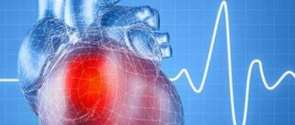
The scar on the heart is presented as a focus of necrotic tissue . Dead cells form tissue that is no longer able to perform its functions. The heart muscle cannot recover, which means that the cells that have died will be replaced by connective tissue , which heal the site of necrosis, but do not perform the functions of cardiac tissue. A scar forms after a myocardial infarction, and failure to take measures for treatment can lead to it. In addition, any disturbances in the functioning of the heart muscles affect the functioning of the cardiovascular system and can lead to serious complications and even death.
The formation of a heart muscle scar is similar to the formation of a skin scar . When tissue or muscle ruptures or dies, cells are replaced by new ones. In order for the healing rate to be high, the protein fibrin is included in the work, which provokes the development of connective tissue at the site of the lesion.
Development mechanism
At the time of the development of an acute infarction, a sharp disruption of the blood supply to the myocardium occurs for the following reasons:
- Rupture of an atherosclerotic plaque under the influence of a sharp jump in pressure, increased heart rate and acceleration, and accelerated blood flow through the coronary vessels.
- Blockage of blood vessels due to blood thickening (acceleration of platelet aggregation, activation of the coagulation system, decreased rate of blood clot lysis).
- Spasm of the coronary artery (vasoconstriction).
I often observed patients in whom several factors were identified as the cause of the disease with myocardial damage. In young patients, vasospasm is often the basis of pathological disorders, which is not possible to determine after the start of treatment.
Symptoms of arrhythmia
Arrhythmias may not appear. A doctor can detect an arrhythmia before it shows any signs during a routine clinical examination. But more often, heart rhythm disturbances cause noticeable changes in the condition, which include signs:
- Feeling of palpitations and chest pains
- Very fast heartbeat
- Extremely slow heartbeat
- Chest pain
- Shortness of breath
- Dizziness
- Loss of consciousness or feeling close to fainting
Even such significant symptoms of ill health do not always indicate a serious problem. Very often, people who experience an arrhythmia do not suffer from severe heart disease, while a person with a life-threatening arrhythmia may not have any complaints at all.
Normal heartbeat
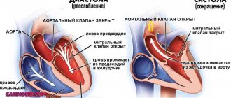
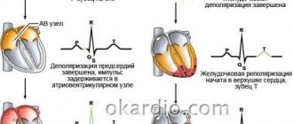
The heart consists of 4 cavities. On each side, on the right and left, there are two pumps: on top of the atrium and below - the ventricles.
During cardiac contraction, chambers with a thin muscle layer and smaller sizes contract, helping to fill the relaxed ventricles with blood. Contraction begins when the sinus node—a small group of cells in the right atrium—sends an electrical impulse that causes both atria to contract. The impulse then travels to the atrioventricular node, located in the very center of the heart and lying at the junction of the atria and ventricles. Leaving the atrioventricular node, the impulse passes to the ventricles. As a result, the latter contract and push blood to all organs.
In a healthy heart, this process occurs evenly and constantly with a heart rate of 60-100 per minute in a calm state. In athletes, especially athletes at rest, the heart rate is usually less than 60, since their heart is much more trained than that of an ordinary person and has great muscle strength, pushing out a large volume of blood per contraction. In children, on the contrary, the pulse is normally more than 100 beats per minute, and in infancy it is 140-160 beats per minute.
Expert advice
I strongly recommend starting treatment in a hospital immediately after an acute attack, since only in this case is it possible to limit the further spread of necrosis and minimize irreversible changes in the myocardium.
The study of histological samples confirms the destruction of the cardiac myocyte 20 minutes after the development of ischemia. After 2-3 hours of lack of oxygen, their glycogen reserves are depleted, which marks their irreversible death. Replacement of myocarditis with granulation tissue occurs within 1-2 months.
As my practice and the observations of colleagues show, the scar on the heart is finally consolidated after six months from the moment the first symptoms of acute infarction appear and is a section of coarse collagen fibers.
Classification
Heart scars can be classified according to their location and extent of distribution.
They can be located along the coronary vessels:
- Impairment of blood flow in the anterior interventricular artery leads to ischemia with the subsequent appearance of a scar in the area of the septum between the ventricles, involving the papillae and lateral wall, as well as on the anterior surface and apex of the left ventricle.
- The infero-posterior and lateral part is affected when the left circumflex coronary artery is blocked.
- Problems with the blood supply to the myocardium in the right artery results in irreversible changes in the right ventricle and can affect the posterior inferior part of the left ventricle and the septum. But such a violation is extremely rare.
According to the type of distribution, scars can be local (focal), which can be compared to a scar on the body, or diffuse (multiple). Experts call the second option dystrophic changes in the myocardium.
Surgical care for scars
In extremely severe cases, an operation is used in which a pacemaker or a cardioverter-defibrillator is installed to maintain the normal rhythm and conduction of the heart. Several more types of surgical intervention that are used for cardiosclerosis:
- Living heart transplantation from a donor. The operation is performed on people under 65 years of age without a history of serious diseases of the internal organs (which is rare, given coronary artery disease or atherosclerosis).
- Bypass surgery is an expansion of the lumen of narrowed coronary vessels. The operation is performed for severe atherosclerosis.
- Removal of the aneurysm. The bulge most often forms in the posterior wall or in the area of the left ventricle. The operation takes place under general anesthesia and consists of truncation of the protruding area.
Surgery is used as both conventional and palliative treatment. The scar after the intervention usually does not pose a threat compared to existing fibrosis.
How does a scar manifest itself?
The acute period of a heart attack is characterized by a variety of clinical manifestations. The main symptom is pain, which can be relieved exclusively with narcotic analgesics and can be observed from an hour to 2-3 days. Then the pain syndrome disappears and the formation of an area of necrosis begins, which takes another 2-3 days. Then comes a period of replacing the affected area with loose connective tissue fibers.
If the correct treatment tactics are used, the following symptoms are noted:
- development of compensatory hypertrophy;
- rhythm disturbance (which often accompanies the acute period) is eliminated;
- tolerance to stress gradually increases.
If a scar that appears on the heart crosses the conduction paths along which the impulse travels, a conduction disorder is recorded, such as a complete or partial blockade.
In the case of successful recovery after a primary small-focal infarction, I did not notice any significant disturbances associated with the functioning of the heart in my patients.
If patients have formed a large scar or many small ones, the following deviations are observed:
- dyspnea;
- increased heart rate;
- the appearance of edema;
- enlargement of the left chambers of the heart;
- pressure fluctuations.
Symptoms of a cardiac aneurysm
Clinical manifestations depend on the size and location of the aneurysm.
Acute cardiac aneurysm corresponds to the following symptoms:
- weakness;
- shortness of breath with episodes of cardiac asthma and pulmonary edema;
- prolonged fever;
- increased sweating;
- heart rhythm disturbances (bradycardia, tachycardia, extrasystole, atrial and ventricular fibrillation, blockade).
Subacute cardiac aneurysm presents with rapidly progressing symptoms of circulatory failure.
Chronic cardiac aneurysm is characterized by:
- pronounced signs of heart failure (shortness of breath, angina at rest and exertion, a feeling of interruptions in the work of the heart, in the later stages - swelling of the veins of the neck, edema, etc.)
- fibrous pericarditis, causing adhesions in the chest cavity.
With chronic cardiac aneurysm, thromboembolic syndrome may develop (most often the iliac and femoropopliteal segments, brachiocephalic trunk, arteries of the brain, kidneys, lungs, and intestines are affected.
Dangerous complications of chronic cardiac aneurysm include gangrene of the limb, stroke, kidney infarction, pulmonary embolism, and repeated myocardial infarction.
In some cases, rupture of a chronic cardiac aneurysm . It occurs 2-9 days after myocardial infarction and is instantly fatal. Clinical manifestations of a ruptured cardiac aneurysm: sudden sharp pallor (which is quickly replaced by cyanotic skin), cold sweat, overflow of the neck veins with blood, loss of consciousness, cold extremities, noisy, hoarse, shallow breathing.
How dangerous is this?
The most dangerous is the development of a scar as a result of large-focal or transmural infarctions, as well as several repeated violations in different basins of the coronary vessels with diffuse multiple lesions.
In the case of a large area of damage or widespread cardiosclerosis, the remaining healthy cells cannot fully compensate for the work of damaged cardiomyocytes. The frequency and strength of contractions increases in order to provide organs and tissues with oxygen and necessary substances.
As a result, tachycardia develops; with its appearance, the load on the heart becomes even greater, which leads to dilatation of the left ventricle and atrium. As it progresses, blood stagnation appears in the right side with the development of heart failure.
I also observed another type of complication: a scar on the heart after a heart attack with extensive and deep damage to all layers of the organ caused the formation of an aneurysm due to the thinning of its wall.
The reasons for the appearance of such a defect are:
- transmural lesion;
- increased blood pressure;
- increased blood pressure inside the ventricle;
- excessive physical activity of the patient, refusal to comply with the regimen.
An aneurysm leads to the rapid development of heart failure, the formation of a parietal thrombus, and pronounced stagnation in the systemic circulation. Often complicated by severe rhythm disturbances leading to death (paroxysmal tachycardia and ventricular fibrillation).
Treatment of cardiac aneurysm
It is impossible to eliminate a cardiac aneurysm using conservative treatment methods, and when the first signs of heart failure appear, the question of surgery is raised. The main treatment method for cardiac aneurysm is surgical excision and suturing of the defect in the heart wall. In some cases, the aneurysm wall is strengthened using polymer materials.
In the preoperative period, cardiac glycosides, anticoagulants, antihypertensive drugs, oxygen therapy, and oxygen barotherapy are prescribed. Patients are advised to strictly limit physical activity.
The cardiology department of MedicCity has all the necessary equipment to carry out comprehensive diagnostics of a wide range of cardiac diseases. Reception is conducted by highly qualified cardiologists who have completed professional internships in Russia and abroad.
Diagnostics
In order to establish a diagnosis, I conduct a survey and study the medical history (mainly, it includes ischemic heart disease with a history of heart attack). External examination usually reveals increased respiratory rate, weakening of heart sounds during auscultation, the presence of edema, and various rhythm disturbances. I will definitely take a blood pressure measurement.
Then I send you to the following research:
- general and biochemical blood test, coagulogram (will help establish concomitant diseases, cholesterol levels and clotting time);
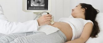
- EchoCG or ultrasound of the heart helps to establish the presence of localized or diffuse areas of connective tissue, allows you to clarify the location and extent of distribution;
- MRI helps to visualize and reliably assess the affected area;
- scintigraphy is required to determine dysfunctional areas of the myocardium.
With the help of an ECG after a transmural and large-focal infarction, it is possible to clarify where the scar is located on the diseased heart.
It is determined by the presence of a Q wave in different leads, as can be seen in the table.
| ECG leads | Localization of post-infarction scar in the left ventricle |
| V1-V3 | Anterior wall with septal involvement |
| V3-V4 | Anterior wall and apex |
| I, aVL, V5-6 | Anterolateral sections |
| I, aVL, V1-6 | Entire front wall |
| II, III, aVF | Posterior wall and diaphragmatic region |
| V7-8 | Posterobasal area |
The increase in the T wave and ST segment, which are characteristic of the acute period, is no longer observed. ST returns to the isoline, T decreases. With a small area and depth of the lesion, no signs of a scar remain; it is leveled, since the function of conductivity and contractility is taken over by neighboring cells. A nonspecific manifestation of dystrophic changes can be a smoothed or moderately negative T wave.
Diagnosis of cardiac aneurysm
In 50% of patients, pathological precordial pulsation is detected.
ECG signs are nonspecific - a “frozen” picture of acute transmural myocardial infarction is revealed, there may be rhythm disturbances (ventricular extrasystole) and conduction (left bundle branch block).
ECHO-CG allows you to visualize the aneurysm cavity, determine its size and location, and identify the presence of a parietal thrombus.
The viability of the myocardium in the area of chronic cardiac aneurysm is determined by stress echocardiography and PET .
Using chest x-ray, it is possible to detect cardiomegaly and congestive processes in the blood circulation.
1 ECG - a method for diagnosing cardiac aneurysm
2 ECHO-CG - a method for diagnosing cardiac aneurysm
3 Chest X-ray
ventriculography, MRI and MSCT are also used to determine the size of the heart aneurysm and detect thrombosis of its cavity.
For the purpose of differential diagnosis of the disease from coelomic pericardial cyst, mitral heart disease, mediastinal tumors, probing of the heart cavities and coronary angiography may be prescribed .
Treatment
Treatment of dystrophic changes includes the use of medications, diet, and lifestyle changes.
Drug therapy
The choice of medications for the appearance of scars depends on the person’s condition.
Typically, after a heart attack and the formation of scar tissue, the following groups of drugs are used:
- statins to lower cholesterol levels (Atorvostatin, Rosuvastatin);
- Beta blockers and ACE inhibitors (Bisoprolol, Ramipril) help reduce the load on the LV and increase its resistance to ischemia
- diuretics allow you to remove swelling and relieve the systemic circulation (Indapamide, Torasemide).
Surgical intervention
When the scar causes complete blockade of the conduction system, treatment consists of installing a pacemaker (pacemaker). An operation (coronary artery bypass surgery) allows you to restore blood flow, when a section of a damaged coronary artery is replaced by another vessel.
In cases of severe degenerative processes and the development of heart failure incompatible with life, an organ transplant is required.
Traditional methods
It is impossible to treat cardiosclerosis and scar changes in the myocardium using traditional methods. They only slightly alleviate a person’s condition, complementing general therapy. However, neither mumiyo nor celandine, as the “healers” write, are capable of ridding the patient of a scar, much less restoring the functioning of the heart.
Myocardial infarction. What it is?
The myocardium is the heart muscle. Blood flows to it through arteries called coronary arteries. If any of these arteries is blocked by a blood clot, then the area of the heart that it supplies is left without blood supply, and therefore without oxygen. “On a starvation diet,” myocardial cells can survive for 20-30 minutes at best. Then they die - this is a heart attack, an area of necrosis in the tissue of the heart. A scar remains on the affected area.
Why does this happen?
The main cause of the disease is atherosclerosis, which almost all of us have. In addition, we will name the circumstances of life (both those that depend on us and those that do not) in which the likelihood of getting sick is highest: male gender; for women, the dangerous age comes after 50 years; heredity (IHD) heart attack, cerebral stroke, in at least one of the direct relatives: parents, grandparents, brother, sister, especially if the disease began before the age of 55); elevated blood cholesterol (more than 5 mmol/l or more than 200 mg/dl); smoking (one of the most significant risk factors!); excess body weight and sedentary lifestyle; increased blood pressure (more than 140/90 mm Hg at any age); diabetes mellitus.
The presence of at least one of these factors actually increases the risk of “acquaintance” with myocardial infarction. Moreover, the addition of each new risk factor increases the likelihood of getting sick exponentially. They also say that baldness in men is a kind of harbinger of a heart attack, since one of the factors in the appearance of baldness is an increased level of androgens, and in the case of hormonal fluctuations, the body reacts to changes in hormone levels by raising blood pressure pressure and increased cholesterol levels in the blood.
What happens?
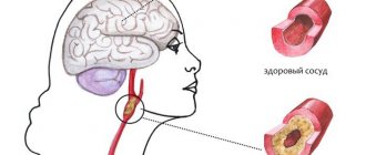
Atherosclerosis is a process in which certain fats (cholesterol and other lipids) are deposited in the walls of large arteries if they are found in excess in the blood. Those places on the vascular wall where there are especially many lipid accumulations are called atherosclerotic plaques.
Plaque is the most vulnerable place in the vascular wall. Especially if it is “young” and calcium has not yet had time to be deposited in it. At the most unexpected moment, the wall of the plaque, and therefore the inner lining of the artery of the heart, can crack and tear. This is an alarm signal for the body. He tries to heal the crack with a blood clot. Therefore, blood immediately begins to clot at the damaged area. The formation of a blood clot resembles a snowball rolling down a mountain. If nothing interferes with it, the thrombus grows very quickly until it closes the entire lumen of the artery. Then the blood flow through it stops, cell death begins and myocardial infarction develops. The larger the artery that the thrombus has closed, the more myocardial cells will die.
The integrity of the atherosclerotic plaque can be disrupted by rapid heartbeat and increased blood pressure. A heart attack can begin during severe physical or emotional stress, but often it develops for no apparent reason, as if out of nowhere. Perhaps even in a dream. But his most “favorite” time is early morning. Depending on the size, infarctions are divided into large-focal, in which necrosis spreads throughout the entire thickness of the heart muscle, and small-focal. Large focal infarctions of the anterior wall of the myocardium are more dangerous. With an infarction of the posterior or lateral walls, especially a small-focal one (not of the full thickness), its consequences are not so traumatic. The scar on the heart muscle remains for life. It cannot resolve, and the heart always “remembers” the heart attack.
Symptoms and diagnosis of heart attack
The first sign to suspect a heart attack is usually severe pain behind the sternum, that is, in the middle of the chest. Usually at rest; presses, burns, squeezes, can radiate to the arm, shoulder, back, jaw, neck. With (angina pectoris, similar pain occurs during exercise, and with a heart attack it is more severe and often begins at rest and does not go away after taking 3 nitroglycerin tablets one after another (1 tablet under the tongue every 5 minutes). If these signs are present, call immediately "Ambulance". The ability to endure in this case is a dangerous enemy. Sometimes the disease manifests itself with vomiting or discomfort in the stomach, interruptions in the heart or difficulty breathing, loss of consciousness or... nothing.
Yes, this also happens: a person suffers a heart attack without even noticing it. The so-called painless form of myocardial infarction is more often observed in people with diabetes. The changes that occur during a heart attack are clearly visible on the electrocardiogram. To clarify the area and extent of the lesion, an ultrasound of the heart, echocardiography, which makes it possible to see structural changes, may be prescribed, the doctor may recommend scintigraphy.
Treatment
To play it safe, at the slightest suspicion of a heart attack, doctors send the person to the intensive care unit of the hospital. And the sooner the better. After all, only during the first few hours, by introducing special drugs, can a “fresh” blood clot be dissolved and blood flow in the coronary artery restored. The formation of new blood clots should then be prevented. For this purpose, medications that slow down blood clotting are used. One of the most reliable remedies is acetylsalicylic acid, that is, regular aspirin. It reduces the number of complications and prolongs the life of people who have had a heart attack. Beta blockers are often used in treatment. These drugs reduce the myocardium's need for oxygen, which means they save heart muscle cells from death and reduce the size of necrosis. At the same time, they make the heart work more economically, which is very important during a heart attack. In recent years, not only medications have been used to treat heart attack. Angioplasty is indicated when drug therapy is ineffective. In another case, a cardiac surgeon may suggest surgery (coronary artery bypass grafting). In the first days, strict bed rest is required. At this time, the damaged heart may not withstand even minimal stress. Previously, a person who had a heart attack did not get out of bed for several weeks. Today, the period of bed rest has been significantly reduced. But all the same, at least three days after a heart attack you need to lie in bed under the supervision of doctors. Then you are allowed to sit, later get up, walk... Recovery begins, adaptation to a new, “post-heart attack” life.
Rehabilitation; after a heart attack
Is it possible to return to normal life after a heart attack? It is difficult to say firmly “yes” or “no”. After all, everyone has their own: one works as a loader, and the other spends his days at a desk. It will not be easy for the first to engage in usual activities, and for the second there will be little to interfere. In addition, complications, and the heart attacks themselves, are different for everyone. Recovery after a heart attack takes several months. After all, the disease is not a joke, it requires you to reconsider your lifestyle and change something in it. Rehabilitation begins in the hospital, where the intensity of physical exercise is gradually increased along with medications and physiotherapeutic procedures. (Particular attention to the word “gradually”). Do not force things under any circumstances. Exercise therapy, walking first on a flat surface, then on stairs... By the way, stairs are a good test of readiness for an active life. If the patient can climb to the fourth floor at an average pace without feeling chest pain or shortness of breath, then recovery is successful. For a more accurate assessment of the condition, a dosed load test is often used. It is usually performed on a special bicycle - a bicycle ergometer or on a treadmill.
How long should I take the medicine? There is only one answer: all your life! Even if you feel great. That’s why it’s wonderful, because medications taken constantly help the heart.
Is it possible to have sex after a heart attack? Feel free to ask your doctor this question. Everything will depend on how you tolerate physical activity (and sexual intercourse is a load, and a considerable one). But there are also rules that are common to everyone, which must be remembered so that sexual contact ends in pleasure and not in a new heart attack. Firstly, it should take place with a familiar partner, secondly, in a familiar environment, and thirdly, when choosing a position for sexual intercourse, you should prefer one in which the load is minimal, for example, on your side.
Prevention
There is no escape from some risk factors (gender and heredity). But everyone else is quite amenable to our efforts! Maintain normal blood pressure, and also monitor the weather - for example, heat and geomagnetic storms are dangerous for people with high blood pressure. Normalize your blood sugar levels.
Move more! It is not at all necessary to run, it is enough to walk in the fresh air at least 5-6 kilometers a day. It is very important to quit smoking - this is one of the most “aggressive” risk factors. Try to lose excess weight. What should be a normal weight? Let's do the math: divide your weight in kilograms by your height in meters squared. The resulting number is called the body mass index. We must strive to ensure that it does not exceed 26. The diet should contain a minimum content of animal fats and cholesterol. More vegetables and fruits. Instead of fatty pork, use white poultry, replace butter with sunflower oil, and lard with fish. This diet allows you to reduce not only your cholesterol levels, but also your costs.
Diet
The diet after a heart attack and scar formation depends on the degree of damage to the heart muscle.
General nutritional requirements are:
- maximum salt restriction;
- reducing fluid consumption (up to 1.5 liters);
- eliminating foods high in cholesterol;
- mandatory use of products with potassium;
- if you are overweight, you should reduce the total number of calories;
- Avoid caffeinated drinks completely.
It should be noted that currently there are no effective ways to completely get rid of a heart scar. Repeated attempts to restore tissue using stem cells have so far been unsuccessful.
Case from practice
A patient came to me for a consultation with complaints of shortness of breath, fatigue, and swelling in the ankles.
History of large-focal myocardial infarction. On examination, the skin is pale and pasty (mild swelling) of the legs. Auscultation - heart sounds are slightly muffled. Blood pressure is within the age norm (130 to 75 mm Hg). The ECG shows signs of dystrophic changes in the posterior wall of the LV, post-infarction scar in the anteroseptal region. After examination, a moderate change in the myocardium and mild hypertrophy of the left ventricle were discovered. A biochemical blood test showed an elevated level of low-density cholesterol.
Diagnosis: IHD. Post-infarction cardiosclerosis, heart failure I. Received treatment with beta blockers, prescribed Aspirin Cardio in a minimal dosage, and a diuretic. She recommended lifelong use of statins (Atorvastatin) under the control of cholesterol levels. She prescribed a diet with a low amount of salt, limiting fluid to 1.5 liters per day, stopping smoking and drinking alcohol. After 2 weeks of treatment, pastiness is not detected, shortness of breath has decreased, and exercise tolerance has increased.
The effect of coronavirus on blood vessels
As noted above, the SARS-CoV-2 virus binds to ACE2 receptors in the endothelial cells lining human blood vessels. Since these cells are involved in the control of blood clots, scientists hypothesize that if the endothelium is damaged by the SARS-CoV-2 virus, the regulation of the blood clotting system is disrupted, resulting in the formation of blood clots .
Damage to the walls of blood vessels can also provoke inflammation and blood clots, which can lead to a heart attack, stroke or pulmonary embolism (PE) . Due to increased blood viscosity, thrombus formation, and vascular damage, acute myocardial infarction may occur. The risk of an attack remains even after discharge from the hospital.
In addition to large vessels, coronavirus also affects small vessels, in which hemorrhages occur from blood clots blocking them.
In patients with a history of chronic diseases of the cardiovascular system before infection with the SARS-CoV-2 virus, there are already changes in the walls of blood vessels (plaques, blood clots, thickenings). COVID-19 worsens the condition of blood vessels, which in turn can lead to a blood clot breaking off and leading to a subsequent heart attack.
Thus, as a result of the action of the SARS-CoV-2 virus, an excessive immune reaction, and side effects from the use of medications, some patients with coronavirus may experience pathologies of the heart and blood vessels after coronavirus, such as:
- tachycardia (rapid heartbeat);
- arrhythmia (rhythm disturbance);
- heart failure;
- inflammation of the myocardium (myocarditis);
- thrombosis;
- pulmonary embolism (PE);
- stroke;
- myocardial infarction, atypical heart attack.
Symptoms of dysfunction of the cardiovascular system:
- pain in the heart area;
- shortness of breath, feeling of lack of air;
- back pain between the shoulder blades, in the shoulder or left arm;
- discomfort in the chest;
- pallor, bluish tint to the cheeks, nose, lips, limbs;
- a cough that is not associated with a cold or flu and does not go away after taking an expectorant; increased heart rate;
- swelling in the limbs, which intensifies towards the end of the day;
- interruptions in heart rhythm;
- absent-mindedness, fatigue, constant anxiety for no reason;
- sleep problems;
- lack of appetite;
- hyperemia;
- increased sweating.
If you have had COVID-19 and are bothered by one or more of the above symptoms, contact your cardiologist immediately for advice. The doctor will select treatment and, if necessary, refer you to a phlebologist or cardiac surgeon for consultation.