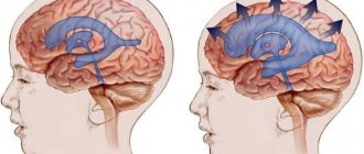
The anatomy of the cranium is designed in such a way as to protect the main organ of the human body. In addition to the dense bone shell, the brain is protected from shocks by the liquor fluid that fills the free space inside the skull. The same substance is found inside the brain, in capsules called ventricles. In situations where the mechanism for the production or natural outflow of cerebrospinal fluid is disrupted, the pressure in the head increases, an anomaly occurs, which in clinical practice is called intracranial hypertension (ICH).
Causes of the disease
The internal cavity of the skull is a closed space. If cerebrospinal fluid accumulates excessively in this area, there is nowhere for the tissue to move. Areas of the brain are compressed, displaced relative to their original position, and blood vessels are compressed, which leads to critical changes in living fibers and cessation of normal blood flow. Internal pressure is considered normal from 7.5 to 15 mmHg. Exceeding the specified values by up to 30 mm RS can lead to death.
The following factors lead to the development of ICH:
- congenital developmental anomalies;
- extensive infection, previous meningitis;
- tumor formations in the brain;
- stroke;
- delay in normal fluid outflow (hydrocephalus);
- penetration of the encephalitis virus into tissue;
- post-traumatic hematomas;
- internal hemorrhages;
- high level of blood clotting;
- diseases in the cervical spine;
- large body weight;
- intoxication with alcohol or medications.
Establishing diagnosis
Only a neurologist, after thorough examinations, can make a diagnosis of high intracranial pressure. The main difficulty is that there is no generally accepted standard for ICP indicators. The experience of many years of research allows us to conclude that pressure values from 70 to 220 mm of water can be considered normal. pillar
Papilledema detected by an ophthalmologist may be a sign of ICP. X-rays of his skull show areas of thinning of his bones. A successful diagnostic method was lumbar puncture. When a needle is inserted into the cerebrospinal fluid space, cerebrospinal fluid flows out in strong jerks.
The use of electronic sensors requires craniotomy and therefore becomes an extremely undesirable way to make a diagnosis. This method is useful in severe cases of ICP to monitor pressure dynamics in the brain. Among the instrumental methods, MRI and CT of the head, ultrasound of the vessels of the head, biopsy of intracranial tumors are most often used; in children, neurosonography is performed through the fontanel.
Signs of pathology
Intracranial hypertension has no specific manifestations. Symptoms of the anomaly are characteristic of many brain diseases, but they help doctors suspect pathological changes inside the skull and serve as a reason for targeted diagnosis. Symptoms of ICH are represented by the following list:
- systematic headaches;
- a feeling of heaviness in the head, especially at night and in the morning;
- nausea not associated with food, vomiting that occurs “for no reason”;
- loss of mood and strength, nervousness, inability to perform prolonged physical and mental stress;
- dark circles around the eyes, resulting from the expansion of small vessels in this area;
- dependence on meteorological conditions.
Since in childhood the sutures of the skull still retain mobility, the signs of the disease have features that differ from those in adults:
- visual enlargement, protrusion of the eyes;
- a large head that is incommensurate with the norm;
- convexity of the fontanel zone;
- rolling the eyeballs upward with the eyelid half lowered;
- frequent restlessness or drowsiness;
- paroxysmal vomiting.
Prevention and prognosis
The outcome of liquor-hypertensive syndrome depends on the underlying disease, pressure level and adequate surgical therapy. In severe cases, the patient's death is possible. ICP of unknown origin responds well to therapy. If a child remains in this state for a long time, the most severe complication may be mental retardation.
Preventive measures that prevent the development of intracranial hypertension include: current diagnosis and treatment of intracranial neoplasms, dyscirculatory disorders, regular use of blood pressure-normalizing drugs in the chronic course of the disease, adherence to a daily routine, proper management of pregnancy and obstetric care.
Diagnostic methods
When the first signs of an abnormality appear, a neurological examination and history taking are performed. If ICH is suspected, invasive measurement of intracranial pressure is prescribed. A needle connected to a pressure gauge is inserted into the spinal canal or cranial cavity. This manipulation is painful, as it involves instrumental penetration.
An alternative that can cope with the diagnosis of ICH is magnetic resonance imaging. The hardware method is absolutely painless and does not involve violating the integrity of living tissues. The patient is placed in a machine that takes pictures of the internal structures. MRI allows not only to detect the main abnormality - intracranial pressure, but also to assess the degree of damage to the central nervous system.
MRI scans reveal the volume of fluid in the cavities, the condition of the vascular network and large arteries, the extent of displacement of brain areas, and areas with impaired blood circulation. Thanks to high resolution and maximum clarity, specialists can detect the root cause of the disease: hematomas, tumors, cysts, foci of inflammation, infection, etc.
NORMOTENSIVE HYDROCEPHALUS
Normal pressure hydrocephalus (NPH) is associated with impaired CSF absorption, and dilatation of the cerebral ventricles develops in the presence of normal intracranial pressure.
EPIDEMIOLOGY
The prevalence of NG is 1-2 cases per 1,000,000 people. In the practice of a neurologist specializing in patients with extrapyramidal diseases, as a rule, no more than a dozen patients are recorded per year. This type of hydrocephalus is more common in older people. The condition should be excluded in persons over 60 years of age with a combination of cognitive impairment (dementia), pelvic organ dysfunction (usually urinary incontinence) and walking impairment (lower body parkinsonism) - the Hakim-Adams triad. Due to the fact that in a significant proportion of cases, bypass surgery in the early stages leads to improvement in walking, it is important to suspect and confirm this condition in a timely manner.
CLINIC
The structure of cognitive impairment is dominated by frontal-subcortical disorders: decreased activity, apathy, and behavioral disorders. Walking disorders have been described as magnetic gait, gait apraxia, and frontal ataxia. Patients experience the greatest difficulty when starting to walk. The area of support is increased, the length and height of the step is reduced, the smoothness of movements is impaired, and there is a progressive slowdown in walking with each step. Patients always have postural instability; often, upon questioning, you can find out that there have been falls before.
DIFFERENTIAL DIAGNOSIS
However, other diseases with cognitive and motor impairments may have a similar pattern of impairments. When carrying out differential diagnosis, the following are considered: vascular dementia (discirculatory encephalopathy stage III), Parkinson's disease with dementia and dementia with Lewy bodies, Alzheimer's disease. The main diagnostic method is MRI of the brain. The examination reveals the expansion of the lateral ventricles, the rounded shape of their anterior horns, and the smoothness of the relief of the cerebral cortex. It is important to use the Evans ventricular-hemispheric index, which is the ratio of the distance between the most distant points of the anterior horns of the lateral ventricles to the largest internal diameter of the skull. Ventriculomegaly is diagnosed if the index exceeds 0.31. NG is characterized by changes in the periventricular white matter, similar to leukoaraiosis (see above). Their severity correlates with the degree of cognitive impairment].
Figure 2 MRI signs of cerebral atrophy (A) in Alzheimer's disease and (B) normal pressure hydrocephalus
At first glance, the images are quite similar, but the image on the right shows the rounded shape of the horns of the lateral ventricles and the smoothness of the sulci of the cerebral hemispheres.
Figure 3 Gliotic and atrophic changes after traumatic brain injury, replacement hydrocephalus
Treatment options
Treatment tactics are structured as follows: eliminating the root cause of the disease, accelerating the natural outflow of cerebrospinal fluid, relieving symptoms. If hypertension is a temporary complication of any other disease, manual therapy is prescribed to help restore normal blood circulation. At the same time, the drinking regime is regulated and a set of exercises is prescribed to relieve pressure inside the head.
More complex anomalies require a course of medications aimed at accelerating the absorption of fluid and its removal to the outside. The basis of drug therapy is diuretics. In some cases, long-term or permanent (lifelong) use of such drugs is required.
In critical cases, when the disease poses a direct threat to life, surgical treatment is performed. If there is a tumor blocking the outflow of cerebrospinal fluid, the possibility of its removal is considered. To urgently reduce pressure, shunts are inserted into the skull to drain excess fluid.
Intracranial hypertension in children
Increased HF in infants and children of the first years of life can be caused by: unfavorable course of pregnancy (infectious diseases of the woman during this period, intrauterine hypoxia), difficult birth accompanied by birth injuries, oxygen starvation during childbirth, congenital anomalies/malformations ( hydrocephalus ), prematurity, neuroinfections, inflammatory diseases of the membranes of the brain ( encephalitis , meningitis ).
As a rule, ICH in children of the first years of life is manifested by regurgitation, general anxiety, autonomic disorders (increased sweating , marbling of the skin, etc.), sleep disturbances, rapid increase in head circumference, pulsation/tension of the large fontanel, tremor of the chin/hands, weather sensitivity, symptom Graefe (when moving the gaze downward, a lag of the upper eyelid from the iris is noted). If such symptoms are present, parents should contact a pediatric neurologist.
Monitoring the effectiveness of treatment
After surgical treatment, it is important for doctors to assess the state of the central nervous system and determine the likelihood of relapse. To monitor the quality of treatment, hardware diagnostics - MRI - are also used. It is absolutely safe to carry out such screening several times in a row, since during the examination the body is not exposed to x-ray radiation. The magnetic fields produced by the MR installation do not have a destructive effect on the body.
The scan can be performed in any medical institution that has the necessary technical facilities and trained functional radiologists. In Moscow, hundreds of diagnostic centers located in every district of the city receive services. To quickly find the nearest MRI medical facility, you can use a single city service.
When you click on the link, a search page will open where you can select a diagnostic service and the area where the clinic is located. In response to the request, a complete list of medical institutions will appear, distributed by rating and prices for procedures. Study the information, read reviews, mark the best offers. To make an appointment for a tomography, just call the phone number listed on each page of the site. All service guests who have booked a scanning time through the portal receive a guaranteed discount of 1,000 rubles on any type of diagnosis.
Stepped therapy to reduce ICP
Typically, treatment to reduce elevated ICP is gradual. It is assumed that the patient is already undergoing basic measures aimed at reducing ICP: artificial ventilation, sedative therapy, body temperature, electrolyte composition and blood gases are normalized.
Any disruption of the patient’s synchronization with the ventilator, coughing during sputum suction, is accompanied by an abrupt increase in ICP. To avoid these issues, muscle relaxants and/or deep sedation should be used. Rarely, the patient can remain spontaneously breathing. The next stage of action is started if the previous actions are ineffective.
Hyperosmolar therapy
Traditionally, mannitol or hypertonic solutions (3-20%) sodium chloride (HS) are used. In some cases, doctors may want to consider giving these drugs alternately. Mannitol, given its diuretic effect, is preferable in patients with normo- or hypervolemia. It is believed that a dose of mannitol less than 0.5 g/kg has little effect on intracranial pressure, and a dose of more than 2 g/kg increases the likelihood of kidney damage. In case of hypovolemia, hyponatremia, hypotension, a hypertonic sodium chloride solution is recommended for the patient.
In most cases, hypertonic sodium chloride solutions were more effective than mannitol in reducing ICP. Firstly, the effect of reducing ICP lasts for 4-8 hours after the administration of sodium chloride, versus 2-5 hours when using mannitol. Second, the blood-brain barrier is less permeable to sodium chloride (reflectance 1.0) compared to mannitol (reflectance 0.9). Therefore, when using sodium chloride, the rebound phenomenon does not appear; the “working range” is significantly expanded - up to an osmolarity of 350 mOsm/l.
Mannitol (Mannitol) is administered over 15-20 minutes at the rate of 1 g/kg body weight. After this - 3-4 times a day at the rate of 0.25-0.5 g per 1 kg of the patient’s body. Sodium chloride 7.5% is prescribed at the rate of 3-4 ml/kg (in 20-30 minutes), then 1-1.5 ml/kg 3 times a day. When blood osmolarity is more than 320 mOsm/L, mannitol is not recommended. When using hypertonic solutions (3-7.5%) sodium chloride, blood osmolarity should not be higher than 340 mOsm/L.
If it is not possible to control ICP, the administration of osmotically active drugs should be limited, in most cases, to one to two days. The blood sodium level during this period of treatment should be maintained within the upper limit of normal - 144-155 mmol/l.
Hyperventilation
The use of mechanical ventilation in hyperventilation mode (PaCO2 <30-32 mm Hg) makes it possible for some patients to reduce the degree of ICH for 1-2 hours, and thereby gain time for other methods of combating ICH.
Sodium thiopental
Sodium thiopental is prescribed if there is no effect from the above treatment. In case of hypotension and hypovolemia in humans, this method is contraindicated. The first (loading dose) of sodium thiopental is 10-15 mg/kg (administered over 30 minutes). In the next 3 hours, if the patient's hemodynamics allow, continue administration of sodium thiopental at a rate of 3-6 mg/kg/hour. And then - throughout the day, 2-4 mg/kg/hour.
Use of cerebrospinal fluid drainage
Liqueur drainage through a ventricular catheter is prescribed for hydrocephalus, but it is not always feasible and increases the risk of purulent complications.
Moderate hypothermia
Moderate hypothermia (33-34°C), performed for 1-2 days, is quite effective in reducing ICH.
Decompressive craniotomy
This is an effective but difficult intervention to reduce treatment-resistant ICH.