15.06.2021
Heart valve replacement surgery is a very complex process that requires close attention and high precision. Fortunately, modern medicine allows us not only to perform operations on such an important organ, but also to do it with high efficiency and minimal risk for the patient. Not only older people, but also younger people may need heart valve replacement.
Anatomy of the heart and reasons for replacing its valve
Our heart has four valves - aortic, mitral, tricuspid and pulmonary valve. They ensure the correct passage of blood through the four chambers of the heart during the heartbeat.
Disturbances in structure and, as a consequence, functioning can occur at birth, as birth defects that will affect later life. Also, destruction of valve tissue can result from certain diseases, for example, rheumatic fever or infective endocarditis.
Unfortunately, like the rest of the body, heart valves are also subject to aging. Regardless of the reason, the valve can either weaken (begin to let blood flow in the opposite direction), or vice versa - heal and become more rigid. This will cause the valve to not open completely. Incomplete, incorrect opening of the valve is called "stenosis". When the valve is weakened or stretched, it is called regurgitation or regurgitation.
When is valve replacement required?
The heart is responsible for delivering blood and oxygen to all tissues of the body. Any disturbances in the functioning of the organ inevitably have a negative effect on the body and require treatment. Often the problem can be solved conservatively, that is, without resorting to invasive intervention. But this does not always happen, and in some cases surgical replacement of the aortic valve cannot be avoided. The main problems of the aortic valve include:
- Congenital and acquired defects. This category includes any changes that interfere with normal blood flow and increase the workload of the heart.
- Valve stenosis, in which the ejection of blood into the aorta is impeded.
- Rheumatic diseases, streptococcal infection.
- Aneurysm, aortic dissection.
- Change in shape, valve damage, calcification, sagging valves.
- The valve does not close tightly, blood flows in the opposite direction.
- Inflammation.
Make an appointment with a specialist without queues, at a convenient time
Sign up
+7
When the pathology is at an early stage of development, it can often be eliminated without surgery. In serious cases, when medications cannot solve the problem, surgery is used. An artificial valve made in the laboratory, as well as an element of the patient’s own tissues, can be installed. The second option is cheaper, since you do not need to purchase an expensive prosthesis.
How is open heart valve replacement surgery performed?
If you have been scheduled for surgery on one of your heart valves, you should go to the hospital at least one day before the operation. During this time, the medical staff will carry out all the necessary procedures (CT, ECG) and collect the necessary tests. You will also have time to meet and talk with your doctor. He will tell you all the details of the upcoming operation and also answer any questions you may have.
It's normal to feel nervous before heart surgery, so don't be afraid to express your concerns to your doctor. Be sure to describe exactly what medications you take on an ongoing basis, this may be important.
About an hour before surgery you will be given the first pain medications. The operation itself is performed under general anesthesia, which guarantees deep sleep and absence of pain. As a rule, surgery lasts 3-3.5 hours.
Your relatives may be in a special room waiting for you. When the surgery is completed, the doctor will tell them the results. For all people, depending on age, body weight and gender, anesthesia takes place over different periods of time, which is usually limited to a few hours.
Indications and contraindications for surgery
Indications:
- congenital heart defects;
- lack of results of drug treatment for chronic heart failure accompanying mitral valve stenosis;
- fainting with aortic stenosis;
- aortic valve stenosis, manifested after coronary artery bypass grafting;
- valve calcification.
Contraindications:
- heart attack;
- stroke;
- extreme degree of heart failure with mitral valve stenosis;
- endocarditis.
The first hours after heart surgery
Typically, after heart surgery, a person spends about a week in the hospital. During this time you will be in the intensive care unit (ICU). Family members will be allowed into your room approximately 60 minutes after surgery. Subsequent visits can be arranged in accordance with the rules of the medical institution.
When you wake up, you will be connected to many tubes, catheters and electrodes. Specifically, you will have a breathing tube in your mouth, which will interfere with your ability to speak. Don't worry, there will be a nurse nearby who will understand what you need.
The breathing tube will be removed a couple of hours after surgery. Also, thin tubes will be connected to your arms or neck, through which medications and nutrients will be introduced into your body. These tubes are needed to take blood samples and monitor your blood pressure. Thin wires may protrude from the chest. They are attached to the heart and, if necessary, allow you to quickly connect a temporary pacemaker. There will also be several tubes coming out of your chest that pump out fluid from the tissues surrounding the heart. A tube in the bladder is needed to remove urine, and various electrodes taped to the chest are used to monitor the heart rhythm.
After surgery, you may experience discomfort and soreness in your chest muscles where the incision was made. Don't worry, the wound is stitched up securely, there is no danger. You will be given painkillers for the first few days. As you recover, the need for them will disappear.
2.Stages of operation
At the first stage
the patient is connected to cardiac monitors, the chest is processed, breathing tubes are inserted from an artificial respiration apparatus, which is connected only after the anesthesia begins to take effect (i.e., the only discomfort for the patient may be some soreness and soreness in the throat after the operation).
The anesthesiologist puts the patient under general anesthesia. From this moment on, the patient does not feel anything.
An ultrasound device is placed through the esophagus into the heart area, transmitting an image of the heart to a monitor throughout the operation.
Second phase
- This is the opening of the chest. After marking, the heart surgeon makes an incision on the chest from the top of the rib cage to the navel. With the minimally invasive method, the incision length is two-thirds shorter.
The purpose of the third stage
is connection to a machine that provides artificial blood circulation. During the operation, the blood is enriched with oxygen outside the lungs, then it returns to the aorta and moves through the systemic circulation.
At this stage, the surgeon stops the heart, washes it and places it in a special solution that maintains its viability outside the circulation.
Fourth and fifth stages
- This is actually replacing the valve. The aorta is cut and the diseased valve is removed. If part of the aorta is also affected, it is also removed and a graft is placed. The valve hole is measured to select a new valve of the required size. The new valve is sewn on, then it is necessary to check that there are no leaks through it.
Sixth stage
- this is a disconnection from the artificial blood circulation machine. The aorta is sutured, and the trapped air is removed from the heart cavity. The heart begins to beat under the current of its own blood. If an uneven rhythm is observed, an electric shock is applied, which restores an even pulse.
Seventh stage
- chest closure. The bones and tissues are stitched together, and the sternum is closed with a suture that remains visible for life.
The entire operation usually lasts from 2 to 5 hours.
is important for the outcome of treatment .
It starts in the intensive care ward. Discharge from the hospital occurs 5-9 days after surgery. Postoperative rehabilitation continues on an outpatient basis.
Visit our Cardiology page
Postoperative period
After 1-2 days, depending on how the recovery goes, you will be able to sit up in bed. Then get up and move short distances with the help of others. After a few days you will be able to wash yourself, but under the supervision of a nurse. Once the air tube is removed, you can drink. You may not have an appetite, but adequate nutrition is essential for a speedy recovery. Your doctor will tell you when you can switch from liquid to solid foods.
Every day you will get better and better, however, fatigue and emotional downturns are possible. These are normal consequences of the operation. When the doctor is completely sure that you have recovered sufficiently, you will be discharged from the hospital to go home. It is advisable for one of your relatives to meet you and take you. As a last resort, take a taxi. Taking public transport or driving a week after open heart surgery is not a good idea. Please note that during the first days you should be under the supervision of loved ones or a caregiver.
Causes of heart valve defects
Heart valve defects in adults are diseases that are based on disorders of the valve apparatus (valve leaflets, annulus fibrosus, chordae, papillary muscles), congenital or developed as a result of diseases and injuries, disrupting proper blood flow through the valves and leading to serious complications, such such as severe heart failure, syncope, arrhythmias, sudden cardiac death, etc.
Stenosis is a narrowing of the valve opening for various reasons, as a result of which the heart has to work excessively to overcome this obstacle.
Valve insufficiency is a malfunction of the valve leaflets, leading to their incomplete closure and the return of blood back into the heart chamber, which leads to the heart working with excess blood volume.
Recovery after heart surgery
When you find yourself at home, try to quickly return to your usual sleep and wakefulness routine. It is advisable to find time for a short nap after lunch. You should weigh yourself regularly for several weeks and record your weight. You most likely lost weight while you were in the hospital. You will gain weight, but be sure to tell your doctor if you are gaining weight quickly. Accelerated weight gain may indicate that fluid is accumulating in the body, and this is dangerous!
The rest is normal rehabilitation. If your doctor allows it, take short walks, preferably accompanied by loved ones or a caregiver. You can also do low-intensity exercise on an exercise bike. Try to do a little more every day than you did yesterday. However, don't overdo it. If you are tired, rest, if you are rested, exercise.
Within six months you will feel better almost every day. Regarding daily activities, you should not lift or move objects weighing more than 2.5 kg. You can do simple housework, go to the movies, cafes, and attend social events. If you drive a car, you can move around in it. It will be useful to climb the stairs, but you need to start small, preferably under the supervision of loved ones.
Rehabilitation of patients after heart valve implantation
After heart transplant and valve replacement surgery, the patient is transferred to the intensive care unit, where medical staff monitors important body indicators in real time. Subsequently, the patient gradually returns to normal life.
During rehabilitation, a person should follow the following rules:
- mandatory medication use. Installation of donor implants requires the patient to take lifelong medications that suppress the immune system, which prevents the rejection of foreign tissue. Another mandatory medication after valve replacement surgery is anticoagulants that prevent the formation of blood clots.
- Moderate physical activity. Cardiology clinics abroad offer people after heart surgery to undergo a recovery course with a specialist in physical therapy. An individually designed course of physical exercises will help the patient return to their normal lifestyle as soon as possible.
- Regulating diet. To prevent the development of coronary heart disease after surgery, doctors recommend limiting the consumption of animal fats and carbohydrates. It is also advisable for the patient to give up coffee and salt.
Nutrition and medications during the rehabilitation period after heart surgery
During your stay at the facility, you will be given detailed information about the diet you will be following. In short, nutrition should be healthy. Most likely, you should limit salt, sugar, and try to reduce the amount of trans fats and saturated fatty acids.
In addition, you will have to change your drug therapy. You should only take medications that your doctor prescribes. Without consulting a specialist, do not take medications that were taken before surgery. If you have been prescribed anticoagulants, then approach this very responsibly. Don’t miss a dose, and if you still couldn’t drink it on time, consult your doctor. No need to take a double dose next time!
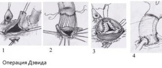
Method of mitral heart valve replacement, effectiveness of the operation and prognosis
Mitral valve replacement is an operation to replace one (several) valves with a prosthesis (biological and artificial). The procedure significantly prolongs the life of patients with heart pathologies and minimizes the manifestation of symptoms of the disease. This is a good alternative to heart transplant surgery instead of the long wait for a donor.
A defective bicuspid heart valve must be replaced, the reason why is calcification, heart failure, fibrous condition of the valves. Cardioplegia or AIC is used, during which long-lasting mechanical (tissue) valves are installed. They can reduce the risk of developing thromboembolism and endocarditis.
Varieties
The most durable prosthesis with excellent survival rate and a low threshold for the development of infection is the pulmonary valve, the installation of which is often performed in young patients aged 20-25 years, who can easily survive a complex operation.
In older patients, surgeons often install the patient's own heart valve instead of the aortic one.
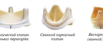
Substitutes used:
- disc prostheses;
- hinged bicuspid dentures;
- from pork heart with a high risk of thrombosis.
8
24/7
The disadvantage of outdated disc models is the high risk of complications: ischemic stroke, femoral thrombosis and pulmonary embolism, thrombus formation in the valve leaflets.
- Mechanical valves, made of synthetic semi-circular structures, move in one direction, are durable, strong and wear-resistant. Patients after surgery require lifelong anticoagulant therapy.
- Biological artificial models made from pig hearts wear out quickly. The service life does not exceed 12 years, so for elderly patients after 65 years of age, the best prosthetic option is to install their own pulmonary valve. The risk of developing re-inflammation remains.
Manufacturers guarantee a service life of mechanical valves of up to 15 years, although they may wear out faster. Often, after 7-8 years, patients need repeat surgery. In the case of mitral valve replacement, the price depends on the type of prosthesis.
Indications for prosthetics
Mitral valve replacement is associated with certain risks and is considered a technically difficult procedure. Doctors of various profiles, most often cardiac surgeons, take part in it. With a defect, a large load is placed on the heart, which can complicate the surgical procedure.
Mitral valve replacement is fraught with complications, although to this day the operation remains in demand when drug treatment is ineffective.
Main indications for prosthetics:
- mitral valve insufficiency;
- fibrosclerosis;
- stenosis;
- deposition of calcium salts;
- wrinkling or shortening of the gate;
- narrowing of the valve in the hole;
- impossibility of eliminating the defect by cutting the valves;
- sclerosis of the tendinous chord.
Surgical correction is required when there are structural changes in the mitral part of the valve, when blood flow is clearly impaired, or when blood flows in only one direction.
Contraindications
Mitral valve replacement may become dangerous and ineffective if the patient has:
- blood clotting disorder;
- chronic diseases of internal organs;
- severe form of heart disease.
Preparing for surgery
The preparatory stage before mitral valve replacement consists of examining the patient:
- chest x-ray;
- general urine analysis;
- blood test for biochemistry;
- blood clotting test;
- Ultrasound of the heart and blood vessels;
- electrocardiography;
- coronary angiography for medical reasons.
Patients need to consult a therapist, cardiologist, and anesthesiologist.
The main preparatory procedures are taking a contrast shower and the last meal 8 hours before surgery. You need to get a good night's sleep and calm down.
Prosthetic methods
Mitral valve replacement on the heart is performed using an open or endovascular method.
Open intervention
Open surgery under anesthesia (anesthesia) involves the surgeon working on the anterior surface of the chest with dissection of the sternum in the longitudinal direction, opening the pericardial cavity.
The patient is connected to a heart-lung machine to disconnect the valve from the systemic circulation. An implant is installed on a non-functioning heart to avoid myocardial hypoxia. Also, throughout the entire procedure, the affected organ is treated with cold saline solution.
The open method of surgery is highly traumatic, the recovery period is long.
When suturing the surgical wound, sensors can be applied for temporary cardiac stimulation, and wire sutures can be applied to the edges of the sternum for rapid tissue fusion.
8
24/7
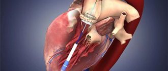
Endovascular intervention
The technique does not require anesthesia. It is performed on a beating heart and without connecting a heart-lung machine. A catheter is inserted into the heart cavity and an implanted valve is inserted. The process of implanting a valve into a damaged area is often quick and easy, since the implant is quite flexible and plastic. Additionally, if necessary, stenting of the coronary vessels is performed.
The endovascular method is minimally invasive. If the vessels and the mitral valve are simultaneously affected, and patients have atherosclerosis, then doctors can eliminate 2-3 pathologies at once in one operation.
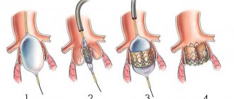
Inaccessible method
The method is minimally invasive, since an operation is performed to replace the mitral valve with an incision of the anterior chest wall of only 2.5 cm. A catheter is inserted into the affected area of the valve through the projection of the apex of the heart. Then, by analogy, as with the endovascular technique, the surgeon performs the implantation of the implant.
The choice of type of prosthesis and technique directly depends on the technical equipment of the clinic, the patient’s well-being, and existing contraindications. Open intervention on the mitral valve is considered the most dangerous and traumatic. The endovascular technique is more advanced, but expensive. Although the advantages are obvious, it is allowed to be performed on both young and elderly patients.
For young patients, doctors can offer vulvoplasty as a safe method of mitral valve replacement, reviews of which confirm its effectiveness. The operation is carried out without installing an artificial prosthesis, solely due to the expansion of the lumen of the artery of its own valve during surgical manipulations.
Period after surgery
The operated patient is placed in the intensive care unit for a day. If the condition on the second day is satisfactory, then he is transferred to a regular ward.
If the mitral valve replacement surgery was performed in an open manner, daily treatment of the sutures with antiseptics is necessary for 7-10 days. If an endovascular technique was performed, then after 3-4 days the patient goes home.
After the intervention, unpleasant sensations may be observed for a month: burning and chest pain. The open method of surgery leads to nagging pain in the heart area, redness and swelling at the suture sites.
If the wound has festered, you should definitely see a doctor for antiseptic treatment.
The rehabilitation period averages 6-8 months. Additionally, patients are prescribed drug therapy:
- anticoagulants for blood thinning;
- painkillers;
- antibiotics if there is a high probability of wound infection after surgery;
- beta blockers, calcium antagonists;
- ACE inhibitors;
- diuretics if patients have hypertension, angina, arrhythmia.
Blood clotting tests are performed regularly, as is monitoring of INR levels. The examination helps prevent stroke and bleeding, which can be caused by a foreign object in the heart.
Against the background of the operation, the following conditions are not uncommon:
- emotional lability;
- insomnia;
- apathy;
- decreased vision;
- depression.
Installing a prosthesis on the heart necessarily requires a radical revision of lifestyle and nutrition, and giving up bad habits. For the first time after surgery, you should come to see a cardiologist in a month. Donate blood and urine. If the indicators are normal, then the next time you will have to visit the doctor in a year.
It is important to review your diet and exclude fried, smoked, salty and fatty foods. Give preference to vegetables, lean fish and meat.
Mitral valve replacement is a restoration of ventricular function and cardiac contractility, and a reduction in intraventricular pressure.
Even a successfully performed surgical intervention to replace the mitral heart valve does not provide a 100% cure prognosis.
It is worth understanding that even the best dentures are not eternal and have a limited service life. After mitral valve replacement, a repeat operation may be required. That is why it is extremely important to identify malfunctions of the implant in a timely manner, because the individual characteristics of the heart do not always coincide with the artificially installed valve. Reduced pressure inside organs remains in patients for life and requires regular attention from a cardiologist.
8
24/7
Mitral valve problems
The mitral valve has the same problems as other heart valves. These problems may develop slowly and without symptoms initially, but sometimes deficiency is rare.
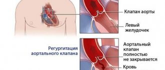
- Mitral regurgitation. Regurgitation is a condition in which the valve leaks and blood flows back out of the left ventricle. This occurs when the valve fails to close completely (mitral valve prolapse). Due to the leakage, the pressure in the pulmonary veins increases, which causes congestion in the lungs. In the initial stages this may result in palpitations, but severe regurgitation can lead to heart failure.
- Mitral stenosis is a thickening of the walls of blood vessels, which makes the valve difficult to open. Stenosis is most often caused by rheumatoid arthritis, and can develop without symptoms for decades and require no treatment. In advanced stages, the pressure in the left atrium increases, causing the heart chamber to enlarge and fluid to accumulate in the lungs.
Both mitral stenosis and mitral regurgitation in advanced stages may require surgery to repair or replace the valve.
Causes of aortic valve disease
There are many pathological conditions that over time can lead to degenerative changes, calcium deposition on the aortic valve leaflets, and changes in its function.
The human heart is able to compensate for poor circulation for some time. Sooner or later, clinical manifestations occur: dizziness, shortness of breath, fainting, palpitations, angina syndrome. Conservatively (with the help of medications) it is possible to partially compensate for the patient’s condition, but it will not change it radically and will not improve the quality of life.