Platelets
(TC)
– the most important element of blood clotting. They store the protein thromboplastin in themselves and, if necessary to urgently stop bleeding, they explode and throw it into the blood. A lack of TC indicates a possible increased bleeding, a threat of bleeding, an excess indicates a tendency to thrombosis. Both are bad. The norm ranges from 180 thousand to 320 thousand. The number of TC in the analysis strongly depends on the state in which the person took the test - nervous, smoked before donating blood, or took alcohol the day before. A deviation from the norm of 10% is acceptable.
Neutrophils
They belong to leukocytes. These are “warrior” cells. They live up to 6 days and are busy guarding the “borders” - the mucous membranes. They are divided into 2 types: segmented-nuclear (Xia) and band-nuclear (Pya). The first are “veterans”, experienced fighters with bacilli, the second are “recruits”, freshly synthesized, inexperienced and weak. Normally, Xia should be 10 times greater than Pia. A change in this ratio towards Pya indicates that there is inflammation, the immune system is tense and throws immature cells into battle. Most often, this picture is observed in prolonged inflammatory conditions.
Lymphocytes
(Lts)
- cells of the immune system. This is the “officer corps”, among which there are both “executors” and “performers”. But Lc from the general analysis is all lymphocytes, without division into types. In order to analyze them in more detail, an immunological analysis is required, which is more labor-intensive, expensive and takes up to 3-4 days. A decrease or increase in Lc indicates the activity of the immune system: a decrease indicates its weakening, and an increase (with a simultaneous decrease in Xia and Pya of neutrophils) indirectly indicates that there is a viral infection in the body.
Negative effects of elevated neutrophil levels
Too much neutrophil activity can increase the release of agents toxic to cells (cytotoxic) and over-activate parts of the immune system. Under normal circumstances, this helps protect your body from infection or injury. However, prolonged and unregulated release of these compounds can cause damage to body tissue. (, )
Diseases of the cardiovascular system
Research shows that neutrophils increase inflammation in plaque in hardened arteries (atherosclerosis). ()
After a heart attack, neutrophils clear away dead cells and debris around the damaged area in the heart. After the neutrophils die, macrophages release growth factors that cause inflammation and tissue formation, which creates scarring in the heart (cardiac fibrosis). ()
A high neutrophil-to-lymphocyte ratio (NLR) is a predictor of cardiovascular disease. It is associated with an increased risk of developing irregular heartbeats (arrhythmias) and death in patients undergoing treatment for cardiovascular disease (percutaneous coronary intervention) [,].
A study of 4,625 people found that higher neutrophil counts were associated with an increased risk of developing cardiovascular disease. () Similar results were obtained in another study with 4,860 men. ()
Another study of 18,558 people found that high levels of neutrophils increased the risk of cardiovascular disease such as ischemic stroke and myocardial infarction. ()
Cancer
In general, neutrophils may play a role in the initiation of cancer , tumor formation , and the spread of metastases to other sites in the body. ()
Neutrophils promote inflammation by releasing reactive oxygen species (ROS). Research shows that they also suppress the antitumor immune response (by stimulating TGF-β to release iNOS or ARG1, which suppresses cytotoxic CD8+ T cells that fight cancer cells). ()
Neutrophils contribute to the development of cancer. (source)
The neutrophil to lymphocyte ratio ( NLR = neutrophil/lymphocyte) is a blood marker that scientists have used to try to predict the outcomes of various types of cancer (colon, lung, head, neck, ovarian, etc.). Numerous studies have shown that elevated NLR values are associated with poor prognosis in colon cancer, pancreatic cancer, gastric cancer, and lung cancer . (, , , )
In a review of more than 100 studies and 40,000 patients, an NLR value greater than 4 was associated with worse overall cancer survival rates. ()
Interpretation of the NLR depends on the clinical context. However, we can roughly imagine how this indicator can be assessed:
- Normal NLR level is in the range of 1-3
- An NLR of 6-9 suggests mild inflammatory stress (eg, in a patient with uncomplicated appendicitis)
- Severely ill patients often have an NLR of ~9 or higher (sometimes reaching values close to 100)
Monocytes
(M)
like neutrophils, they participate in cleansing the body of various aggressors; they usually live on the mucous membranes and in the submucosal layer. Participate in the formation and regulation of the immune response. Their behavior is reminiscent of police officers, because they grab the bacillus and carry it to the lymph node, where “fingerprints” - membranes - are taken from it, and then, based on these data, the Lc produces antibodies - proteins that destroy aggressors. Normally, there are few monocytes in the blood. Their increase indicates inflammation; absence is acceptable if there is no inflammation.
ESR
(Erythrocyte sedimentation rate)
is the only functional indicator that reflects the activity of an inflammatory or autoimmune process in the body. ESR depends on the concentration in the blood of special substances that enter the blood from foci of inflammation. These substances enhance the cohesion (adhesion) of Er, Lc and Tc. Normal ESR for men is no more than 10 mm/hour, for women – no more than 15 mm/hour. These substances are constantly present in the body in small quantities, but in the presence of infection they become more numerous. To keep your blood viscosity normal, you should regularly take blood thinners, such as Trombo ACC, which reduces the stickiness of blood cells. Blood is divided into red and white. Red blood cells, hemoglobin, color index, reticulocytes and platelets are red blood. Leukocytes, neutrophils, lymphocytes – white. Red blood (RB) is responsible for transporting oxygen. A decrease in its parameters indicates a lack of oxygen in the tissues. The synthesis of CK is regulated by the kidneys.
Clinical significance of a blood test (excerpt from a lecture by Prof. E.B. Vladimirskaya)
Hematology:
12.03.2009
Doctor of Medical Sciences, Prof. E.B. Vladimirskaya Research Institute of Pediatric Hematology, Ministry of Health of Russia
Blood, being the internal environment of the body, carries within itself the stigmata of the vital functions of various organs and systems, the study of which is of undoubted clinical significance and is necessary for the diagnosis, prognosis of the course and monitoring of therapy of almost all internal human diseases.
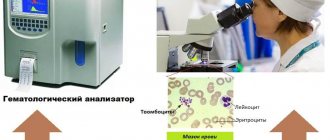
The most accessible is the study of the morphological composition of blood; its results are included in the diagnostic algorithm for most pathological processes.
Since the first studies of blood under a microscope without the use of staining (the middle of the last century), blood cells have been divided into red - erythrocytes (based on the color of hemoglobin) and white - leukocytes. Leukocytes, in turn, are divided into cells containing specific granularity in the cytoplasm, and according to the ratio of this granularity to color, they are divided into (neutrophils, eosinophils and basophils) and those without - lymphocytes and monocytes. Based on the shape of the nucleus, the former are often called polymorphonuclear cells, and the latter are called mononuclear cells.
In recent years, clinical blood tests have increasingly been performed on automatic counters, which significantly increases the accuracy of calculations, but, however, does not negate the value of data obtained “manually” using light-optical microscopy. A comparison of the results of these two methods, together with the reference values of the indicators used, is presented in Table 1.
Table 1
| Automatic counting | Units _ | Normal limits | Manual counting |
| hgb -hemoglobin | g/liter | M: 132 - 173 F: 117 - 155 | Hb |
| rbc- red blood cells | 1012 /liter | M: 4.3 – 5.7 F: 3.8 – 5.1 | er. |
| hct-hematocrit | % | M: 39 – 49 F: 35 – 45 | ht |
| mcv - average volume red blood cell | 1 µm3 = 1 femtoliter | 80,0 – 95,0 | Spherical index (3,2-3,4) |
| mch - average hemoglobin content in an erythrocyte | picograms 1 g = 1012 picograms | 27,0 – 31,0 | Color index (0,85 – 1,0) Color.= Nb (g/l) x 3_____ Er (first three digits) |
| mchc – average concentration of HB in 1 erythrocyte | g/dl | 32,0 — 36,0 | |
| rdwwidth of distribution of red blood cells by volume | width histograms | 11,5 – 14,5 | No analogue |
| plt – platelets | *109 /l | 150 — 400 | Platelets |
| wbc- leukocytes | *109 /l | 4,5 –11,0 | Leukocytes |
| neu - neutrophils | *109 /l % | 1,8 – 5,5 47,0 – 72,0 | Neutrophils |
| lym – lymphocytes | *109 /l % | 1,2 – 3,0 19,0 – 37,0 | Lymphocytes |
| mon – monocytes | *109 /l % | 0,1 – 0,9 3,0 – 11,0 | Monocytes |
| eos – eosinophils | *109 /l % | 0,02 – 0,3 0,5 – 5,0 | Eosinophils |
| bas – basophils | *109 /l % | 0,0 – 0,07 0,0 –1,0 | Basophils |
When commenting on the data presented in the table, it is necessary to note:
- the vast majority of automatic counters do not detect young forms of leukocytes, normoblasts and reticulocytes - this data can only be obtained “manually”;
- normative values are never expressed in one figure; there is a limit of permissible fluctuations (it is presented in the table for all indicators), which fits 99.9% of the norm.
Let's analyze the clinical significance of individual blood test indicators.
Red blood counts.
Anemia is a decrease in hemoglobin below 120 g/l: mild - 110-120 g/l; average degree – 90-110 g/l; severe degree – below 90 g/l.
Based on the ratio of red blood indicators, 3 types of anemia are distinguished, which is the starting point for further diagnosis.
Microcytic-hypochromic anemia:
MCV < 80 Color.p. <0.85
MCH < 27; MCHC < 32
- Iron-deficiency anemia
- Anemia due to chronic inflammation
- Congenital spherocytic hemolytic anemia.
- Thalassemia.
Normocytic-normochromic anemias:
MCV 80 – 95
MCH = 27-31; MCHC = 32-36 Color.p. = 0.85-1.0
Acute blood loss.
- Anemia in chronic renal failure
- Anemia due to endocrine pathology.
- Anemia in cancer.
- Hemolytic anemias, immune and non-immune.
- Aplastic anemia.
- Myelodysplastic syndrome.
Hyperchromic macrocytic anemia.
MCV > 95
MCH>31; MCHC = 32-36 Color points > 1.0
- Megaloblastic B-12 is a deficiency (pernicious) anemia.
- Megaloblastic folate deficiency anemia.
- Autoimmune hemolytic anemia.
Thus, when detecting a decrease in hemoglobin, one should first determine the nature of anemia: normo-, hypo- or hyperchromic.
Let us dwell in more detail on iron deficiency anemia and anemia due to chronic inflammation, the diagnosis and treatment of which are the prerogative of general practitioners and do not require special hematological studies.
Iron deficiency anemia (IDA) is detected in 50% of women and 40% of men, representing one of the most common human diseases. The most common cause of IDA is a latent form of bleeding: in men – from the gastrointestinal tract and bronchopulmonary system, in women – menopause and metrorrhagia. Pregnancy is also one of the factors in the development of iron deficiency in women. Insufficient dietary intake of meat products is another significant and at the same time socially determined cause of the development of iron deficiency. In children born with a reserve of iron received from the mother in the last month of intrauterine development, further intake of iron occurs only from complementary foods. Thus, the reasons for the development of iron deficiency in young children may be prematurity, multiple pregnancies, late introduction of complementary foods, infections (increased iron consumption by microbial flora and malabsorption).
The clinical picture of IDA consists of the following clinical syndromes:
1. Anemia: weakness, drowsiness, fatigue, shortness of breath, palpitations, functional systolic murmur, floaters floating before the eyes.
2. Sideropenic syndrome:
· dryness, fragility, hair loss, early gray hair;
· flattening and brittleness of nails;
· dry skin, hyperkeratosis;
· painful, non-healing cracks in the corners of the mouth, tongue, fingers, toes, heels;
· perversion of taste and olfactory addictions;
· frequent infections;
· impaired swallowing, muscle weakness, sphincter weakness (urinary incontinence when laughing, coughing)
The diagnostic algorithm for IDA, in addition to a detailed clinical study to identify hidden bleeding, includes the mandatory determination of basic indicators of iron metabolism. The diagnosis of IDA is confirmed by the following values of these indicators:
- serum iron below 12.5 µmol/l
- total iron binding capacity (TIBC) above 64.4 µmol/l
- serum ferritin below 12 µg/l.
Correction of IDA is carried out by long-term oral treatment with iron preparations (6-8 weeks after hemoglobin restoration).
It is necessary to carry out a differential diagnosis between IDA and anemia in chronic inflammation.
Anemia during chronic inflammation in terms of the morphological composition of the blood is no different from IDA and is also accompanied by a decrease in serum iron content. However, its development is not based on exogenous iron deficiency, but on the impossibility of its utilization. Iron treatment for such anemia is contraindicated. Differential diagnosis is based on the study of iron metabolism indicators and is presented in Table 2.
Table 2. Differential diagnosis between IDA and anemia in chronic inflammation.
| Indicators | ZhDA | Anemia due to chronic inflammation |
| Serum iron | Reduced | Reduced |
| OZhSS | Increased | Normal or reduced |
| Serum ferritin | Reduced | Normal or increased |
To summarize the brief analysis of the clinical significance of the morphological parameters of red blood, we should dwell separately on reticulocytes and normoblasts.
A morphological sign indicating the hemolytic nature of the decrease in hemoglobin is an increase in the number of reticulocytes. With normal hemoglobin, the number of reticulocytes does not exceed 0.5-1.5%. The expected reticulocyte response to hemolytic anemia with intact hematopoiesis is presented in Table 3.
Table 3. Expected reticulocyte response to hemolytic anemia.
| Hematocrit, % | 45 | 40 | 35 | 30 | 25 | 20 | 15 |
| Hemoglobin, g/l | 130 | 120 | 115 | 100 | 83 | 66 | 50 |
| Reticulocytes, % | 0,5 | 1,5 | 5 | 10 | 15 | 20 | 30 |
Dynamic monitoring of reticulocyte levels is also necessary to assess the expected effectiveness of treatment for IDA and pernicious anemia. An increase in reticulocytes is naturally observed on days 5-8 of treatment with iron for IDA and is especially pronounced (up to 60%) on days 5-8 of treatment with vitamin B-12 for pernicious anemia. Such a hematopoietic response to the therapy of these diseases can also be considered as confirmation of the corresponding diagnosis of exjuvantibus.
Normoblastosis in peripheral blood is rare and always indicates a serious pathology. Its appearance is naturally observed in severe forms of hemolytic anemia and in patients who have undergone splenectomy. The detection of normoblasts in the blood of patients who do not suffer from this pathology should be a reason to search for oncological pathology.
about erythrocytosis when the following blood parameters are present: red blood cells above 5.7x10*12/l in men and 5.2x10*12/l in women, hemoglobin above 177 g/l and 172 g/l, respectively, hematocrit above 52% and 48 % respectively.
Primary erythrocytosis is considered to be rare genetically determined familial erythrocytosis and erythremia.
Secondary erythrocytosis, caused by increased formation of erythropoietin in response to arterial hypoxia or in certain tumors, is much more common.
Secondary erythrocytoses can be divided into the following groups:
1. Arterial hypoxia
- Altitude sickness
- Chronic pulmonary failure
- "Blue" heart defects
2. Tumors that produce erythropoietin
- Kidney tumors, hypernephroma
- Adrenal tumor
- Cerebellar hemangioma
- Ovarian cancer
3. Local renal ischemia
- Cyst
- Hydronephrosis
- Renal artery stenosis
4. Harmful production
- Cobalt poisoning
Treatment of secondary erythrocytosis requires elimination of their cause, but can also be symptomatic due to the threat of thrombosis. Symptomatic treatment of erythrocytosis is bloodletting.
White blood counts.
An increase or decrease in the number of different types of peripheral blood leukocytes can be judged only by changes in the absolute number of these formed elements.
Neutrophilia is an increase in the number of neutrophils more than 6x10*9/l.
Less commonly, neutrophilia is a manifestation of chronic myeloid leukemia, accompanied by clinical and hematological features specific to it (enlarged spleen, lymph nodes, blood rejuvenation, anemia, hyperthrombocytosis, bone marrow hyperplasia, the presence of a Ph chromosome and the chimeric c-abl-bcr gene).
Much more often, neutrophilia is a blood reaction to inflammation, the result of exposure to bacterial endotoxin and the release of inflammatory cytokines and chemokines by tissues. Neutrophilic leukocytosis can accompany any inflammation, bacterial, fungal and parasitic infections, necrotic tissue changes, hypoxemia, intoxication and tumors of various locations. With prolonged exposure to factors that induce neutrophilia, the bone marrow granulocyte reserve is depleted and young cells of the neutrophil series (band cells, metamyelocytes and myelocytes) begin to enter the blood. This blood condition is called leukemoid reaction of the neutrophil series. Sometimes it becomes necessary to make a differential diagnosis between such a reaction and the initial form of chronic myeloid leukemia. The absence of anemia, hyperthrombocytosis and a high level of alkaline phosphatase in neutrophils is characteristic of the leukemoid reaction.
Neutropenia is a decrease in the number of neutrophils less than 1.8 x 10*9/l.
Agranulocytosis - a decrease in the number of neutrophils less than 0.5 x 10*9/l.
Neutropenia can be primary (congenital and acquired), associated with blood diseases (acute leukemia, aplasia of hematopoiesis, cyclic neutropenia), and secondary, accompanying diseases, during which destruction and increased consumption of neutrophils occurs.
Secondary, reactive neutropenia includes immune and neutropenia in severe infections. Neutropenia in sepsis is usually accompanied by a rejuvenation of the leukocyte count and is a poor prognostic symptom, indicating depletion of hematopoiesis.
It is necessary to dwell on constitutional, so-called harmless neutropenias. About 4% of people have normal blood composition with a low content of neutrophils. This feature is associated with the genetically determined rapid movement of neutrophils into tissues, where they carry out their inherent protective functions. People with this blood composition are usually less susceptible to intercurrent infections and recover from them faster. However, often such patients, unfortunately, are the subject of close attention of doctors, are subjected to many unnecessary invasive studies, and they develop iatrogenic pathology. Thus, neutropenia, not accompanied by other blood changes and any clinical symptoms, does not require immediate intervention. Such patients require dynamic monitoring.
Separately, I would like to touch upon redistributive neutrophilia and neutropenia. The circulation of neutrophils has its own characteristics: half of the cells circulate with the blood (these cells are to be counted), while the other half is in the “marginal position” at the walls of the vessels. Irritation of the sympathetic system and vasospasm increase the number of circulating cells, while irritation of the parasympathetic system, on the contrary, reduces their number. Hence, stressful conditions contribute to transient neutrophilia (for example, neutrophilia in small children when screaming), and vagotonia - neutropenia.
Eosinophilia – an increase in the number of eosinophils above 0.4 x 10*9/l.
An increased release of eosinophils into the blood occurs under the influence of IL-4 and IL-5, which are formed in increased quantities during immunological tissue damage. Recently, the killer effect of eosinophils has been proven in some helminthiases and parasitic infections. Eosinophilia is a characteristic feature of collagenosis, allergies, many helminthic and parasitic infestations, immunodeficiency, especially hyper-IG-E syndrome, and some tumors.
Monocytosis – the number of monocytes is higher than 0.8 x 10*9/l.
Conditions often, but not always, associated with monocytosis include:
- Infections (especially tuberculosis, endocarditis, syphilis).
- Fever of unknown origin
- Various forms of neoplasia and myeloproliferative diseases.
- Chronic inflammation (especially cholecystitis and rheumatoid polyarthritis)
- Condition after splenectomy.
Lymphocytosis – an increase in the number of lymphocytes more than 4.0 x 10*9/l
Among malignant lymphoproliferative diseases with high lymphocytosis, the most common is chronic lymphocytic leukemia, a disease of people over 45 years of age. The distinctive feature of this lymphocytosis is its monoclonal nature and B-cell origin.
Secondary, reactive lymphocytosis , which is polyclonal in nature, accompanies many viral infections and some inflammatory and immune complex diseases. These include:
1. Lymphotropic viral diseases:
— infectious mononucleosis (atypical mononuclear cells, characteristic clinical picture);
- infectious lymphocytosis (asymptomatic epidemic form in young children - up to 20-30 thousand)
2. Cytomegalovirus infection (atypical mononuclear cells, characteristic clinical picture).
3. Childhood infections: whooping cough, chickenpox, scarlet fever prodrome.
4. Other viral infections: rubella, hepatitis, some respiratory adenoviral infections in the convalescent stage.
5. Inflammatory and immune complex diseases: thyrotoxicosis, ulcerative colitis, Crohn's disease, vasculitis.
Lymphocytopenia - a decrease in the number of lymphocytes below 1.2 x 10*9/l.
It is observed relatively rarely, most often with corticosteroid therapy. May also accompany AIDS, lymphogranulomatosis and various chronic infections (for example, tuberculosis, disseminated lupus erythematosus, sarcoidosis).
Hyperthrombocytosis is considered to be an increase in the number of platelets more than 400.0 x 10*9/l.
Primary hyperthrombocytosis accompanies myeloproliferative diseases and is a consequence of tumor transformation of the megakaryocytic lineage of the bone marrow.
Secondary reactive hyperthrombocytosis is observed:
- After surgical interventions (about 2 weeks).
- After splenectomy (up to 1 year).
- For malignant tumors
- For acute posthemorrhagic and hemolytic anemia.
- For certain inflammations (tuberculosis, acute rheumatism, ulcerative colitis, osteomyelitis).
Thrombocytopenia - a decrease in the number of platelets below 100.0 x 10*9/l most often occurs with tumor diseases of the blood, aplastic anemia and immune thrombocytopenic purpura. Thrombocytopenia is an obligatory component of the hypersplenism syndrome with splenomegaly. It should be borne in mind that a serious threat of bleeding occurs when the platelet count drops below 20.0 x 10*9/l.
Reactive thrombocytopenia is rare and can accompany any immune pathology and disseminated intravascular coagulation.
ESR - erythrocyte sedimentation rate is a nonspecific reaction. Normally, it is 2-15 mm per hour in men under 50 years old, and 2-20 mm per hour in women under 50 years old. After 50 years, men have up to 20 mm per hour, and women – up to 30 mm per hour.
The rate of erythrocyte aggregation depends on their number (it accelerates when their number decreases) and the amount of coarse proteins (inflammatory proteins, fibrinogen, antibodies, gamma globulin, etc.) adsorbed on erythrocytes and accelerating their sedimentation. Based on this, it is clear that there is a wide range of pathologies in which ESR acceleration can be detected.
Thus, analysis of the morphological composition of blood is of great clinical importance, and sometimes is a leading sign in the diagnosis and choice of therapy for many human diseases.
However, it should be remembered that the most important link in such an analysis is the integral assessment of all blood parameters and the mandatory correlation of changes in the blood with the history and clinical manifestations of the disease.
Literature:
1. Guide to Hematology, ed. A.I. Vorobyov, Moscow, 1985
2. Hematology, ed. by WSBeck, London, 1991
3. Manual of Clinical Hematology, ed.by J. Mazza, NY, 1995
Tags: Laboratory, Blood
« Back to article list Share on
Important Additions
Comments are added to any analysis. For example, terms such as “anisocytosis”, “poikilocytosis”, “toxogenic granulation” indicate exposure to various toxins (including antibiotics, chemotherapy drugs, analgesics that are taken for too long) and essentially indicate the presence of damaged, altered bone cells brain (BM) - in both red and white blood. The appearance of so-called Joly bodies and Cabot rings in the analysis indicates possible pathological processes in the BM associated with the development of blood cancers. The presence of these structures in itself does not mean anything - only that a more detailed examination of the blood, bone marrow, and leukocytes is required. The article was published in the weekly magazine TV 7 / Your Health, No. 43 2009.
Elevated neutrophil levels
A test result that shows a slightly elevated neutrophil count may not be medically significant because the number often changes from day to day. Your doctor will interpret the test, taking into account your medical history and other tests, and repeat it if necessary.
Abnormally high levels of neutrophils in the blood are called neutrophilia . ()
Causes of elevated neutrophil levels
Infections and inflammation
Infections caused by bacteria, fungi, viruses and parasites increase the number of neutrophils. () Any inflammatory conditions also increase the number of neutrophils. (, , )
In patients with inflamed appendicitis, the number of neutrophils is significantly higher than normal. However, neutrophil counts do not indicate disease severity and are not sensitive enough to be a marker of disease. ()
Smoking
Research shows that smoking increases the number of neutrophils. () Smoking is usually associated with increased white blood cell counts and neutrophilia (25% increase in these cells), which can last up to 5 years after smoking cessation. The exact mechanism of smoking-induced leukocytosis is unknown.
Psychological stress
The stress response stimulates neutrophils, likely by increasing inflammation . () Stress-associated neutrophilia appears to be associated with the redistribution of neutrophils from the marginal pool (located in the body organs) to the circulating pool (in the blood), probably due to a decrease in neutrophil adhesion during the release of the stress hormone, adrenaline .
Lots of physical activity
Strenuous exercise can increase neutrophil counts in a short period of time. () Exercise-induced neutrophilia is likely due to a combination of elevated adrenaline
in the blood plasma and changes in cardiac output, which leads to the redistribution of neutrophils from the marginal pool to the circulating one.
Lack of sleep
Research has shown that insufficient sleep (insomnia) increases the number of neutrophils, increasing the level of inflammation in the body. ()
Damage to body tissue
Tissue damage due to injury or surgery increases the number of neutrophils. (, )
Depression
A study that measured neutrophil counts in 41 patients with depression found that their neutrophil counts were elevated compared to healthy controls. ()
Medicines and toxins
Some medications (glucocorticoids, adrenaline, lithium, etc.) and poisoning (lead, mercury, insect poison, etc.) can also cause neutrophilia. (, , , , )
Bone marrow problems
Bone marrow disorders, such as leukemia , can increase neutrophil production. (, )
Genetic disorders
A mutation in the CSF3R gene promotes the formation of neutrophils. This may lead to higher susceptibility to hereditary chronic neutrophilia. ()
Obesity
Chronic mild neutrophilia has been observed in obesity. Adipose tissue is capable of releasing inflammatory cytokines, creating a state of prolonged low-level inflammation manifested by elevated levels of C-reactive protein.
A) Obesity is characterized by an increase in adipose tissue and chronic inflammation in which adipocytes secrete adipokines such as tumor necrosis factor alpha (TNF-alpha), IL-6 and IL-8, which are associated with increased peripheral neutrophil activity. B) In mice on a high-fat diet (HFD), there is an increase in neutrophil recruitment to adipose tissue and neutrophil elastase (NE) activity.
Leptin , a hormone produced by adipocytes (fat cells), can act on the membrane protein CD34, leading to increased differentiation of neutrophils. More recently, it was discovered that chronically inflamed visceral adipose tissue in obesity mimics the action of hematopoietic progenitor cells to proliferate and expand, causing both neutrophilia and monocytosis through the production of the cytokine IL-1β.
In an obese person with chronic mild neutrophilia and no other obvious health problems, it can be assumed that fat cells are the cause of the increased neutrophil count.