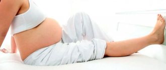
As you know, pregnancy is a risk factor for the development of varicose veins: often, it is during the period of bearing a baby that “snakes” of dilated veins or spider veins appear on the legs. However, it is problematic for expectant mothers to treat this disease - they can only take measures to prevent the progression of the disease. But after childbirth, it’s time to give the disease a worthy rebuff, because now a woman has the opportunity to use the entire arsenal of therapeutic agents for this.
The word "varicose veins" comes from the Latin word "varix" - bloating. Varicose veins are a chronic disease accompanied by various disturbances in the outflow of blood in the veins associated with pathological changes in the venous valves, which often causes changes in the entire vascular system.
The problem of varicose veins has been known since the times of Hippocrates and Avicenna, who devoted entire sections of their works to the description and treatment of vein diseases. In Russia, more than 35 million people suffer from varicose veins, while women are exposed to a much larger number of risk factors compared to men. The problem of varicose veins is especially relevant for women during pregnancy and in the early postpartum period, especially if the disease arose before pregnancy.
How do varicose veins occur?
In order to deal with the problem of varicose veins, it is necessary to understand how the venous system of the legs is structured and how the outflow of venous blood occurs.
The venous system of the lower extremities consists of superficial and deep. Superficial veins are located directly under the skin, deep veins are located in the muscle layer of the leg and thigh. The connection between these two systems is provided by communicating (perforating) veins. In each vein there are valves that ensure blood flow in only one direction - from the periphery to the center.
The flow of venous blood to the heart is ensured by the contraction of the heart muscle, leg and thigh muscles (muscle pump). Another necessary factor is the full functioning of the venous valves, which, similar to heart valves, allow blood to flow in only one direction. When the muscles of the leg and thigh contract, the deep veins of the limb are compressed, and blood from them flows into the veins of the leg and thigh, as well as into the veins of the pelvis. Unmodified vein valves prevent blood from entering the superficial venous system. If the deep vein valves work normally, when the muscles relax, there is no return of blood from the pelvic veins.
An increase in pressure in the veins of the pelvis due to the growth and development of the fetus leads to the gradual destruction of the valves of the veins, as a result of which, during muscle contractions, blood under high pressure flows from the deep venous system to the superficial one. Increased pressure in the superficial veins leads to their expansion. This condition is called varicose veins. A separate group of manifestations of this disease includes the so-called spider veins, which arise due to the expansion of venous capillaries of the skin and subcutaneous tissue. They can be congenital or acquired, appearing anywhere on the human skin. But if they appear on the skin of the legs and are actively increasing, then they should be regarded as a symptom of incipient varicose veins.
Diagnostics
First of all, the doctor conducts an examination with palpation - already at this stage the disease can be detected. Additionally, the following studies can be carried out:
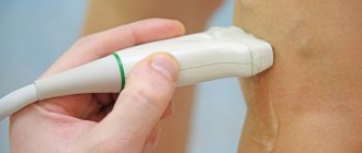
- Ultrasound examination of veins in combination with duplex or triplex scanning based on Doppler sonography. The procedure allows the doctor to examine the veins and valves in detail and determine the stage of the disease.
- Contrast X-ray venography. It consists of introducing a contrast agent into the body and monitoring the entire vascular network using x-rays.
- Functional tests to identify varicose veins, with which you can determine the volume of affected veins and the feasibility of surgical treatment.
These types of diagnostics allow you to obtain comprehensive information about the condition of the veins and the degree of development of the disease, thanks to which the doctor can make an accurate diagnosis and prescribe effective treatment.
Symptoms of varicose veins
Manifestations of varicose veins depend on the stage of the disease. At the beginning of the disease, when irreversible changes in the vessels and capillaries have not yet occurred, the only symptom may be “worms” of dilated veins in the legs. As the disease progresses, fatigue, a feeling of heaviness in the legs, bloating, cramps in the calf muscles (especially in the evening, and sometimes at night), numbness, and a tingling sensation appear. Swelling usually occurs in the evening, especially after prolonged standing or sitting, and disappears completely after a night's rest. Over time, as varicose veins progress and the valves of the veins are destroyed, trophic disorders appear (they are associated with a deterioration in the supply of oxygen and other substances to the tissues) - most often they occur on the inner surface of the lower third of the leg. These are induration (hardening, thickening of the skin), pigmentation, dermatitis (the appearance of areas of inflammation on the skin), then a trophic ulcer, which is difficult to treat.
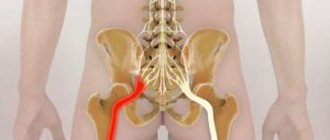
Varicose veins are a common disease of the vascular system, occurring in 50-96% of women during pregnancy [1-4]. The influence of the hormonal factor on the venous wall occurs already in the first trimester of pregnancy [5, 6]. From the second trimester, an increase in the size of the pregnant uterus contributes to an increase in intra-abdominal and venous pressure in the lower extremities and pelvis. The latter is aggravated by the presence of a large fetus, multiple births and polyhydramnios [6-8]. This influence is especially pronounced in the last weeks before childbirth. As a result, various variants of varicose veins account for 5.6% of all extragenital pathology during pregnancy [7-10].
Varicose veins sharply reduce the quality of life of pregnant women, complicate the course of gestation, leading to such complications and consequences as varicose veins of the pelvic organs (50-60%), phlebitis, phlebothrombosis (20%), bleeding during childbirth and the early postpartum period (15 -17%) [7-11].
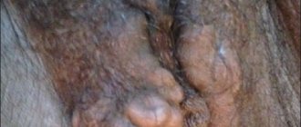
There are two possible variants of varicose veins of the pelvis: transformation of the veins of the vulva and perineum and pelvic venous congestion syndrome [6]. Moreover, in more than 50% of cases, varicose veins of the perineum and vulva are accompanied by impaired outflow from the pelvis and vice versa [6, 11]. Varicose veins of the perineum and vulva are registered in 30% of pregnant women (ICD-10. Code O22.1 - Varicose veins of the genital organs during pregnancy), in 2-10% of women it remains after childbirth [4-6, 10, 12] .
The purpose of the study is to evaluate the effect of varicose veins of the vagina, vulva and perineum on the course and outcome of pregnancy and childbirth.
Material and methods
A retrospective study of the course of pregnancy and childbirth was conducted in 64 women with varicose veins of the vulva, vagina and perineum, who delivered in 2011-2012. in the clinical maternity hospital No. 1 of Omsk. These patients made up the main group.
The control group was formed using the case-control method: it included pregnant women without varicose veins of the external genitalia and perineum, who were registered at the dispensary immediately after the pregnant women of the main group at up to 12 weeks of pregnancy. As a result, the control group included 64 pregnant women who gave birth during the same period of time. The age of women in both groups ranged from 20 to 45 years.
Criteria for inclusion in the main group: the presence of varicose veins of the vagina, vulva, perineum, detected during pregnancy and at the time of delivery.
Exclusion criteria from the main and control groups: preeclampsia, concomitant severe extragenital diseases requiring early delivery.
All women underwent a general clinical examination, including the study of somatic and obstetric-gynecological history, gynecological examination, determination of blood group and Rh factor, group and anti-Rh antibodies, clinical blood and urine tests, and biochemical blood tests. The study of hemostasis included the identification of thrombophilia, the level of natural anticoagulants, assessment of the platelet component of hemostasis, and determination of direct markers of activation of intravascular coagulation.
Varicose veins of the vulva and perineum were detected during examination and palpation on the gynecological chair and in the standing position of the pregnant woman (legs shoulder-width apart). Varicose veins of the vagina, more often observed on the lateral walls, were diagnosed during examination using speculum.
The echographic examination included fetometry, placentometry and Doppler ultrasound using Sonoace 3200, AlokaSSD-500 devices. Fetal health was determined by measuring heartbeat and cardiotocography.
Statistical analysis was carried out using the Statistica 6.0 program.
Results and discussion
The formed groups were comparable in age: among women in the main group, the average age was 30±4.9 years, in the control group - 29±1.6 years (p<0.05).
In women of the main group, the clinical picture of the disease consisted of varicose veins of the vulva, vagina and perineum progressing as pregnancy progressed. Starting from the 9-12th week of pregnancy, itching, a feeling of heaviness and bursting pain in the perineum, and swelling of the external genitalia appeared. 11 (17.2%) women had dysuric disorders.
In the main group, varicose veins of the vulva were present in 15 (23.4%) women, veins of the vagina - in 23 (35.9%), veins of the perineum - in 18 (28.1%), combined lesions of the veins of the vagina, vulva and perineum - in 8 (12.5%) women.
There were more primiparous women in the main group (Table 1),
than in the control group (62.5 and 45.3%, respectively).
However, among the primigravidas of the main group, 34.4% of women were multigravidas (there was a history of repeated abortions and amniocentesis). In the control group, there were almost 3 times fewer multigravida primigravidas than in the main group. There were 7.8% multiparous women (from 3 to 7 births in history) in the main group, and 3.1% in the control group.
The frequency of gynecological diseases in the anamnesis in patients of the main group was higher - 98.4%, than in women in the control group - 21.9% (Table 2).
The leading place in the main group was occupied by infectious and inflammatory diseases of the genitals. There was a history of adnexitis and endometritis in 10.9% of mothers in the main group and in 3.1% of women in the control group. Mycoplasma and ureaplasma infections were diagnosed in 40 (62.5%) women in the main group. A predominance of streptococci (32.3%), especially group B (19.0%), over pathogenic staphylococcus (13.3%) was noted. Associations of B-streptococci with ureaplasma were clinically significant (48.4%).
Bacterial vaginosis (37.5%) and specific vaginitis (15.6%) detected during pregnancy were also observed more often in the main group than in the control group (7.8 and 1.6%, respectively).
Extragenital pathology occurred in 100% of women in the main group (Table 3)
and in 58.7% of women in the control group.
The most common disease in the main group was kidney pathology, observed 4 times more often than in the control group. In second place in frequency in the main group was chronic iron deficiency anemia (32.8%), in third place was cardiovascular disease (26.6%), while arterial hypertension and neurocirculatory dystonia were observed equally frequently [13]. In the control group, iron deficiency anemia was diagnosed in 26.6% of those examined, cardiovascular diseases - in 9.4%. Hereditary thrombophilias and impaired hemostasis were more often detected in the study group (25%) than in the control group (3.1%).
Among complications of gestation in women of the main group, placental insufficiency predominated, diagnosed 13 times more often (Table 4),
than in the control group. There was a threat of termination of this pregnancy in 26.6% of women in the main group. In the control group, this complication occurred 2.8 times less frequently (9.4%).
Every fifth (18.75%) woman in the main group had fetal growth retardation, which was absent in the control group. Polyhydramnios, confirming the presence of intrauterine infection, was detected in 17.2% of women in the main group and only in 1.6% in the control group.
Every sixth pregnant woman in the main group had one or another manifestation of gestosis: the edematous variant - in 12.5% of those observed, classic symptoms (triad) of gestosis - in 6.3%. In the control group, only 1 (1.6%) patient had an edematous variant of gestosis.
In the main group, 53 (82.8%) pregnant women were delivered through the natural birth canal, cesarean section was performed in 11 (17.2%). In the control group, natural birth occurred in 92.2%, abdominal delivery in 7.8%. Data on routes of delivery are presented in Table. 5.
In the main group, premature rupture of water was observed 2.8 times more often. First-degree perineal ruptures (posterior commissure, labia, vulva, vagina) during delivery were observed 15 times more often, second-degree ruptures 10 times more often in the group of women with varicose veins. Vaginal hematoma and hemorrhages into soft tissues formed during delivery in 18.75% of women in the main group and in 3.1% in the control group.
Blood loss exceeding physiological (more than 400 ml) was observed in 7.8% of women in labor in the main group (primarily due to external bleeding from ruptured varixes), while in the control group - in 1.6% of women. External bleeding from ruptured varixes during childbirth in all women was stopped by stitching (Table 6).
When managing childbirth in women of the main group, digital expansion of the vulvar ring was more often used to avoid bleeding, and a perineal incision was used less often. Due to the risk of bleeding, episiotomy was performed in 7.8% of cases in the main group for more stringent indications, which were a large fetus in combination with a threatening rupture of the perineum and threatening fetal asphyxia. In the control group, episiotomy was performed in 12.5% of women.
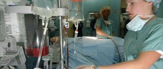
Caesarean section was performed in 17.2% of women in the main group for the following indications: premature abruption of a normally located placenta, progressive fetal hypoxia - 7.8%, severe varicose veins of the vulva and vagina - 7.8%, placenta previa - 3.2%.
During abdominal delivery, varicose veins of the small pelvis were detected in all women of the main group. In this regard, a feature of performing a cesarean section in such pregnant women was to minimize trauma to the woman and the fetus, which was achieved, among other things, by choosing the location of the incision and its sufficient size, as well as by using blunt dilation of the uterine muscles after a two-centimeter transverse incision in the lower segment.
Varicose veins in women are often associated with pregnancy and childbirth [1, 6]. Our observations confirm the literature data [6, 13] on the influence of repeated pregnancy, childbirth and complicated obstetric history on the risk of developing varicose veins of the vulva, vagina and perineum. It is noted that the development and progress of this pathology during pregnancy is accompanied by a significantly greater number of complications and more often causes fetal suffering and obstetric trauma. Disorders of fetoplacental hemodynamics, which develop against the background of pathology of the veins of the external genitalia, changes in hemostasis, systemic inflammatory response in combination with infectious diseases, contribute to total damage to the endothelium and the formation of gestational complications [10, 13].
The work revealed that women with varicose veins of the vulva, vagina and perineum are more susceptible to diseases of the pelvic organs, primarily inflammatory ones. In this regard, it can be assumed that some patients had disturbances in venous blood flow, causing regional immune deficiency and aggravating the course of infectious and inflammatory diseases of the genitals, before pregnancy. Noteworthy is the high incidence of gestational disorders with the threat of miscarriage and fetal growth disorders in women of the main group. It is important to note that all pregnant women in the main group had certain complications during natural delivery. The most dangerous thing was bleeding. In connection with this, almost every fifth (17.2%) woman had a caesarean section. At the same time, a combination of varicose veins of the vulva, vagina and perineum with varicose veins of the small pelvis was detected intraoperatively in all women.
The results of the retrospective study confirmed the relevance of identifying this group of pregnant women due to the additional risks they introduce for the development of pregnancy complications, fetal development and delivery.
conclusions
1. Varicose veins of the vulva and perineum are more often observed in multi-pregnant women with a complicated obstetric history and multiparous women.
2. Gynecological diseases, combination with other extragenital diseases, pregnancy complications were observed more often in the group of women with varicose veins of the vulva, vagina and perineum.
3. Varicose veins of the vulva, vagina and perineum during delivery can affect the frequency of untimely rupture of amniotic fluid, first and second degree perineal ruptures, bleeding as a result of obstetric trauma.
4. In the presence of varicose veins of the vulva, vagina and perineum, preparation for delivery should be carried out jointly with a phlebologist.
Conflict of interest:
The authors declare no conflict of interest.
Author contributions:
Concept and design of the study - Yu.Ts., E.K.
Collection and processing of material - E.K., I.K., V.V.
Text writing - E.K.
Editing - Yu.Ts.
Possible complications of varicose veins
The greatest danger is not varicose veins itself, but its complications, which include:
- thrombophlebitis - inflammation of the veins;
- trophic ulcers resulting from a sharp deterioration in blood supply to the skin;
- thromboembolism - the formation of blood clots in veins of different diameters, blockage of the lumen of the veins, including large pulmonary veins.
As mentioned above, these complications most often occur in people with increased intra-abdominal pressure, which includes pregnant women. However, it must be remembered that an increase in intra-abdominal pressure in pregnant women is a temporary phenomenon and it occurs gradually.
Why do varicose veins develop after pregnancy or during pregnancy?
The body of a pregnant woman undergoes various fairly pronounced changes regarding the endocrine and reproductive systems, as well as the anatomical structure and position of the abdominal and pelvic organs, associated with their displacement of the increasing size of the uterus. Hormonal changes in a woman’s body affect not only the reproductive system, but also the functional state of the vein walls with their weakening. Also, the enlarged uterus compresses large venous vessels through which blood flows from the legs. This leads to stagnation of blood and increased pressure inside the veins, which accelerates the process of formation of pathological protrusions.
Prevention of varicose veins
In the absence of varicose veins before pregnancy and with correct behavior during pregnancy and after childbirth, the manifestations of the disease disappear. But, as we already know, it is easier to prevent a disease or identify it in the early stages (compensation stage) than to subsequently treat its advanced forms.
In order to suspect that you have varicose veins of the lower extremities, just look at your legs and listen to your feelings. In the stage of compensation for varicose veins, when the deep vein system is not affected, works as usual and there are manifestations only from the superficial veins, you can notice a slight expansion of the saphenous veins: they are soft, easily collapse, the skin over them is not changed. In the stage of decompensation (at this stage there are already changes in the valve apparatus and deep veins), a moderate or sharp expansion of the saphenous veins is determined; they are dense, sometimes painful, the skin over them acquires a pigmented tint (red, bluish or purple-brown).
If you have discovered at least one of the listed signs before pregnancy, you need to visit a phlebologist or vascular surgeon. And if this possibility does not exist, then contact the surgeon at the clinic at your place of residence.
Varicose veins can be prevented by knowing about the risk factors that contribute to its occurrence and development:
- hereditary predisposition;
- features of the anatomical structure and blood supply of the legs;
- the use of old-generation hormonal contraceptives (drugs with a high content of hormones that predispose to varicose veins are not currently sold);
- hard physical labor;
- prolonged stay in a sitting or upright position;
- decreased physical activity during pregnancy;
- chronic intestinal diseases, constipation;
- chronic lung diseases with prolonged debilitating cough.
Thus, among the risk factors there are those that we cannot influence (for example, heredity), but there are also those that we can completely eliminate.
For example, you should not stand or sit in one position for a long time. There is no need to stand in front of a bathtub or basin, washing your baby’s diapers and undershirts: this process can be completely entrusted to a washing machine.
Walking with your child will serve as feasible physical activity and exercise. But at the same time, you don’t have to walk until exhaustion or, conversely, sit for hours with a book, comfortably sitting on a bench.
Since high heels are also a risk factor for the progression of varicose veins, you should, if possible, avoid them.
After childbirth, a woman may continue or develop the problem of constipation: its solution also applies to measures to prevent varicose veins.
Diagnosis of the disease
This pathology has characteristic manifestations, which are characterized by the appearance of small nodular protrusions, which have a different consistency and bluish coloration. Therefore, a preliminary diagnosis is possible after examination by a surgeon. The first surgical department of the hospital, if necessary, conducts additional laboratory, instrumental and functional studies (rheovasography, hemogram, ultrasound examination), with the help of which the degree of damage to the walls of the veins, the causes of the disease, as well as the stage of its course are determined. These studies can also be carried out if varicose veins appear after childbirth.
Diagnosis of varicose veins
Currently, there is a huge arsenal of methods for diagnosing varicose veins. They are available and do not cause any discomfort. The most common and reliable methods include:
- Doppler ultrasound is the simplest and safest method, when ultrasound is used to determine the speed of blood flow in the superficial and deep venous systems;
- angioscanning - the method is similar to Doppler ultrasound; it allows you to more accurately determine the degree of venous insufficiency in small-caliber vessels and capillaries;
- Contrast X-ray phlebography - allows you to identify at what level the damage to the valve apparatus of the superficial and deep veins occurred, by introducing contrast (a special drug) and its movement along the vascular bed.
The simplest and least time-consuming measures that can be taken after childbirth if you have varicose veins during pregnancy are:
- wearing elastic stockings or bandages during the day;
- compliance with the regime of physical activity and rest;
- complex of therapeutic gymnastics.
It is necessary to wear elastic knitwear throughout the entire period of pregnancy and about a month after childbirth, which will prevent disruption of the vein valves.
How are varicose veins treated after childbirth?
You should consult a doctor immediately after the appearance of varicose veins, swelling, and heaviness in the lower extremities. The doctor, a phlebologist, conducts clinical and ultrasound examinations of the lower extremities. In the process of ultrasound duplex scanning, the picture of pathologically altered venous blood flow is determined in detail.
Diagnosis of varicose veins after childbirth
The doctor formulates a treatment regimen, its stages and timing of optimal implementation. Most often, radical treatment of varicose veins after childbirth is recommended to begin after the end of the breastfeeding period. Until this point, it is recommended to limit yourself to wearing compression hosiery and a regime of certain restrictions. Baths, saunas, other thermal effects, as well as increased physical activity are contraindicated. Hiking, cycling, swimming are shown.
Complications of varicose veins in pregnant women:
Nature made sure that during childbirth the mother did not lose a lot of blood. During pregnancy, under the influence of hormones, blood becomes thicker. Thick blood and venous congestion increase the risk of developing blood clots (thrombosis), resulting in the following complications:
- dermatitis, eczema of the skin over altered veins
- bleeding from dilated veins
- thrombophlebitis (inflammation of superficial veins with the formation of blood clots); manifested by redness, soreness and thickening along the vein, increased temperature
- deep vein thrombosis of the lower extremities; characterized by severe pain, severe swelling and enlargement of the affected limb, increased temperature
- BODY (pulmonary embolism) – a blood clot from varicose veins enters the branches of the pulmonary artery. It appears depending on the degree of damage. In severe cases: chest pain, increased pulse, drop in blood pressure, increased breathing, sudden loss of consciousness. The skin of the face takes on a bluish tint, and the veins in the neck swell. In mild cases: pain when breathing, cough, possible rise in temperature.
According to statistical data, thrombosis and BODY in pregnant women is observed 5-6 times more often than in non-pregnant women, and after childbirth 3-6 times more often than before childbirth. The risk is especially high after major blood loss during childbirth and caesarean section.
Diagnosis and treatment of varicose veins during gestation
Unfortunately, the symptoms of varicose veins will only increase during pregnancy. To the visual manifestations will be added pain and a disturbance in the intensity of blood flow, which can cause oxygen starvation of the uteroplacental system. Only a phlebologist can make an accurate diagnosis of varicose veins after a detailed examination of the patient and ultrasound diagnostics.
The problem is complicated by the fact that during pregnancy not all remedies and medications can be used, not to mention surgical intervention. That is why varicose veins are considered one of the most insidious and risky diseases. Let's consider the most effective and safe recommendations for the treatment of varicose veins in pregnant women:
- Moderate physical activity - slow walking, swimming, yoga, aerobics for pregnant women. Each specific case has its own contraindications, so a phlebologist and gynecologist will help you choose the optimal set of exercises;
- Special compression jersey. During pregnancy, it is important to properly distribute the load on the musculoskeletal system. Bandages, compression tights and stockings help normalize blood circulation and support the abdomen;
- Taking herbal phlebotonics. This group of drugs helps to increase the tone of the vein walls and reduces their permeability. During treatment, the condition of the blood vessels improves and the symptoms are alleviated. Experts recommend taking phlebotonics from the 2nd trimester, the dosage is determined by the attending physician.
Clinical studies have shown that the French drug Phlebodia 600, which is actively used as part of complex therapy to get rid of the symptoms of varicose veins, will help strengthen the walls of blood vessels. You can find out about the effectiveness of the drug and get a doctor’s consultation on our website in a special form.