Published: 07/07/2021 11:15:00 Updated: 07/07/2021
Thrombosis is a complete or partial blockage of the lumen of a vessel by a parietal or mobile thrombus. A thrombus is a dense blood clot that appears as a result of a change in its fluidity. Normally, thrombus formation is a protective mechanism. Damage to the vascular wall leads to a slowdown in blood flow and accumulation of platelets around the damage. The thrombus literally “darns” the wall of the vessel.
The classic causes of thrombosis are described by Vikhrov’s triad: damage to the vascular wall, slowing of blood flow and changes in blood properties [3]. Some blood clots (they are called emboli) are able to move to narrower areas of the vessel, which are completely or partially blocked. Every year, about 25 million people die from thrombosis, and even more face trophic disorders caused by blood clots [3].
Types of vascular thrombosis
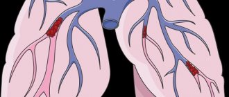
The most common thrombosis of the lower extremities, however, the greatest danger is pulmonary embolism - PE - and disseminated intravascular coagulation syndrome - DIC syndrome.
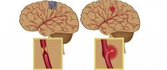
Arterial thrombosis develops when its lumen is blocked by a thrombus or embolus. Clinical signs are determined by the location where the blockage occurs, an organ or tissue that has little or no blood supply. If blockage with impaired vessel patency occurs slowly, “spare” collateral vessels open, which mitigates the clinical symptoms of arterial thrombosis [3]. Arterial thrombosis occurs more often in middle-aged and elderly men [7].
Vein thrombosis varies depending on the location of the lesion into deep or superficial vein thrombosis and pulmonary embolism. Among all cardiovascular pathologies, venous thrombosis ranks third in frequency of occurrence, second only to ischemic heart disease and atherosclerosis. The third place in the structure of causes of mortality is occupied by pulmonary embolism. Starting at age 40, the risk of developing venous thrombosis doubles every 10 years [5].
Two variants of damage to the veins of the lower extremities are described: phlebothrombosis (primary thrombosis, the thrombus is not firmly fixed) and thrombophlebitis (secondary thrombosis due to inflammation of the vessel wall, the thrombus is firmly fixed) [6]. Thrombophlebitis is more often associated with superficial vein thrombosis [2]. The larger the vein affected by thrombosis, the more pronounced its clinical manifestations. The surrounding tissues are compressed by stagnation of blood, since the blood stays at the site of occlusion, but does not move towards the heart. Venous blood clots tend to break off and travel through the bloodstream (thromboemboli). When they enter vital organs, life-threatening conditions develop [3].
What causes blood clots?
Thrombi, in the form of blood clots, form in blood vessels in response to damage to the vascular wall, slowing of blood flow and changes in blood composition. “Thrombosis does not occur out of nowhere; there must be some prerequisites. This is facilitated by the presence of varicose veins in the lower extremities. If it is, you should pay attention to compactions that may appear in the veins,” says phlebologist, surgeon Fedor Shpachenko .
According to the phlebologist, compactions may indicate that blood clots have already formed in the veins. “Often, these blood clots may not occur in the superficial veins that we see, but in deep veins, and it is impossible to visualize them without special research methods,” warns Shpachenko.
Risk factors for thrombosis
Internal:
- arterial hypertension [7];
- pregnancy, childbirth, postpartum period [3];
- biochemical changes in blood [2,3,5,7];
- vasculitis [2];
- age over 40 years [5];
- congenital thrombophilia, thrombosis, varicose veins of the lower extremities [3,5];
- congestive heart failure [5, 6];
- malignant neoplasms, radiotherapy and chemotherapy [3];
- strokes [3, 6];
- myeloproliferation [2, 5, 7];
- nephrotic syndrome and renal failure [5, 6, 7];
- obesity (BMI over 30) [3];
- myocardial infarction [6];
- diabetes mellitus [6, 7];
- systemic lupus erythematosus [2];
- chronic pulmonary diseases [3];
- enterocolitis [5].
External:
- heroin addiction [2];
- hormonal therapy [3,5];
- dehydration due to vomiting, diarrhea, increased sweating, direct lack of fluid [6];
- immobilization [3];
- travel by plane, bus or in a seated car [3];
- infectious diseases, including COVID-19 [1, 3, 5, 9];
- catheterization of central and peripheral veins [2, 5];
- smoking [6, 7];
- sedentary lifestyle [3];
- operations [3];
- fractures of large bones, other injuries [3];
- taking oral contraceptives [5];
- taking Diazepam, Amiodarone, Vancomycin [2];
- sclerotherapy and thermal ablation [2];
- condition after joint replacement [3];
- holding an awkward position [3].
Who is at risk?
Those who are most likely to develop thrombosis are people who are obese and have varicose veins, women who take oral contraceptives, and those who take many long flights. But a high risk of blood clots occurs when all these factors are combined.
“Patients with obesity and varicose veins are at risk. As for medications, in particular oral contraceptives, now most of them are well balanced and we rarely see the development of thrombosis and thromboembolism in women taking these drugs. People with a sedentary lifestyle, impaired hemostasis, namely blood clotting disorders, also fall into this group. If the patient has all these provoking factors, then the risk of developing thrombosis increases. Age also makes it worse. Over the years, the vessels lose elasticity and become brittle, plus a number of concomitant diseases cause swelling of the lower extremities,” says Shpachenko.
Thrombosis Clinic
Symptoms of thrombosis can be general, regardless of location, or specific.
Common symptoms include pain with movement and at rest, limited mobility, and decreased function of the affected organ or tissue. Symptoms of arterial obstruction (acute thrombosis, or gradual obstruction of vessel patency):
- asymmetry of blood pressure when measured on both arms [7];
- pallor of the skin, turning into cyanosis [7];
- pain at rest at night [7];
- pain when moving in the thigh, buttock, lower leg, foot, shooting or aching [7];
- sleep disorders [7];
- numbness, coldness of the limb [7];
- absence of peripheral pulsation [7];
- necrosis (necrosis) of affected tissues, trophic ulcers, gangrene [7];
- intermittent claudication [7].
Symptoms of venous thrombosis:
- pain [6];
- swelling, soft and asymmetrical [6];
- blue discoloration of the skin (skin cyanosis) [6];
- increased skin temperature of the extremities [6];
- increased sensitivity and compaction in the projection of the superficial veins [2];
- post-inflammatory hyperpigmentation [2];
- dilated saphenous veins [6];
- erythema [2].
Sometimes the only symptom of venous thrombosis is PE [6].
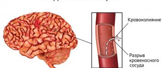
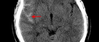
Consequences of cerebral hemorrhage in adults
The most dangerous thing with a cerebral hemorrhage is the consequences that can develop.
Among them, Polina Petrosyan notes: “The consequences can lead to disability, but may not affect a normal lifestyle and ability to work,” says Dr. Polina Petrosyan. – After hemorrhage, one-sided weakness in the limbs, decreased sensitivity in them, and various speech disorders may persist. Full recovery is possible if rehabilitation measures are taken in a timely manner and if the patient independently puts effort into rehabilitation measures.
Diagnosis of thrombosis
Primary diagnosis is based on a detailed history and anthropometry (calf or thigh circumference).
The Wells scale is used to diagnose acute thrombosis and diagnose pulmonary embolism [8,9]. Instrumental diagnostics include compression or duplex scanning of veins, Doppler ultrasound with compression of veins, impedance plethysmography, pulmonary angiography, radiocontrast or MRI venography [6,9], CT and MRI angiography [7,9].
To diagnose arterial thrombosis, physical tests are used (6-minute walk test, treadmill test), determination of pulsation of superficial arteries (arteries of the dorsum of the foot), duplex scanning of the arteries of the extremities, angiography (X-ray of a vessel filled with a radiopaque substance) and measurement of transcutaneous oxygen tension [ 7].
Tests for thrombosis
Laboratory indicators play a significant role in the timely diagnosis of thrombosis.
Thus, guidelines for the management of patients with a new coronavirus infection provide for stratification of the risk of coagulopathy in patients with COVID-19 based on simple laboratory tests: D-dimer, prothrombin time, platelet count, fibrinogen level [1,9]. A clinical blood test can detect inflammation. It also determines the level of platelets, that is, the very substrate of thrombosis.
Additionally, the level of inflammation in the blood and the risk of thrombosis is indicated by an increased level of C-reactive protein.
Biochemical analysis primarily demonstrates blood glucose levels. It can be used to judge the presence of diabetes, one of the most serious risk factors for thrombosis.
Also, a biochemical analysis can determine the level of protein C, which also characterizes the severity of the risk of thrombosis.
Elevated levels of homocysteine in the blood are also a currently proven risk of thrombosis, leading to miscarriage and cardiovascular events (heart attacks and strokes).
D-dimer is a laboratory marker of fibrin formation [8]. It also indicates the presence of inflammation, just like C-reactive protein. The level of D-dimer is a control indicator of COVID-19 and its complications, including those associated with thrombosis.
You can take tests under the comprehensive Thrombosis program, which includes determining the levels of Antithrombin-III, D-dimer and genetic factors of cardiac diseases and platelet levels. This program allows you to determine the fact of thrombosis occurring somewhere in the body, as well as determine the genetic predisposition to it. This program, like other tests, is offered by the CITILAB network of clinics.
Additional determination of homocysteine and C-reactive protein levels will help determine the biochemical risk of thrombosis.
Causes of cerebral hemorrhage in adults
There are many reasons why blood vessels rupture and blood spills into the brain tissue. Among the most common are:
- arterial aneurysms (thinning of the wall, the formation of a sac with blood that overflows and bursts);
- vascular malformations (birth defects, thinning, tortuosity of the walls);
- ruptures of blood vessels during a hypertensive crisis due to the prohibitive load on the walls;
- head injuries with vascular ruptures;
- tumors that grow and damage arteries;
- taking blood thinning medications (if dosages are not followed);
- the development of certain systemic diseases in which the walls of the arteries are affected (for example, amyloidosis).
At a young age, the leading causes of hemorrhage are injuries and congenital vascular anomalies. In the elderly – damage to blood vessels by atherosclerosis and their rupture due to hypertension, tumor processes.
Treatment and prevention of thrombosis
Treatment of thrombosis includes anticoagulant and antiplatelet therapy, thrombolytic therapy, installation of an inferior vena cava cava filter, and surgical removal of the thrombus [5].
Complications of anticoagulant therapy must be kept in mind: major bleeding, heparin-induced thrombocytopenia and warfarin-induced skin necrosis [5]. To reduce the risk of continued thrombus formation, NSAIDs are used [2]. For the purpose of secondary prevention, small doses of heparin are prescribed. Non-drug treatment methods are also prescribed - elastic bandaging, compression hosiery, local hypothermia and exercise therapy [2, 4].
Prevention of thrombosis includes a number of measures used in situations of increased risk of thrombosis.
Primary prevention of atherothrombosis:
- systematic physical activity in the form of walking or morning exercises;
- blood pressure control, maintaining working blood pressure below 140/90 mmHg;
- control of blood sugar levels (less than 6 Mmol/l), early detection and treatment of diabetes mellitus;
- weight loss, body mass index less than 25 kg per m2;
- a diet limited in cholesterol and high-density fat (total cholesterol less than 5 mmol/l), fruits and vegetables;
- smoking cessation [3,7].
Primary prevention of venous thrombosis:
- compression underwear;
- bandaging with elastic bandages;
- drinking plenty of fluids, especially after surgery;
- regular exercise, walking, especially when traveling;
- prohibition of taking alcohol and sleeping pills in large doses;
- prohibition of the use of compressive shoes and clothing [2,5,6].
Sometimes, during periods of particular risk, anticoagulants are prescribed several days before the flight. There is no point in taking aspirin in such cases [5].
Briefly about the treatment method
For ischemic stroke, the most effective and reliable method of removing blood clots from the arteries of the brain is thrombectomy. There are several options for the procedure: - Aspiration thrombectomy. During the operation, a micro-incision is made through which a special catheter is inserted. Through it, saline solution flows under pressure into the affected vessel. Under the influence of the solution and salt, the thrombus softens, is divided into pieces and aspirated (suctioned out) using a syringe. Thanks to this method, the number of injuries to the internal walls of blood vessels is reduced. — Percutaneous mechanical thrombectomy. A catheter with a stent is inserted through a large artery. With the help of a stent, the clot is captured and removed through an incision in the vessel. This method is most often used in the treatment of stroke caused by blockage of blood vessels in the brain.
Bibliography
- Consensus position of experts of the Eurasian Association of Therapists on some new mechanisms of the pathogenesis of COVID-19: focus on hemostasis, issues of blood transfusion and the blood gas transport system / G.P. Arutyunov, N.A. Koziolova, E.I. Tarlovskaya, A.G. Arutyunov, N.Yu Grigorieva, etc. // Cardiology. 2020;60(6). DOI: 10.18087/cardio.2020.5.n1132.
- Thrombophlebitis (thrombosis of superficial veins): modern standards of diagnosis and treatment / V.Yu. Bogachev, B.V. Boldin, O.V. Jenina, V.N. Lobanov//Hospital-substituting technologies: Outpatient surgery. 2016.- 3-4 (63-64)- P.16-23.
- Modern problems of thrombosis of arteries and veins / I.N. Bokarev, L.V. Popova // Practical Medicine, 2014. - No. 6 (82) – P13-17.
- Treatment of thrombophlebitis. Current recommendations and clinical practice / P.F. Kravtsov, K.V. Mazaishvili, S.M. Markin, H.M. Kurginyan // Thrombosis, hemostasis and rheology, 2020 No. 2 – P 68-72.
- Venous thrombosis: modern treatment / P.S. Laguta // Atherothrombosis, 2015 - No. 2 - P. 7-16.
- Deep vein thrombosis of the lower extremities / A.K. Lebedev, O.Yu. Kuznetsova // Russian family doctor, 2015.
- National recommendations for the diagnosis and treatment of diseases of the arteries of the lower extremities, Association of Cardiovascular Surgeons of Russia, Russian Society of Angiologists and Vascular Surgeons, Russian Society of Surgeons, Russian Society of Cardiology, Russian Association of Endocrinologists, M, 2021.
- Diagnosis and pharmacotherapy of acute venous thrombosis / N.V. Sturov, G.N. Kobylyanu// Difficult Patient, 2013., No. 12., T. 11. –P.19-22.
- Principles of management of patients with venous thromboembolism during the COVID-19 pandemic // V.Ya. Khryshchanovich // Surgery News, 2021.- t28 No. 3 – P329-338.