Examinations and operations on the main arteries of the lower extremities are successfully performed in the clinic of the Tver Medical University. Examinations and operations can be performed free of charge for all patients with a compulsory medical insurance policy, and you can be registered in any region of Russia and even be a foreign citizen.
If you need surgery on the femoral artery, or consult a doctor, make an appointment by calling:
- 8 (Research Institute “Phlebology”, Tver, 2nd Krasina St. 49)
- 8 (University Clinic, Tver, Peterburgskoye Highway, 115 building 2)
Which leg arteries are most often affected?
The arteries supplying blood to the lower extremities can be divided into 3 groups or floors, depending on location:
- aorta and iliac arteries
- femoral arteries
- arteries of the leg and foot
The surgeon most often deals with operations on the femoral arteries. These include the common, superficial and deep femoral arteries.
How dangerous is a gap?
Many patients, unaware of the presence of an aneurysm, continue to lead a normal lifestyle. The formation and growth of this formation can last more than 3 years, accompanied by minor symptoms. Under the influence of physical overload, during pregnancy or childbirth, with a sharp increase in pressure, a rupture of the wall of the femoral artery can occur. If the patient is not operated on in a timely manner, intense bleeding is life-threatening.
In addition to rupture, the presence of an aneurysm increases the risk of the following complications:
- blockage of an artery by a blood clot;
- movement of parts of a blood clot with embolism of the branches and gangrene of the lower extremities;
- suppuration of a hematoma (false aneurysm) with phlegmon of surrounding tissues;
- trophic disorders (dermatitis, ulcers) due to lack of blood flow.
What operations are performed on the femoral arteries?
Operations are divided
By way of execution:
- Open (prosthetics, bypass surgery, angioplasty, etc.). A skin incision is made, the vessel is exposed, and the surgeon performs the operation under visual control. Most often, bypass surgery involves replacing a section of an artery with the body’s own vein or a synthetic prosthesis. In this case, the blood bypasses the affected area.
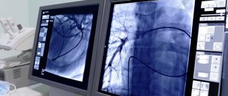
- Endovascular (balloon angioplasty, stenting, etc.). Instruments are inserted into the artery through a puncture of the skin, and under X-ray control using an angiograph machine, the surgeon performs an operation: a special balloon expands the lumen of the artery, and, if necessary, places a stent. The image is displayed on the monitor.
According to the urgency of implementation:
- Emergency – performed for acute pathologies (thrombosis, vessel injury, etc.) within several hours after the onset of the disease.
- Planned – for chronic diseases (atherosclerosis, arterial aneurysm, etc.) are performed as planned.
History[edit]
Teaching illustrations of knee anastomosis, such as the one shown in the side box, appear to have been taken from an idealized image first created by Gray's Anatomy in 1910. Neither the 1910 illustration nor any subsequent version was drawn from an anatomical dissection, but rather from the work of John Hunter and Astley Cooper, who described knee anastomosis many years after ligation of the femoral artery for a popliteal aneurysm. [11] Knee anastomosis has not been demonstrated even with modern imaging techniques such as X-ray computed tomography or angiography.[11]
What operations are performed for atherosclerosis?
Atherosclerosis is the most common cause of chronic ischemia of the lower extremities. Atherosclerosis leads to narrowing (stenosis) or blockage (occlusion) of a vessel (for more details, see the section “Atherosclerosis”). Surgeries for atherosclerosis are aimed at restoring blood flow:
Endovascular: under X-ray control, the lumen of the vessel is expanded using special balloons; if necessary, a stent is placed, which acts as a cylindrical frame and ensures patency of the artery.
During open operations, vascular remodeling occurs under visual control.
Femoral artery replacement or bypass surgery. The name of the prosthesis or shunt depends on the method of its application (suturing). In this case, the essence is the same - the artery is replaced with one’s own vein or a synthetic prosthesis.
Angioplasty is the remodeling of blood vessels using the artery’s own tissue, as well as using a patch from one’s own vein or synthetic material. The most common procedure performed is profundoplasty – remodeling of the deep femoral artery.
Main branches
A series of connections extend from the main vessel. Each of them provides blood supply to a separate area and performs certain functions:
- Superficial epigastric artery. Transports blood to the external oblique muscle of the abdomen and the skin of the anterior wall of the peritoneum. It goes from the bottom of the inguinal ligament up the anterior abdominal wall to the umbilical ring. Near the navel it connects with the superior epigastric artery.
- Superficial femoral. Responsible for nourishing the groin muscles, lymph nodes and skin. Departs from the epigastric or from the outer wall of the femoral artery. It runs along the inguinal ligament to the anterior iliac spine.
- External genital arteries. Their number varies from 2 to 3. They are directed medially, bending around the anterior and posterior periphery of the femoral vein. They also include a large number of smaller branches that are located in the scrotum in men, the labia in women and above the pubis.
- Inguinal branches. Provides a flow of nutrients and blood to the lymph nodes and skin. They originate from the external genital arteries in the form of small stems. Then they pass through the fascia lata of the thigh.
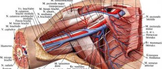
- Deep artery of the femur. The largest of all branches, which consists of a whole network of vessels. It starts 3-4 cm below the inguinal ligament and ends in the lower third of the thigh, between the long and large adductor muscles. Arteries depart from it - lateral, medial, perforating, as well as small capillaries. They promote normal blood circulation in muscles, joints, and deep layers of the epidermis.
- Descending knee. A long vessel that can arise either directly from the femoral artery or from the lateral one. It ends in the thickness of the knee muscles and the capsule of the knee joint. It has branches - articular and subcutaneous.
Since the deep femoral artery is the main element of the blood circulation of the femoral artery, the peculiarities of its structure should be taken into account. Several more vessels depart from each of its branches:
- Medial artery. Its continuation is the ascending, transverse, deep branches and the branch of the acetabulum.
- Lateral. It arises from the outer wall of the deep artery and divides at the intersection with the trochanter of the femur. There the ascending, descending and transverse branches depart from it.
- Perforated arteries. Located at different levels from the main artery. In the area where the adductor muscles attach to the femur, they move to the back of the thigh. They supply the adductor, semimembranosus, semitendinosus, and biceps muscles.
Disruption of blood flow in at least one channel is fraught with serious consequences for the entire vascular system. Ligaments, external genitalia, and lower limbs also suffer due to lack of oxygen and nutrients.
The Scarpian or femoral triangle is formed by the superficial epigastric, superficial and genital arteries. Its height is 15-20 cm.
Which surgery is better?
Each operation has its own indications and contraindications. The method of surgical treatment is chosen by the doctor based on many different factors, taking into account not only ultrasound and angiography data, but also the patient’s health status, the presence of concomitant diseases, age, etc. Some patients may require a combination of different techniques, such as stenting one section of the artery and replacing another. Such operations are called “hybrid”.
Surgery on the great vessels is one of the most common operations in the world, which has saved the lives and preserved the health of millions of people.
Online consultations (Skype, Viber, Whats App)
Sign up for a consultation
Links[edit]
- Schulte, Eric; Schumacher, Udo (2006). "Arterial blood supply of the thigh". In Ross, Lawrence M.; Lamperti, Edward D. (ed.). Thieme's Atlas of Anatomy: General Anatomy and the Musculoskeletal System
. Time. p. 490. ISBN 978-3-13-142081-7. - Jacob, S. (1 January 2008), Jacob S. (ed.), "Chapter 6 - Lower Limbs", Human Anatomy
, Churchill Livingstone, pp. 135-179,. Doi: 10.1016/b978-0-443-10373-5.50009-9, ISBN 978-0-443-10373-5, retrieved January 18, 2021 - ^ a b Mikael Häggström (2019). "Subsartorial vessels as a substitute name for the superficial femoral vessels" (PDF). International Journal of Anatomy, Radiology and Surgery
: AV01 – AV02. - Snell, Richard S. (2008). Clinical anatomy by region (8th ed.). Baltimore: Lippincott Williams and Wilkins. pp. 581–582. ISBN 978-0-7817-6404-9.
- Bundens, W.P.; Bergan, JJ; Halasz, N. A.; Murray, J; Drehobl, M. (1995). “Superficial femoral vein. Misnomer, potentially life-threatening." JAMA
.
274
(16):1296–8. DOI: 10.1001/jama.1995.03530160048032. PMID 7563535. - Hammond, I (2003). "Superficial femoral vein." Radiology
.
229
(2): 604, discussion 604-6. DOI: 10.1148/radiol.2292030418. PMID 14595157. - Kitchens CS (2011). "How to treat superficial vein thrombosis". Blood
.
117
(1):39–44. DOI: 10.1182/blood-2010-05-286690. PMID 20980677. - Thiagarajah R, Venkatanarasimha N, S Freeman (2011). "Use of the term 'superficial femoral vein' in ultrasound." J Clin Ultrasound
.
39
(1): 32–34. DOI: 10.1002/jcu.20747. PMID 20957733. S2CID 23215861. - Deakin, Charles D.; Low, J. Lorraine (September 2000). "Accuracy of advanced trauma life support guidelines for predicting systolic blood pressure using carotid, femoral, and radial pulses: an observational study". BMJ
.
321
(7262):673–4. DOI: 10.1136/bmj.321.7262.673. PMC 27481. PMID 10987771. - McPherson, D.S.; Evans, D.H.; Bell, P.R.F. (January 1984). "Doppler waveforms of the common femoral artery: comparison of three objective analysis methods with direct pressure measurements." British Journal of Surgery
.
71
(1):46–9. DOI: 10.1002/bjs.1800710114. PMID 6689970. S2CID 30352039. - ^ abc Sabalbal, M.; Johnson, M.; McAllister, W. (September 2013). "Absence of knee arterial anastomosis, as is usually indicated in textbooks". Annals of the Royal College of Surgeons of England
.
95
(6):405–9. DOI: 10.1308/003588413X13629960046831. PMC 4188287. PMID 24025288.
Risks of Angioplasty and Stenting
Complications can occur with any medical procedure. Here are some of the possible complications of carotid angioplasty and stenting:
- Stroke or mini-stroke (transient ischemic attack or TIA) . During angioplasty, blood clots that may form on the catheters can break free and travel to your brain. You will receive blood thinners during the procedure to reduce this risk. A special trap filter is used for these procedures, but the risk is associated with placing it after the catheter has passed the narrowing site.
- New narrowing of the carotid artery (restenosis) . The main disadvantage of carotid angioplasty is the likelihood that your artery will narrow again within a few months of the procedure. Special drug-eluting stents have been developed to reduce the risk of restenosis.
- Blood clots . Blood clots can form in stents even weeks or months after angioplasty. These clots can cause stroke or death. It is important to take aspirin, clopidogrel (Plavix), and other medications exactly as prescribed to reduce the chance of clots forming in your stent.
- Bleeding . You may have bleeding at the injection site. Usually this simply results in a hematoma, but sometimes serious bleeding occurs and blood transfusions or surgical procedures may be required.
Additional images[edit]
- Structures passing behind the inguinal ligament. (The femoral artery is indicated at the top right.)
- Transverse section showing the structures surrounding the right hip joint.
- The shell of the femur is opened to show its three compartments.
- Femoral artery.
- The spermatic cord in the inguinal canal.
- Anterior part of the right thigh with markings on the surface of the bones, femoral artery and femoral nerve.
- Femoral artery and its main branches - right thigh, anterior view.
- Illustration showing the main arteries of the leg (front view).
- Femoral artery - deep dissection.
- Femoral artery - deep dissection.


