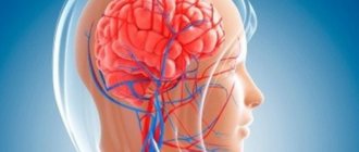
The existing first-choice immunomodulatory therapy for multiple sclerosis (MS) (interferons, Copaxone) is aimed at reducing inflammation in the central nervous system, the frequency of exacerbations and, to a lesser extent, the rate of disease progression [11]. The effectiveness of such therapy decreases as the duration of the disease and the severity of the neurological deficit increase. This is due to both a gradual decrease in the number of functioning axons due to neurodegeneration and an increase in metabolic disorders in brain tissue. Currently, there are no effective methods for inhibiting the interrelated processes of neurodegeneration and chronic metabolic damage to the brain and therefore they are not usually considered as a target for therapy. Nevertheless, practitioners, based on their own experience in such cases of MS, often use long-existing metabolic and neurotrophic therapies. Among the most active metabolic drugs with a trophic effect is Cerebrolysin (C) [14]. However, relevant scientific studies are not yet available.
The purpose of this study is to study objective methods of neuroimaging of the metabolic effect of C in MS.
Main problems that can be solved with neuroprotectors
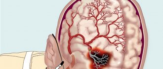
According to statistical estimates, more than 400,000 acute cerebrovascular accidents are diagnosed annually in Russia. The mortality rate reaches 40%, and those who managed to survive a stroke often suffer severe complications in the form of speech and motor impairments, which usually causes disability.
Circulatory disorders in the brain often provoke cognitive disorders, the clinical picture of which is characterized by:
- chronic fatigue, fatigue;
- decreased memory, attention;
- inability to concentrate quickly and for a long time on an object;
- deterioration of mental performance.
The degree of manifestation of these symptoms affects the outcome of rehabilitation measures. It is impossible to improve the patient's quality of life without at least partially eliminating cognitive disorders.
MRS results
In the main group of patients (Table 1, Fig. 1)
As a result of treatment with C, a significant increase in the level of N-acetylaspartate (NAA) was obtained, which indicates an increase in the number of actively functioning neurons.
Figure 1. Changes in metabolite levels in MS lesions in the study group. Explanations in the text. * - p
<0.05, *** -
p
<0.001 when comparing the level of metabolites before and after treatment.
The level of inositol (Ins) increased 5 times, however, due to the low initial level of this metabolite, the changes obtained cannot be caused by increased hypoxia. There was also a 4-fold decrease in lactate (Lac) levels as a result of treatment; its high initial level was interpreted as a manifestation of hypoxia in the area of MS lesions, therefore, a decrease in lactate levels was associated with its decrease (Fig. 2)
. Figure 2. An example of changes in metabolism in MS lesions according to MRS data in patient N. (main group) as a result of treatment. The spectrum on the left reflects the initial state at the time of inclusion in the study, the spectrum on the right shows changes after treatment in the form of an increase in NAA, a decrease in Lac and a slight increase in Ins. There is a tendency for Cr and CrPh levels to increase, which may indicate activation of aerobic oxidation in the energy supply system while limiting the participation of the anaerobic pathway.
In the control group, when analyzing spectroscopic changes in the foci of demyelination before and after treatment, no significant differences were revealed (Fig. 3)
.
Figure 3. An example of changes in metabolism in areas of demyelination in patient P. (control group) as a result of treatment.
In the MRS picture (the spectrum on the left was obtained before treatment, on the right - after), no positive metabolic changes were observed in this patient. There is some negative dynamics in the form of a threefold increase in lactate, a decrease in creatine and creatine phosphate. Results of MRS of perifocal areas of demyelination
.
In the areas of the brain immediately adjacent to the lesions of MS in patients receiving C (the main group), a twofold decrease in Lac was recorded, which indicates a decrease in the severity of hypoxia of brain cells in these areas (Fig. 4, 5)
.
Figure 4. Changes in the level of metabolites in the perifocal area in the main group. Explanations in the text. ** — p
<0.01 when comparing the level of metabolites before and after treatment. Figure 5. An example of changes in metabolism according to MRS data in the area adjacent to the focus of demyelination in patient N. (main group) as a result of treatment. There is a slight decrease in lactate and choline and a tendency toward an increase in NAA.
In the control group, no significant differences were obtained in the areas adjacent to the foci of demyelination (Fig. 6)
.
Figure 6. An example of changes in metabolism in areas of demyelination in patient P. (control group) as a result of treatment. There are no positive metabolic changes in the MRS picture of this patient. There is a slight negative trend in the form of a threefold increase in Lac, a decrease in Cr and CrPh.
Results of MRS in visually intact tissue
In visually intact tissue in the main group, the level of metabolites remained stable, no significant changes were recorded (Fig. 7)
.
Figure 7. Changes in metabolite levels in visually intact brain tissue.
Explanations in the text. In the control group, the level of Cr in undamaged brain tissue significantly increased, which may indicate an improvement in the processes of energy supply to cells. However, due to the small sample and the absence of fluctuations in the levels of other metabolites, this fact does not seem significant (Fig.
.
Figure 8. An example of changes in metabolism in visually undamaged brain tissue in patient P. (control group) as a result of treatment according to MRS data.
Lack of positive metabolic changes, with the exception of an increase in Cr levels, possibly indicating an improvement in energy processes in neuronal axons. MRS results for the delayed comparison group
.
3 months after the end of treatment with C, the most pronounced positive changes were recorded in 5 patients of the main group. MRS showed the stability of the positive changes in brain metabolism achieved during treatment (Table 2)
.
From those presented in table. 2
The data shows that in 3 out of 5 patients, 3 months after cessation of treatment, the positive metabolic changes achieved were maintained, of which in 1 patient the positive changes were stable, despite cessation of treatment. In the remaining 2 patients, positive metabolic changes were partially or completely leveled out 3 months after cessation of treatment.
Groups of neuroprotectors by mechanism of action
Neuroprotective therapy in modern medicine plays an important role in the treatment of neurological diseases of various origins. They are distinguished by their structure and principle of operation. Among the common drugs are combination drugs (like Phezam), as well as:
- Nootropics.
- Vasoactive drugs - anticoagulants, antiplatelet agents, methylxanthines.
- Adaptogens.
- Antioxidants.
Good results are demonstrated by medications that bind free radicals. They are called antioxidants and are used to control the functioning of mitochondria. Thus, the cells begin to absorb oxygen better and are filled with energy.
The most famous and popular neuroprotective drugs of this group are vitamin E, that is, tocopherol, as well as traditional ascorbic acid. But, as has been established through numerous scientific tests, these substances are not able to provide the proper therapeutic effect due to their low adhesive ability to cell membranes.
High-quality neuroprotective therapy involves the use of tissue respiration correctors. This includes medications from different manufacturers. Highlight:
- coenzyme q10, riboflavin;
- cytochrome C, succinic acid and others.
According to scientists and medical experts, the best effect is achieved through combined tactics, when the attending physician prescribes a combination of tissue respiration correctors based on the patient’s age and gender. The presence of chronic pathologies and the clinical picture (the intensity of the manifestation of mental and physical symptoms) are also taken into account.
Neuroprotective therapy can be developed through the use of new medications that began to be used in medical practice relatively recently. They remove the excitotoxicity of glutamate and block the receptors that demonstrate the reaction due to the amino acid.
results
Tolerability of therapy.
In the main group of patients receiving C, in 70% of cases there was a slight increase in blood pressure in the first hours after using the drug, while the upper blood pressure values did not go beyond the normal range in accordance with the criteria of the World Health Organization (140/90 mm Hg) . In 2 patients, in order to relieve discomfort due to increased blood pressure, amlodipine was used once at a dose of 5 mg, which allowed stabilizing blood pressure and improving well-being. Two patients of the main group, after using C within 10-20 minutes after the end of the drug administration, complained of short-term dizziness, which disappeared without special treatment. Patients in the control group did not make any complaints about changes in well-being.
Subjective changes
. In the group of patients receiving C, in 80% of cases there was an improvement in well-being and general mood, in 50% there was an increase in performance, improved memory, normalization of sleep, and increased resistance to physical and psycho-emotional stress.
In the control group, no changes in well-being were detected.
Changes in neurological status
.
In 1 patient from the main group (patient N.
), after treatment for 2-3 months, a significant decrease in neurological deficit was revealed in the form of an increase in the ability to move due to increased strength in the legs, decreased fatigue, the ability to move without outside support or a cane, and a decrease in severity of MS on the EDSS scale by 1.0 points.
At the time of the beginning of the examination, this patient’s neurological status showed bilateral pyramidal-cerebellar insufficiency with a decrease in strength in the right extremities to 3 points, in the left - to 4 points, with pathological carpal (Rossolimo, Zhukovsky) and foot (Babinsky, Oppenheim) reflexes with two sides. He also had decreased vibration sensitivity in his legs down to the level of the iliac crests. The patient could move independently for a distance of no more than 100 m using a cane. After treatment with C and B vitamins, the neurological status began to gradually change for the better with an increase in muscle strength in the right extremities to 4 points, in the left – up to 5 points, however, pathological hand and foot signs, as well as a decrease in vibration sensitivity remained at the same level . An objective assessment of the patient's motor activity showed an increase in the distance that the patient could walk without outside support and relying on a cane to 220 m, which corresponded to a decrease in the severity of MS by 1.0 points on the EDSS scale.
In the remaining patients of the main group and in all patients in the control group, the neurological status did not change.
Changes in neuropsychological tests
were not statistically significant, probably due to the insufficient sample size and time of observation of patients in both the main and control groups.
Features and principles of therapy
According to its concept, neuroprotection has several directions:
- Primary, where the effect is aimed at interrupting the rapid mechanisms that contribute to cell death. This is problematic to implement due to the selective nature and the importance of accurately detecting the receptor that is involved in the process.
- Secondary, aimed at reducing the severity of the consequences of ischemic damage.
Neuroprotective therapy is used not only for acute conditions associated with brain function. In this way, chronic diseases of the nervous system are also treated. Neuroprotection is prescribed for:
- vegetative-vascular dystonia;
- neuroses;
- encephalopathy;
- depressive disorders;
- asthenia;
- neuroinfections.
The scheme for a specific patient is formed individually. An anamnesis is collected and the clinical picture is studied in detail. The course also depends on the general health of the patient.
Material and methods
We examined 15 patients with a relapsing-remitting form of MS who were in remission. The duration of the disease in these cases was from 1 year to 5 years, its severity on the EDSS scale was from 2.5 to 5.5 points.
All patients continued previously prescribed pathogenetic therapy (Copaxone, Rebif, Betaferon).
Of the total number of patients, 10 made up the main group; they were prescribed intravenous infusions of 20 ml of C per 200 ml of saline solution for 10 days and B vitamins (Pentavit tablets) in a standard therapeutic dosage. Patients in the control group - 5 people received only B vitamins (Pentavit tablets) in a standard therapeutic dosage. Patients underwent multivoxel magnetic resonance spectroscopy MRS before the start of the examination and for 3 days after the last infusion.
Based on the results of treatment, 5 patients were selected from the main group (delayed comparison group), in whom the most clear positive changes were established according to the second MRS. Patients in this group underwent a third MRS in order to determine the duration of metabolic changes 3 months after the end of treatment.
MRS was performed on a 1.5 T tomograph. In this case, the focus of demyelination, the perifocal area and morphologically intact brain tissue located at least 1 voxel away from the MS plaques were assessed, with a total of 5 voxels from each region of interest. For statistical processing of the results, proprietary built-in software was used.
Expected results
If neuroprotective therapy is developed by an experienced doctor, and the patient adheres to all recommendations, the outcome is favorable even in severe conditions and cerebrovascular accidents. A psychoneurologist will be able to diagnose the condition and prescribe complex therapy taking into account the activation of the central nervous system. As a result, you will be able to:
- improve memory and attention;
- prevent mental exhaustion;
- eliminate symptoms of constant depression;
- get rid of neuropsychic weakness and chronic fatigue;
- provide mental clarity;
- reduce excitability.
The doctor will definitely select medications so that neuroprotective therapy is as effective as possible for a particular case. Depending on the pharmacological drugs and the active substance, the effect may vary. There are medications that help get rid of insomnia and headaches. Medicines for improving mood are also popular.
Properly selected treatment using high-quality neuroprotectors will prevent dementia and protect the brain from the severe consequences of circulatory disorders at any age.
LITERATURE:
- Boyko A.N. and others // Journal. neuropath. and a psychiatrist. 2007.
- Kadykov A.S. and others. Chronic vascular diseases of the brain. M., 2006.
- Revenok E.V. Cognitive impairments of cortical and subcortical types in vascular diseases of the brain. M., 1999.
The text was checked by expert doctors: Head of the socio-psychological service of the Alkoklinik MC, psychologist Yu.P. Baranova, L.A. Serova, a psychiatrist-narcologist.
CAN'T FIND THE ANSWER?
Consult a specialist
Or call: +7 (495) 798-30-80
Call! We work around the clock!
Neuroprotective therapy for chronic cerebral ischemia
New demographic trends today are of particular relevance in geriatric practice. In the second half of the 20th century, the age composition of the population changed significantly. Improvements in socio-economic conditions and the quality of medical care in industrialized countries have led to a significant increase in life expectancy. The result of this was a significant increase in the number of elderly and senile people. According to the 2010 All-Russian Population Census, about 47 million elderly people live in our country, 71.8% are people over working age [1]. The fastest growing age group in Europe is older people over 80 years of age.
The main group of patients visiting the clinic in our country are elderly people. Thus, for 1 patient aged 50 years and older there are from 1.7 to 3.6 diseases (while for people 70 years and older there are 5–7 diseases). The most common complaints with which elderly patients consult a doctor are associated with manifestations of cerebrovascular disease (insomnia, depression, cognitive disorders), exacerbation of chronic somatic diseases, and side effects from frequent and sometimes uncontrolled use of medications [1].
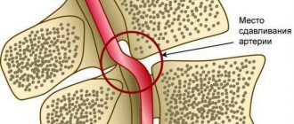
Cerebrovascular diseases (CVD) are one of the main causes of mortality and permanent disability in patients. According to the classification of cerebrovascular disorders adopted in our country in 1958, dyscirculatory encephalopathy (DEP) is the main clinical form of chronic cerebrovascular insufficiency [2–4]. Despite the fact that in the International Statistical Classification of Diseases and Related Health Problems, the IX and X revisions, DEP as a nosology is absent and it was replaced by the term “cerebral ischemia (chronic) (CHI)” - ICD-10, class IX “Diseases of the circulatory system” "(I00-I99), I67.8 - "Other specified cerebral vascular lesions: 1) acute cerebrovascular insufficiency of 5-bromo-2'-deoxyuridine (NOS) and 2) cerebral ischemia (chronic)", the term is still widely used "encephalopathy".
Definition. CCI is a special type of vascular cerebral pathology, caused by a slowly progressive diffuse disorder of the blood supply to the brain with gradually increasing various defects in its functioning [5]. DEP is a chronic progressive form of cerebrovascular pathology, characterized by the development of multifocal or diffuse ischemic brain damage [3, 4, 6, 7]. DEP, like stroke, is a cerebrovascular syndrome [2] - a syndrome of chronic progressive brain damage of vascular etiology, which develops as a result of repeated acute cerebrovascular accidents and/or chronic insufficiency of blood supply to the brain [8].
The clinical picture of DEP is characterized by [4]: 1) a progressive increase in cognitive impairment (decreased memory, attention, intelligence), reaching the level of dementia in the final stages, which is manifested by a combination of pronounced impairment of cognitive functions, personality changes with significant difficulty in normal social activity and the inability to continue work; 2) a gradual increase in emotional impoverishment, loss of interest in life; 3) gradual increase in coordination and walking disorders, destabilization of the tempo and rhythm of movements, tendency to falls; in severe cases, walking becomes impossible, despite the absence of paresis; 4) subcortical syndrome: oligobradykinesia, hypomimia, acheirokinesis, increased muscle tone of the extrapyramidal type (like parkinsonism syndrome); 5) pseudobulbar syndrome of varying severity: dysarthria, dysphagia, forced laughter and crying, symptoms of oral automatism; 6) decreased strength in the limbs, mild paresis with severe brain damage; 7) gradual appearance of disturbances in control of the function of the pelvic organs.
There are three stages of DEP: I - mild or moderate (compensation), II - severe (subcompensation), III - pronounced (decompensation) [9, 10]. For DEP Art. I typical [4]: complaints of increased fatigue, frequent headaches, irritability, moderate memory impairment (primarily operational), moderate decrease in performance, sleep disturbances, which are accompanied by fairly persistent objective disorders in the form of anisoreflexia, discoordination phenomena, mild oculomotor disorders, symptoms oral automatism. There are diffuse neurological symptoms, and the identified disorders are subsyndromal in nature [9]. The early stages of DEP are most characterized by vascular depression and emotional lability, and, as is known, emotional disorders can have an adverse effect on the cognitive sphere [8]. The gradual progression of the disease and worsening symptoms are a powerful factor in social maladjustment [1].
With inadequate treatment, DEP progresses and turns into stage II DEP, in which the noted diffuse neurological symptoms develop into a separate dominant syndrome, which most significantly reduces the patient’s professional and social adaptation [4, 9]. Anxious and depressive reactions appear, cognitive disorders worsen, mental production and volitional activity decrease, professional memory deteriorates, there is a viscosity of thinking, a narrowing of the range of interests, a decrease in criticism and a change in personality. Circadian rhythms are disrupted in the form of daytime sleepiness with poor night sleep. For DEP II Art. characteristic: deepening memory impairment and decreased attention function, increasing intellectual and emotional disorders, and a significant decrease in performance. Somewhat less frequently, there are complaints of chronic fatigue, headache and other manifestations of asthenic syndrome. In some patients, mild subcortical disturbances and changes in gait are detected (it becomes shuffling, mincing). Focal symptoms are more clearly manifested in the form of revitalization of reflexes of oral automatism, central insufficiency of the facial and hypoglossal nerves, coordination and oculomotor disorders, pyramidal insufficiency, and amyostatic syndrome [4].
With DEP stage III. the number of complaints decreases, which is due to a decrease in patients’ criticism of their condition, the severity of intellectual-mnestic and neurological disorders increases, unmotivated actions and inadequate reactions are observed, emotional disorders are characterized by a dysthymic-dysphoric mood background with irritability, dissatisfaction with others and weakness. This stage is characterized by: clearly defined discoordination, amyostatic, psychoorganic, pseudobulbar, pyramidal syndromes. Paroxysmal conditions are more often observed - falls, fainting, epileptic seizures. The main difference between DEP Art. III. from DEP II Art. is that with DEP stage III. In the clinic, several fairly pronounced syndromes are observed, while with DEP stage II. one of them clearly predominates [4, 9].
The relationship between the proportion of complaints and neurological symptoms at different stages of DEP is shown schematically in Fig.
Diagnosis of DEP is possible if the following criteria are met [4]: 1) objectively identified neuropsychological or neurological symptoms; 2) signs of CVD, including risk factors (arterial hypertension, hyperlipidemia, diabetes mellitus, heart rhythm disturbances, etc.), and/or anamnestic signs, and/or instrumentally confirmed signs of damage to cerebral vessels or brain matter; 3) evidence of a causal relationship between (1) and (2); 4) correspondence of the dynamics of neuropsychological and neurological deficits to the characteristics of the course of CVD (a tendency to progression with alternating periods of sharp deterioration, partial regression and relative stabilization); 5) correspondence of changes in the brain substance of vascular origin detected by computed tomography/magnetic resonance imaging with the leading clinical manifestations; 6) exclusion of other diseases that can explain the clinical picture.
Diagnostic criteria for DEP [11]: 1) presence of signs (clinical, anamnestic, instrumental) of brain damage; 2) the presence of signs of acute or chronic dyscirculation (clinical, anamnestic, instrumental); 3) the presence of a cause-and-effect relationship between disorders and the development of clinical, neuropsychological, and psychiatric symptoms; 4) clinical and paraclinical progression of cerebrovascular insufficiency.
Course of DEP and prognosis. There are stable, slowly progressive (with paroxysms and transient cerebrovascular accidents and without vascular episodes), paroxysmal, rapidly progressive course. Stable and slowly progressive are more typical for stage I DEP, which can last for 7–12 years. With a rapidly progressive variant of stage II or III DEP. develop in less than 5 years of illness. Clinical prognosis of stage III DEP. unfavorable, at this stage of the disease patients often need care, and sometimes are completely helpless in everyday life.
Cognitive impairment is a key manifestation of DEP, which largely determines the severity of the patient’s condition. They can serve as the most important diagnostic criterion for DEP and are perhaps the best guide for assessing the dynamics of the disease [2]. At the early stage of DEP, moderate neurodynamic disturbances predominate in the form of slowness, aspontaneity, decreased performance, exhaustion, and weakened concentration. However, such patients generally perform well on timed tests, consistent with mild cognitive impairment [2]. Further progression of the cognitive defect in DEP is associated with the development of dementia, in which cognitive deficit (regardless of motor and other symptoms!) leads to limitation of daily activity and at least partial loss of household independence [2].
Treatment of patients with DEP is a complex medical and social problem and, in fact, is limited to the therapeutic effect on the manifestations of DEP stages I and II. The main directions of management of these patients are reversing the developed decompensation of the pathological process, preventing the progression of the disease, including stroke, reducing the severity of cognitive disorders and neurological deficits [10, 11]. The most effective measure to prevent further progression of the disease is to influence vascular risk factors, primarily proper antihypertensive therapy, correction of carbohydrate and lipid metabolism, changes in vascular tone, increased cerebral perfusion, and improvement of brain tissue metabolism [2].
The basis of therapy aimed at improving the well-being of a patient with DEP and improving his quality of life are drugs that affect cerebral circulation at the microcirculatory level (vasoactive drugs) and drugs that improve metabolic processes in the brain (nootropic drugs). Vasoactive drugs improve blood supply to the brain by dilating the microvasculature. These include: phosphodiesterase blockers (such as pentoxfilline), including those of plant origin (Ginkgo biloba); calcium blockers, the effect of which is most pronounced if the blood flow is impaired in the vertebral arteries supplying blood to the brain stem; α-blockers acting on receptors of the vascular wall. Nootropic drugs can increase neuronal plasticity, thus increasing the adaptive capabilities of nerve cells and reducing their susceptibility to damaging factors by improving metabolic processes in neurons. Nootropic drugs have a positive effect on the higher integrative functions of the brain, facilitate learning processes and memory consolidation.
Today, neurotrophicity, neuroprotection, neuroplasticity and neurogenesis are considered fundamental neurobiological processes involved in the implementation of endogenous protective activity, as well as in attempts to counteract pathophysiological damaging mechanisms and stimulate endogenous repair. The classical concept of neuroprotection involves suppression of a specific pathophysiological mechanism using an appropriate drug [12]. The action of various drugs is aimed at enhancing the oxidation of glucose in mitochondria (Actovegin), inhibiting the oxidation of long-chain fatty acids, which increases the synthesis of adenosine triphosphoric acid (ATP) and neutralization of oxygen radicals, the production of which increases under ischemic conditions [13].
Today, there are several pharmacological groups of drugs with neurometabolic action. They can be roughly divided into classic ones, used for several decades to treat patients with cognitive impairment, and drugs that have relatively recently become widespread in the practice of rehabilitation of patients with vascular diseases of the brain, and were originally proposed for the treatment of Alzheimer's disease. The first group includes Actovegin, piracetam, pyriditol and Cerebrolysin [6]. One of the drugs that has a wide range of pharmacological effects (antihypoxic, antioxidant) is Actovegin, a drug that has a neurotrophic effect. Antioxidant and anti-apoptotic mechanisms of action underlie the neuroprotective properties of Actovegin. It not only improves glucose transport and oxygen absorption, but also stimulates their utilization, which improves oxygen metabolism even under hypoxic conditions.
Impaired cognitive functions (lack of attention, concentration, impaired ability to quickly orientate in a changing environment; decreased memory, especially for current events; slowness of thinking, rapid exhaustion during intense mental work, narrowing of the range of interests) is one of the most common manifestations of CVD. Treatment of cognitive impairment in DEP should, first of all, include measures to prevent further damage to cerebral vessels and brain matter, improve and long-term stabilization of cognitive functions, and correct other clinical manifestations of the disease [2]. On the 1st and 2nd stages. DEP cognitive impairment is present in 88% of cases. A wide range of nootropic and neuroprotective drugs are used to improve cognitive functions [8].
A double-blind, placebo-controlled study was conducted to evaluate the therapeutic effect of the drug Actovegin (tablet form containing 200 mg of the active substance) on mnestic-intellectual abilities in patients with CVD [14]. The study included 120 patients with CVD; the average age of the patients was about 67 years (60–72 years), and the average duration of the disease was 2.5 years. All patients were randomized into 3 groups: group 1 (n = 40), taking Actovegin 3 tablets 3 times a day; group 2 (n = 40) - placebo, of which: 20 patients took 2 tablets 3 times a day, 20 patients took 3 tablets 3 times a day; group 3 (n = 40) - Actovegin 2 tablets 3 times a day. The duration of therapy was 12 weeks, with assessment of the dynamics of the condition before the start of therapy, after 4, 8 and 12 weeks of treatment. To assess the clinical effectiveness of Actovegin, tests were used that assessed mnestic-intellectual functions (memory, synthetic and analytical abilities, concentration, comprehension). The study showed high effectiveness (92%) of 12-week therapy with Actovegin forte on mnestic-intellectual abilities in patients with CVD. Thus, the tablet form of Actovegin can be recommended for long-term outpatient treatment of elderly patients with DEP.
A multicenter study was conducted to evaluate the effectiveness of Actovegin in 1549 elderly patients with impaired cerebral functions [15]. The average age of the patients was 74.1 years. Patients included in the study received treatment according to a standard regimen: 2 weeks of intravenous injections of 10 ml of Actovegin solution, and then 4 weeks of oral administration of Actovegin film-coated tablets (2 tablets 3 times a day). Patients were examined before the start of therapy, two weeks after IV therapy and after 4 weeks of oral therapy. All baseline values were compared with results after 2 and 6 weeks of treatment, and results obtained after 2 weeks were also compared with results after 6 weeks of treatment. After 4 weeks from the start of Actovegin therapy, in 80% of cases, an improvement in the general condition was noted: a decrease in the severity (or cessation) of headaches, dizziness, anxiety and feelings of fear, improvement in memory and concentration (according to psychological testing). In 10.9%, undesirable effects of the drug were noted (feelings of heat and nausea), which were observed in the first, parenteral phase of therapy. The study shows the practical significance of combination therapy: start therapy with iv 10 ml of Actovegin to achieve a quick and good response, followed by continued oral administration of Actovegin forte tablets). The proposed treatment regimen with Actovegin can be recommended for long-term therapy of elderly patients with organic syndrome.
A number of clinical studies have shown that the use of the drug Actovegin has a positive effect on cognitive functions, improves psychological and behavioral reactions, and is also most effective for mild and moderate cognitive impairment. Currently, therapeutic regimens for the treatment of CVD have been developed and are widely used with the prescription of Actovegin: 10 ml (400 mg) per 200 ml of saline solution intravenously for 7–10 days, then 1–2 tablets (200–400 mg) 3 times /day orally for 1-2 months, in the presence of mnestic-intellectual disorders in the elderly - up to 12 weeks, 2-3 tablets 3 times a day. Repeated courses after 6–8 months [16].
In the prevention and treatment of moderate cognitive disorders of vascular origin, neurometabolic drugs have been shown to be highly effective, including the drug Ceraxon (citicoline) [17–19]. The drug has a targeted effect on the key links in the processes of death of nerve cells of vascular, traumatic, toxic and other etiologies. Since Ceraxon is a natural metabolite of biochemical processes in the body and combines neurotransmitter and neurometabolic effects in its spectrum of action. The most important of them is the activation of the biosynthesis of membrane phospholipids of brain neurons, primarily phosphatidylcholine. Citicoline can correct cognitive impairments already at the initial stages of their manifestations in patients with DEP [18]. Its use promotes regression of cognitive impairments and reduces concomitant emotional, affective and behavioral disorders. Cerakson is able to potentiate the effect of other drugs in the treatment of acute cerebrovascular pathology, including thrombolytics, antiplatelet agents and neurotrophics [17]. Prescription regimen for the drug Ceraxon: 1000 mg for 10 days IM or IV once a day, then Ceraxon solution 2 ml orally 3 times a day for 3 months [18] or according to the scheme [20]: daily at a dose of 3 ml 2 times a day for 6 months. The duration of neuroprotection can last up to 12 months.
Conclusion
Moderate and severe cognitive impairment of a cerebrovascular nature can serve as an equivalent to DEP and is detected in 3–16 people over 60 years of age [2]. The main thing in the treatment of a patient with stage I or II DEP. in an outpatient setting is to relieve the developed decompensation of the pathological process, prevent the progression of the disease, including stroke, reduce the severity of cognitive disorders and neurological deficits [21].
A number of studies have shown the high clinical effectiveness of administering the drugs Actovegin and Cerakson as long-term neuroprotection in patients with dyscirculatory encephalopathy.
Literature
- Shavlovskaya O. A. Medical and social aspects of the elderly / Articles of the All-Russian Scientific and Practical Conference with International Participation “Society and Health: Current State and Development Trends”. 2013, vol. 2, p. 365–371.
- Levin O. S. Use of Ginkgo biloba extract (EGb 761) for the treatment of cognitive impairment in dyscirculatory encephalopathy // Breast Cancer. 2009, vol. 17, no. 20, p. 1356–1361.
- Maksudov G. A. Dyscirculatory encephalopathy. In the book: Vascular diseases of the nervous system. Ed. E. V. Schmidt. M.: Medicine, 1975, pp. 501–512.
- Temnikova E. A. The use of the drug Omaron in the practice of a general practitioner when working with elderly patients // RMZh. 2009, vol. 17, no. 20, 1345–1356.
- Putilina M.V. Modern ideas about the treatment of anxiety and depressive disorders in chronic cerebral ischemia // RMJ. 2011, no. 9, p. 569–573.
- Polyakov I. A., Malozemov I. V., Stepanova N. S. Antioxidant therapy in the complex treatment of dyscirculatory encephalopathy // Terra Medica Nova. 2009, no. 4–5, p. 22–24.
- Shtulman D. R., Levin O. S. Neurology. Handbook of a practicing physician. M.: MEDpress-inform, 2008, 1025 p.
- Zakharov V.V., Lokshina A.B. Cognitive impairment in discirculatory encephalopathy // Breast Cancer. 2009, vol. 17, no. 20, p. 1325–1329.
- Dadasheva M.N., Podrezova L.A., Shuchalin O.G. et al. Algorithm for the treatment of dyscirculatory encephalopathy in patients with arterial hypertension in general medical practice // RMZh. 2009, vol. 17, no. 20, p. 1320–1324.
- Kadykov A. S., Shakhparonova N. V. Vascular diseases of the brain. Miklos, 2006, 192 p.
- Damulin I.V., Parfenov V.A., Skoromets A.A., Yakhno N.N. Circulatory disorders in the brain and spinal cord. In the book: Diseases of the nervous system. Guide for doctors. Ed. N. N. Yakhno, D. R. Shtulman. M., 2003. pp. 231–302.
- Shakhparonova N.V., Kadykov A.S. Antioxidant therapy for cerebrovascular diseases // Breast Cancer. 2010, vol. 18, no. 26, p. 1570–1572.
- Astashkin E.I. The influence of Actovegin on the energy metabolism of cells during ischemia. Proceedings of MMA im. I. M. Sechenov. M., 2009: 1–4.
- Jansen V., Bruckner G.V. Treatment of chronic cerebrovascular insufficiency using Actovegin forte tablets (double-blind, placebo-controlled study) // RMZh. 2002. T. 10. No. 12–13. pp. 543–546.
- Letzel H., Schicktiger U. Use of Actovegin in elderly patients with organic syndromes. Multicenter study of 1549 patients // Breast Cancer. 2003. T. 11. No. 25. P. 1428–1431.
- Shavlovskaya O. A. Use of Actovegin in neuroprotective therapy of patients with cerebrovascular diseases // Journal of Neurology and Psychiatry named after. S. S. Korsakova. 2013, v. 113, no. 6, p. 74–76.
- Boyko A. N., Kabanov A. A. Cytocoline: new possibilities of neuroprotection and pharmacotherapy for diseases of the nervous system // Farmateka. 2007, no. 15, pp. 42–48.
- Putilina M.V. Features of combined neuroprotective therapy for acute cerebrovascular accidents // Breast Cancer. 2009, vol. 17, no. 4, p. 261–267.
- Putilina M.V., Shabalina N.I. Possibilities of early correction of mild and moderate cognitive disorders in patients with dyscirculatory encephalopathy // Treating Doctor. 2010, no. 9, p. 100–103.
- Shavlovskaya O. A. Neuroprotective therapy of neurological deficit in cerebrovascular pathology // Practicing doctor today. 2012, no. 3, p. 39–44.
- Shavlovskaya O. A. Efficacy of antioxidant drugs in the treatment of mild and moderate cognitive disorders // Breast Cancer. 2013, vol. 21, no. 10, p. 476–480.
O. A. Shavlovskaya, Doctor of Medical Sciences
GBOU VPO First Moscow State Medical University named after. I. M. Sechenova Ministry of Health of the Russian Federation, Moscow
Contact Information