Depending on the type of hemodynamics and some features of pathogenesis, the following clinical variants of acute heart failure (AHF) are distinguished (Table 1):
1. With stagnant hemodynamics:
- right ventricular (venous congestion in the systemic circulation);
- left ventricular (cardiac asthma, pulmonary edema).
2. With a hypokinetic type of hemodynamics (small output syndrome - cardiogenic shock).
3. Hypoxemic crisis (dyspnea-cyanotic attack).
Symptoms
Signs of heart failure in children:
- dyspnea;
- constant malaise;
- weakness and loss of strength;
- sweating;
- cardiopalmus;
- increase in baking.
A very important symptom of heart failure in children is swelling.
When to see a doctor
First, make an appointment with your pediatrician, and then, if necessary, your pediatrician will make a referral to a pediatric cardiologist.
The listed manifestations, even if they are not evidence of heart failure, are still sufficient reason to seek help as soon as possible. Remember that acute heart failure in children can be fatal.
Our pediatrician will refer you to a pediatric cardiologist. The doctor sees you in the center of Moscow, a 5-minute walk from the Mayakovskaya metro station.
Diagnostics
During the appointment, the doctor will question and examine, analyze, and collect anamnesis. Of course, auscultation is not enough, and the doctor will need the results of electrocardiography, echocardiography, possibly tomography and chest x-ray. Daily monitoring can be informative.
As for laboratory tests, clinical blood and urine tests will be needed.
If there is a concomitant pathology, and this is a common occurrence in children with heart failure, consultation with other specialized specialists is necessary, and if necessary, they will work on the problem in tandem. After all diagnostic measures, treatment of heart failure in children begins immediately.
All tests and studies are carried out in one place; there is no need to travel anywhere else. All our specialists receive appointments at this address. The clinic is located in the center of Moscow, near the Mayakovskaya metro station.
Cardiogenic shock (low cardiac output syndrome)
Shock is an acutely developing, life-threatening pathological process characterized by a progressive decrease in tissue perfusion, severe disturbances in the functioning of the central nervous system, blood circulation, respiration and metabolism.
Cardiogenic shock is a clinical syndrome characterized by arterial hypotension and signs of a sharp deterioration in microcirculation and tissue perfusion, including blood supply to the brain and kidneys (lethargy or agitation, decreased diuresis, cold skin covered with sticky sweat, pallor, marbled skin pattern); sinus tachycardia is compensatory in nature.
In addition to cardiogenic shock, possible causes of shock may include:
— decrease in total blood volume (hypovolemic shock) due to bleeding or dehydration due to losses from the gastrointestinal tract (vomiting, diarrhea), polyuria, burns, etc. The main pathogenetic mechanism is insufficient cardiac preload due to a deficiency of venous inflow;
- deposition of blood in the venous pools (distributive or vasogenic shock) during anaphylaxis, acute adrenal insufficiency, sepsis, neurogenic or toxic shock. The leading pathogenetic mechanism is insufficiency of cardiac afterload.
Low cardiac output (cardiogenic shock) develops as a result of failure of the pumping function of the heart (acute myocardial ischemia, infectious and toxic carditis, cardiomyopathies), as well as as a result of obstruction of the venous inflow to the heart or cardiac output (obstructive shock) in diseases of the pericardium (pericardial tamponade), tension pneumothorax, acute obstruction of the atrioventricular orifice by atrial myxoma, rupture of chords, heart valves, massive pulmonary embolism, etc. Pericardial tamponade and obstruction of the atrioventricular orifice require immediate surgical intervention; drug therapy in these cases can only worsen the situation.
A special clinical variant of cardiogenic shock is arrhythmic shock, which develops as a result of a drop in cardiac output due to tachycardia/tachyarrhythmia or bradycardia/bradyarrhythmia; After stopping the rhythm disturbance, adequate hemodynamics are quickly restored.
With the development of low cardiac output syndrome, a pain syndrome is observed, manifested by severe anxiety in the child, followed by lethargy. A drop in blood pressure, threadlike pulse, tachycardia, “marble” pallor of the skin, collapsed peripheral veins, sticky cold sweat, acrocyanosis, and oligoanuria are noted.
The course of cardiogenic shock is often accompanied by the development of pulmonary edema, mesenteric ischemia, disseminated intravascular coagulation syndrome, and renal failure.
Cardiogenic shock at the prehospital stage is diagnosed based on:
- progressive decrease in systolic blood pressure;
— reduction of pulse pressure to less than 20 mm Hg;
- signs of impaired microcirculation and tissue perfusion: decreased diuresis to less than 20 ml/h, cold skin covered with sticky sweat, pallor, marbled skin pattern, in some cases - collapsed peripheral veins.
Treatment of cardiogenic shock
1. Elimination of the main cause: relief of cardiac arrhythmias and pain. In case of severe pain, administer fentanyl at a dose of 0.01 mg/kg or 1% promedol at a dose of 0.1 ml/year of life intravenously. For children of the first two years of age, prescribe non-narcotic analgesics: baralgin or 50% analgin solution at a dose of 0.1–0.2 ml/year of life. In the presence of psychomotor agitation, prescribe a 0.5% solution of diazepam (Seduxen, Relanium) at a dose of 0.1–0.3 mg/kg intravenously.
2. In the absence of signs of congestive heart failure (shortness of breath, moist rales in the posterior lower parts of the lungs), the patient must be placed in a horizontal position.
3. If the clinical picture of shock is extensive and there are no signs of congestive heart failure, therapy should begin with intravenous fluid administration (infusion therapy to increase preload) under the control of blood pressure, heart rate, respiratory rate and auscultation of the lungs. Rheopolyglucin is administered at a dose of 5–8 ml/kg + 10% glucose solution and 0.9% sodium chloride solution at a dose of 50 ml/kg in a 2:1 ratio with the addition of cocarboxylase and 7.5% potassium chloride solution at a dose of 2 mmol/ kg.
4. An increase in cardiac output is achieved:
- administration of dopamine (6–9 mg/kg/min), which has a positive inotropic effect. Dopamine, an agonist of dopamine receptors, causes excitation of α- and β-adrenergic receptors, increases the release of norepinephrine into the synaptic cleft, increases the strength of heart contractions and cardiac output, the effect of the drug on heart rate is insignificant. The drug promotes the redistribution of total vascular peripheral resistance, causing dilatation of the renal and mesenteric vessels and a vasoconstrictor effect; improved renal perfusion helps to increase diuresis. Dopamine infusion is carried out in the intensive care unit under continuous monitoring using a dispenser for 24–48 hours. The effect occurs after 5 minutes, its peak after 5–7 minutes. Considering the possible tachycardic and arrhythmogenic effects of dopamine, the drug is used in very short courses, only in extremely severe cases and with complete depletion of the sympathoadrenal system, with an increase in AHF to degree III;
- administration of drugs that have a positive chronotropic effect: adrenaline, norepinephrine (0.05-0.2 mcg/kg/min).
5. The lack of effect from dopamine or the inability to use it due to tachycardia, arrhythmia is an indication for the addition of dobutamine or monotherapy with it. This drug, unlike dopamine, has a more pronounced vasodilatory effect and a less pronounced ability to cause an increase in heart rate and arrhythmia. 250 mg of the drug should be diluted in 500 ml of 5% glucose solution (1 ml of the mixture contains 0.5 mg, and 1 drop - 25 mcg of dobutamine); in monotherapy, it is prescribed at a dose of 2.5 mcg/kg/min, increasing every 15–30 minutes by 2.5 mcg/kg/min until an effect, side effect is obtained, or a dose of 10 mcg/kg/min is achieved, and with a combination of dobutamine with dopamine - in maximum tolerated doses. Dobutamine is a β1-adrenergic agonist, has a positive inotropic effect on the heart, moderately increases heart rate, as well as stroke and cardiac output, reduces the total peripheral and vascular resistance of the pulmonary circulation, while systemic blood pressure tends to increase, reduces the filling pressure of the ventricles of the heart, increases coronary blood flow, improves oxygen supply to the myocardium. Increased cardiac output improves renal perfusion and increases sodium and water excretion. The drug is used for decreased renal blood flow and cardiac output, moderate hypotension. Dobutamine is not prescribed if systolic blood pressure (BP) is <70 mmHg.
6. If an increase in the volume of administered fluid does not lead to an increase in cardiac output (with high central venous pressure and no effect of drugs with a positive inotropic effect), the reason for its decrease may be either a large afterload, to reduce which sodium nitroprusside is used (0.5– 0.8 mcg/kg/min), or a decrease in myocardial contractility, which serves as an indication for the administration of cardiac glycosides.
7. If there are signs of congestive heart failure and in the case of using inotropic drugs from the group of pressor amines, the administration of peripheral vasodilators - nitrates (nitroglycerin) is indicated.
8. Administer a 3% solution of prednisolone at a dose of 3–5 mg/kg intravenously.
9. In the absence of contraindications, heparin is prescribed for the correction of microcirculatory disorders, especially in case of long-term intractable shock.
10. Oxygen therapy.
11. Hospitalization in the intensive care unit.
Treatment
First, acute heart failure in children requires emergency care. Treatment in this case involves eliminating hypoxia, improving cardiac contractility, and stabilizing electrolyte disturbances. At this stage, oxygen, vasodilators, diuretics, antispasmodics, cardiac glycosides and other agents are required. Under no circumstances give your child any available funds!
As for milder cases, treatment of heart failure in children is aimed at eliminating tachycardia and removing excess fluid. Cardioprotective drugs are prescribed. Chronic heart failure in children requires periodic monitoring by a specialized specialist.
In some cases, surgical treatment may be necessary when acute heart failure in children is not corrected by conservative methods.
How to make an appointment with a specialist
Acute cardiovascular failure in children can cause serious illness, even death. Do not put off consulting a cardiologist until later. By wasting time, we worsen the prognosis and ourselves reduce the likelihood of a complete cure for the child. Make an appointment at JSC "Medicine" (clinic of Academician Roitberg) in just a few clicks. To sign up right now, click on the feedback button in the upper right corner and leave your details. Or contact us at contact number +7 (495) 995-00-33.
We guarantee a competent approach, focus on results and, of course, special attention to the child.
Even if the child feels better, periodic monitoring is still required. If there has not yet been treatment as such, remember: the faster heart failure in children is brought under control, the better the prognosis and the higher the likelihood of curing the disease forever.
Acute congestive left ventricular failure
Acute left ventricular failure is associated with a sharp decrease in the pumping function of the left ventricle and a rapid increase in blood stagnation in the lungs.
The main causes of acute left ventricular failure:
1. Myocardial diseases in the stage of decompensation (myocarditis, cardiomyopathies of various origins).
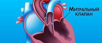
2. Hemodynamic volume overload of the left chambers of the heart with heart defects: atrial and interventricular septal defects; open aortic duct; insufficiency of the aortic and mitral valves, aortic stenosis, coarctation of the aorta, etc.
3. Hemodynamic pressure overload of the left side of the heart with heart defects: coarctation of the aorta; stenosis of the mitral and aortic valves; hypertrophic cardiomyopathy; heart tumors; malignant arterial hypertension.
4. Heart rhythm disturbances (paroxysmal tachycardia, atrial fibrillation).
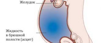
Acute left ventricular failure is characterized by a sequential course of stages: interstitial and alveolar. Clinically, it is manifested by the syndromes of cardiac asthma and pulmonary edema. Cardiac asthma is considered to be the initial stage of pulmonary edema. When it occurs, fluid leaks from the vascular bed into the interstitial (interstitial) tissue, that is, interstitial pulmonary edema occurs. With pulmonary edema, fluid from the interstitial space moves into the alveoli. In this regard, this phase of acute left ventricular failure is called the alveolar stage.
Cardiac asthma is manifested by attacks of suffocation, which are often preceded by physical and emotional stress. Manifests with paroxysmal shortness of breath, painful suffocation and orthopnea, occurring more often at night (predawn) hours; sometimes - Cheyne-Stokes breathing, cough (initially dry, and then with sputum, which does not bring relief), later foamy sputum, often pink in color, pallor, acrocyanosis, hyperhidrosis and is accompanied by excitement, fear of death. The child is restless, complains of tightness in the chest, lack of air. Takes a forced position: sits with legs down. In case of acute congestion, moist rales may not be heard at first, or a meager amount of fine bubbling rales is detected over the lower parts of the lungs; swelling of the mucous membrane of small bronchi can manifest itself as a moderate picture of bronchial obstruction with prolongation of exhalation, dry wheezing and signs of pulmonary emphysema. A differential diagnostic sign that allows one to differentiate this condition from bronchial asthma can be the dissociation between the severity of the patient’s condition and (in the absence of pronounced expiratory dyspnea, as well as “silent” zones) the paucity of the auscultatory picture. Possible acute expansion of the heart to the left, the appearance of a systolic murmur at the apex of the heart, a proto-diastolic gallop rhythm, as well as an emphasis on the second tone on the pulmonary artery and other signs of load on the right heart, up to the picture of right ventricular failure. Blood pressure can be normal, high or low. A constant symptom is increasing tachycardia with a change in the ratio of heart rate and respiratory rate of more than 3: 1. The attack lasts from several minutes to several hours. Cardiac asthma does not always develop into a detailed picture of pulmonary edema, especially if treatment measures are timely.
With the development of cardiogenic pulmonary edema, anxiety is noted at the beginning, children may rush about in bed, and subsequently there may be a disturbance of consciousness. The skin is first pale, and then bluish, covered with cold, sticky sweat. When coughing, foamy, pink sputum is produced and may be streaked with blood. Breathing is noisy, bubbling. A large number of moist, small- and medium-bubble rales are heard over the lungs, appearing first paravertebrally, and then over all other parts of the chest. The development of oligoanuria is possible.
In the interstitial stage, differential diagnosis is carried out with an attack of bronchial asthma, and in the alveolar stage - with non-cardiogenic pulmonary edema (adult respiratory distress syndrome). The latter occurs in pediatric practice much more often than cardiogenic shock, and develops with toxicosis, disseminated intravascular coagulation syndrome, all types of shock, drowning, poisoning with gasoline, kerosene, turpentine, etc.
Treatment of acute congestive left ventricular failure
1. Treatment of acute congestive heart failure begins with giving the patient an elevated position (with an unexpressed picture of congestion - an elevated head end, with extensive pulmonary edema - a sitting position with lowered legs); these measures are not performed in cases of severe arterial hypotension. Ensure patency of the upper respiratory tract by removing mucus from the mouth with a gauze swab.
2. A universal pharmacological agent for acute congestive heart failure is furosemide (Lasix); already 5–15 minutes after administration, due to venous vasodilation, it causes hemodynamic unloading of the myocardium, which increases over time due to the diuretic effect that develops later. Furosemide is administered intravenously as a bolus and is not diluted; the dose of the drug is 2–4 mg/kg.
3. Severe congestion in the pulmonary circulation in the absence of arterial hypotension is an indication for intravenous drip administration of nitroglycerin. The use of nitrate drugs requires careful monitoring of blood pressure and heart rate. Nitroglycerin is prescribed at a dose of 0.1–0.7 mcg/min. Contraindications to the use of nitrates are arterial hypotension and hypovolemia, pericardial constriction and cardiac tamponade, pulmonary artery obstruction, and inadequate cerebral perfusion.
For mild congestion in the lungs, nitroglycerin can be prescribed 1/4–1 tablet under the tongue.
4. Modern methods of drug treatment have minimized the importance of applying venous tourniquets to the extremities, however, if adequate drug therapy is impossible, this method of hemodynamic unloading not only can, but should be used, especially with severely progressive pulmonary edema. Tourniquets are applied to 2–3 limbs (upper third of the shoulder or thigh) for 15–20 minutes, with the procedure repeated after 20–30 minutes. An indispensable condition in this case is the preservation of the pulse in the artery distal to the tourniquet.
5. Continued signs of pulmonary edema with hemodynamic stabilization may indicate an increase in membrane permeability, which requires the administration of glucocorticoids to reduce permeability (prednisolone at a dose of 2–3 mg/kg).
6. A means of combating foaming in pulmonary edema are “defoamers” - substances that ensure the destruction of foam by reducing surface tension. The simplest of these means is alcohol vapor, which is poured into a humidifier (33%), oxygen is passed through it, supplied to the patient through a nasal catheter or breathing mask at an initial rate of 2–3 l/min, and after a few minutes at a rate of 6–8 l /min.
7. For low blood pressure and hypokinetic variant (myocardial failure), cardiotonic drugs are indicated: cardiac glycosides (strophanthin, korglykon, digoxin, etc.), sympathomimetic amines (dopamine 2–5 mcg/kg/min), polarizing mixture (10% glucose solution 5 ml/kg, Panangin 0.5–1.0 ml/year of life, insulin 1 unit per 5 g of dry glucose) IV drip.
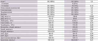
For the treatment of decompensated heart failure with the development of acute left ventricular failure and pulmonary edema, intravenous drip administration of short-acting cardiac glycosides, namely strophanthin or corglycon, is necessary. The drugs are administered intravenously slowly; dosage is presented in table. 3.
8. In case of hyperkinetic type of blood circulation with a persistent increase in blood pressure, cardiac glycosides are not indicated; ganglion blockers are prescribed (5% pentamine solution for children 1–3 years old at a dose of 1–3 mg/kg, over 3 years old - 0.5–1 mg/kg or 2% benzohexonium solution for children 1–3 years old at a dose of 0.5–1 .5 mg/kg, over 3 years - 0.25–0.5 mg/kg), droperidol (0.25% solution at a dose of 0.1 ml/kg). The drugs are used once; a reduction in blood pressure by no more than 40% from the initial level is permissible.
9. In the absence of contraindications, heparin is indicated for the correction of microcirculatory disorders, especially with long-term intractable pulmonary edema.
10. If the condition is severe and there is a risk of cardiac and respiratory arrest, tracheal intubation and transfer to mechanical ventilation are indicated. Urgent hospitalization in the intensive care unit or intensive care unit. Transportation is carried out in a semi-sitting position against the background of ongoing oxygen therapy.