The T wave on the ECG is normal in children and adults
The beginning of the T wave coincides with the repolarization phase, that is, with the reverse transition of sodium and potassium ions through the membrane of heart cells, after which the muscle fiber becomes ready for the next contraction. Normally, T has the following characteristics:
- begins on the isoline after the S wave;
- has the same direction as the QRS (positive where R is dominant, negative when S is dominant);
- smooth in shape, the first part is flatter;
- amplitude T up to 8 cells, increases from 1 to 3 chest leads;
- may be negative in V1 and aVL, always negative in aVR.
In newborns, T waves are low in height or even flat, their direction is opposite to the adult ECG. This is due to the fact that the heart turns in direction and takes a physiological position by 2 - 4 weeks. At the same time, the configuration of the teeth on the cardiogram gradually changes. Typical features of a pediatric ECG:
- negative T in V4 persists up to 10 years, V2 and 3 – up to 15 years;
- adolescents and young adults may have negative T waves in the 1st and 2nd chest leads; this type of ECG is called juvenile;
- height T increases from 1 to 5 mm; in schoolchildren it is 3–7 mm (as in adults).
We recommend reading about how an ECG is performed. You will learn about the principles of operation of an electrocardiograph, preparation for an ECG, methods of conducting, and interpretation of indicators. And here is more information about what myocardial ischemia looks like on an ECG.
ECG changes and their meanings
Most often, changes are suspected of coronary heart disease, but such a disorder may be a sign of other diseases:
- thromboembolism,
- myocarditis, pericarditis,
- tumors, infections and injuries,
- ventricular hypertrophy,
- intoxication, including cardiac glycosides, antiarrhythmic drugs, aminazine, nicotine,
- stress, neurocirculatory dystonia,
- diseases of the endocrine system,
- potassium deficiency,
- decreased blood circulation in the brain,
- osteochondrosis.
Therefore, to make a diagnosis, all clinical signs and changes in the cardiogram are taken into account as a whole.
Two-phase
On the cardiogram, T first decreases below the isoline, and then crosses it and becomes positive. This symptom is called the “roller coaster” syndrome. May occur in the following pathologies:
- left ventricular hypertrophy;
- Hiss bundle branch block;
- increased calcium levels in the blood;
- intoxication with cardiac glycosides.
Biphasic T wave with left ventricular hypertrophy
Smoothed
Flattening of the T wave can be caused by:
- taking alcohol, Cordarone or antidepressants;
- diabetes mellitus or eating a lot of sweets;
- fear, excitement;
- cardiopsychoneurosis;
- hypokalemia;
- myocardial infarction in the scarring stage.
Decrease in indicator
A reduced T is indicated by its amplitude, which is less than 10% of the QRS complex. This symptom on the ECG causes:
- coronary insufficiency,
- cardiosclerosis,
- obesity,
- elderly age,
- hypothyroidism,
- dishormonal cardiomyopathy,
- myocardial dystrophy,
- taking corticosteroids,
- anemia,
- tonsillitis.
The T wave on the ECG is smoothed
The T wave can be smoothed in the same conditions as an absent wave, since both definitions characterize low-amplitude oscillations. It should be taken into account that violation of the rules for ECG registration can also cause smoothing of T. It also occurs in metabolic diseases - low function of the thyroid gland (myxedema, hypothyroidism). It can be found in completely healthy people throughout the day in several cardiac cycles (according to Holter monitoring).
Inversion
Inversion (turning over) of the T wave means a change in its position relative to the isoline, that is, in leads with a positive T, it changes its polarity to negative and vice versa. Such deviations can also be normal - in the right chest leads with a juvenile ECG configuration or a sign of early repolarization in athletes.
T wave inversion in leads II, III, aVF, V1-V6 in a 27-year-old athlete
Diseases that are accompanied by T inversion:
- myocardial or cerebral ischemia,
- influence of stress hormones,
- bleeding in the brain,
- attack of tachycardia,
- violation of impulse conduction along the branches of the Hiss bundle.
Negative T wave
For coronary heart disease, a characteristic sign is the appearance of negative T waves on the ECG, and if they are accompanied by changes in the QRS complex, then the diagnosis of a heart attack is considered confirmed. In this case, changes in the cardiogram depend on the stage of myocardial necrosis:
- acute – abnormal Q or QS, ST segment above the line, T positive;
- subacute – ST on the isoline, negative T;
- in the scar stage, weakly negative or positive T.
A negative T wave in leads V5-V6 (highlighted in red) indicates ischemia.
A variant of the norm may be the appearance of negative T wave with frequent breathing, excitement, after a heavy meal, which contains a lot of carbohydrates, as well as with individual characteristics in some healthy people. Therefore, the detection of negative values cannot be considered a serious illness.
Pathological conditions that are accompanied by negative T waves:
- heart disease - angina pectoris, heart attack, cardiomyopathy, inflammation of the myocardium, pericardium, endocarditis, mitral valve prolapse;
- violation of hormonal and nervous regulation of cardiac activity (thyrotoxicosis, diabetes mellitus, diseases of the adrenal glands, pituitary gland);
- pulmonary heart;
- after paroxysmal tachycardia or frequent extrasystoles;
- subarachnoid hemorrhage.
Subarachnoid hemorrhage is accompanied by negative T waves
Absence of T wave on ECG
The absence of T on the ECG means that its amplitude is so low that it merges with the isoelectric line of the heart. This happens when:
- drinking alcohol;
- against the backdrop of excitement, anxiety;
- cardiomyopathy in patients with diabetes mellitus;
- neurocirculatory dystonia (with a sudden change in body position or after rapid breathing);
- insufficient intake of potassium or its loss through sweat, urine, intestinal contents (diarrhea);
- scarring of myocardial infarction;
- use of antidepressants.
High rate
Normally, in those leads where the highest R is recorded, the maximum amplitude is noted; in V3 - V5 it reaches 15 - 17 mm. Very high T can occur when the influence on the heart of the parasympathetic nervous system, hyperkalemia, subendocardial ischemia (first minutes), alcoholic or menopausal cardiomyopathy, left ventricular hypertrophy, and anemia predominate.
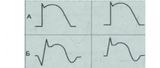
Changes in the T wave on the ECG during ischemia: a - normal, b - negative symmetrical "coronary" T wave, c - high positive symmetrical "coronary" T wave, d, e - biphasic T wave, f - reduced T wave, g - smoothed T wave, z - weakly negative T wave.
Flat
A slightly inverted or flattened T may be either a normal variant or a manifestation of ischemic and dystrophic processes in the heart muscle. It occurs with complete blockade of the conduction pathways in the ventricles, myocardial hypertrophy, acute or chronic pancreatitis, taking antiarrhythmic medications, and hormonal and electrolyte imbalance.
Coronary
When the heart muscle is hypoxic, the fibers located under the inner membrane, the endocardium, are most affected. The T wave reflects the ability of the endocardium to maintain a negative electrical potential, therefore, in case of coronary insufficiency, it changes its direction and becomes this shape:
- isosceles;
- negative (negative);
- pointed.
These signs characterize the ischemic wave, or it is also called coronary. Manifestations on the ECG are maximum in those leads where the greatest damage is localized, and in mirror (reciprocal) leads it is sharp and isosceles, but positive. The more pronounced the T wave, the deeper the degree of myocardial necrosis.
Rise of the T wave on the ECG
Moderate physical stress, hyperkalemia, infectious processes in the body, thyrotoxicosis, and anemia lead to an increase in the amplitude of T waves. Elevated T without changes in well-being can occur in healthy people, and can also be a symptom of vegetative-vascular disorders with a predominance of vagal tone.
Depression
A reduced T wave can be a manifestation of cardiomyodystrophy; it occurs with pneumonia, rheumatism, scarlet fever, acute inflammatory process in the kidneys, cor pulmonale and hypertrophic increase in the muscular layer of the myocardium.
The T wave is positive
Normally, T waves in leads should be positive: first, second standard, aVL, aVF, V3-V6. If it appears where in healthy people it is negative or close to the isoelectric line, then this indicates a lack of blood flow through the arteries of the heart (myocardial ischemia), blockade of the branches of the His bundle. Temporary changes are caused by stress, an attack of rapid heartbeat, and intense exercise in athletes.
Heart ST depression
Depression of the ST segment, or its decrease relative to the isoline by more than 1 mm, is a clinically significant sign. Such a change can be caused by both nonspecific and specific reasons, which are discussed below.
Non-cardiac and physiological causes include:
- errors on the part of medical staff when taking an ECG - incorrect application of electrodes or their poor contact with the patient’s skin;
- water and electrolyte disturbances that occur when drinking sea water in large quantities, dehydration, alcohol abuse, salt-free diet, anorexia, heavy physical activity, use of diuretics, as well as certain pathologies (diseases of the kidneys, gastrointestinal tract, thyroid gland, burns, significant blood loss);
- drinking very cold water;
- hyperventilation (increased air exchange in the human body).
What does ECG mean in decoding?
Nonspecific ST-T changes on the ECG in the form of its decrease relative to the isoelectric line are primarily associated with cardiac ischemia, as the most dangerous condition that can lead to death.
However, this change is nonspecific and can also occur with other disorders of the heart. To correctly interpret this deviation, differential diagnosis is necessary.
Why is the ST segment reduced in adults and children?
A decrease in the ST area on the ECG in adults and children can occur in the following cases:
- Myocardial infarction with absence of ST elevation. In this case, depression of the segment is observed in two or more leads (by lead is meant the difference in biopotentials between two points of the body, for example, right hand - left hand). This type of coronary insufficiency is less common than elevation of the segment infarction (about 28% of all cases), but in recent years there has been an increase in incidence. The cause of this disorder is most often blockage of the coronary artery by a thrombus, which leads to a significant deterioration in the blood supply to the heart muscle and the development of necrosis in the tissues. The development of this condition can be triggered by stress, anemia, and infectious and inflammatory pathologies.
- Unstable angina (58% of all cases) is an intermediate condition in coronary heart disease, when the risk of developing myocardial infarction is high. The most common cause is atherosclerotic plaques in the coronary vessels of the heart, closing their lumen by ¾ or more. Risk factors are arterial hypertension, tachycardia, physical and emotional stress, consumption of heavy food and alcohol, which increases the load on the heart and causes a lack of oxygen.
- Inflammatory lesions of the heart muscleappearing against the background of infectious diseases (including influenza and respiratory pathologies).
- Toxic effects of ethanol in alcohol abuse. In this case, there is an accumulation of fatty tissue in the heart muscle and an expansion of all the cavities of the heart. At the initial stage, this disorder can be reversible, that is, giving up alcoholic beverages contributes to a significant improvement in function.
- Changes in hormonal levels during menopause in women. The main factor in menopausal cardiomyopathy is a decrease in estrogen production, which contributes to the formation of protein deficiency in the myocardium.
- Dilated cardiomyopathy , in which stretching of the cavities of the heart develops. At the same time, heart failure progresses, the rhythm and conductivity of this organ are disrupted.
- Enlargement of the right chambers of the heart due to embolism (blockage) of the pulmonary artery, severe attack of bronchial asthma, pneumonia, accumulation of air in the pleural cavity.
- Intoxication with cardiac glycosides. Heart failure does not occur immediately, but as the drug accumulates in the blood.
- Paroxysmal tachycardia , detected in elderly people.
In pregnant women, disorders associated with the ST segment are most often caused by physiological reasons - changes in the anatomical position of the heart. Therefore, if such a condition is not accompanied by complaints or a history of cardiac pathologies, then treatment is not required. In doubtful cases, the doctor may prescribe an echocardiogram.
Types of segment reduction
There are 3 types of ST depression:
- obliquely ascending, occurring against the background of tachycardia and being a variant of the norm;
- horizontal, often serving as a sign of ischemic processes in the myocardium;
- oblique, also a symptom of cardiac muscle dysfunction.
To accept the above-described phenomena as normal, the doctor must first exclude possible pathological causes.
Symptoms of deviation
Patients may experience the following symptoms as subjective symptoms:
- Myocardial ischemia:
- severe pain, feeling of pressure, heaviness behind the sternum;
- in women, pain can often radiate to the neck and arm;
- dyspnea;
- arrhythmia;
- general weakness;
- cough;
- in more rare cases - nausea, vomiting, bloating, dizziness, darkening of the eyes, cold sweat, swelling.
- Unstable angina:
- painful attacks lasting 1-10 minutes, similar in nature to the previous case;
- increased symptoms when walking, physical and emotional stress;
- signs subside when taking nitroglycerin.
- Alcoholic cardiomyopathy:
- heartache;
- dyspnea.
- Menopausal cardiomyopathy:
- pressing pain in the upper region of the heart;
- At the same time, other symptoms occur - facial redness, sweating, feeling hot;
- increased symptoms are not associated with physical activity and are not relieved by nitroglycerin.
- Intoxication with glycosides:
- nausea, vomiting, diarrhea;
- severe arrhythmia;
- tachycardia.
Differential diagnosis
Nonspecific ST-T changes require differential diagnosis taking into account medical history, additional studies and analysis of other indicators on the ECG.
To detect acute myocardial infarction, rapid determination of cardiac markers in the blood (globular protein troponin, oxygen-binding protein myoglobin, creatine kinase-MB) is used. For angina pectoris, this test shows negative results.
With alcoholic cardiomyopathy, making a diagnosis usually does not cause difficulties even after interviewing the patient. Painful attacks usually occur the day after alcohol abuse.
Pulmonary embolism is accompanied by a sharp increase in pressure in the right parts of the heart and their expansion, coronary insufficiency occurs, and on the ECG graph the electrical axis of the heart deviates to the right.
In case of an overdose of cardiac glycosides, the ECG shows a rarer heart rhythm, short electrical systole, and a trough-shaped appearance of the ST segment.
Emergency (first) aid for depression
As medical statistics show, about 70% of patients with acute myocardial infarction die in the first 6 hours.
First aid consists of the following activities:
- Call an emergency medical team.
- If the patient is conscious, give him 1 tablet of nitroglycerin under the tongue.
- Provide the patient with peace and fresh air, unfasten his clothes.
- If the patient is alone in the house, open the door to provide free access to the ambulance team, sit down and cough forcefully to induce blood flow to the heart.
Treatment of conditions accompanied by ST depression
Treatment of the most severe and dangerous condition - myocardial infarction - is carried out using the basic means indicated in the table below.
| Drug name | Dosage per day | Average price, rub. |
| Painkillers, narcotic analgesics, intravenously (dispensed strictly according to prescription) | ||
| Morphine | 3-10 mg | 120 |
| Fentanyl | 0.05-0.1 mg | 300 |
| Moradol | 1-2 mg | 250 |
| Thrombolytic agents (dissolution of blood clots) | ||
| Streptokinase | 150 million IU | 1500 |
| Urokinase | 2 million IU | 1400 |
| Anticoagulant therapy (preventing blood clots) | ||
| Heparin | 5 ml (25 thousand units) | 260 |
| Agents for limiting foci of necrosis in the heart muscle (cardioselective beta blockers) | ||
| Metoprolol | 15-50 mg | 25 |
| Atenolol | 10-50 mg | 20 |
All patients also undergo oxygen therapy - oxygen supply through a mask or catheter, which is inserted into the nasal passages. This procedure allows you to saturate the blood with oxygen and reduce the degree of ischemic damage to the myocardium.
Nonspecific T wave changes
Nonspecific changes in the T wave include all its deviations from the norm that cannot be associated with any disease. There are such ECG descriptions:
- variant of the norm;
- with strong compression of the limbs with electrode cuffs;
- after taking cardiac glycosides, diuretics, and some drugs to lower blood pressure;
- with frequent and intense breathing;
- due to abdominal pain;
- associated with an imbalance of the main blood electrolytes (sodium, potassium, calcium, magnesium) with vomiting, diarrhea, dehydration, and drinking alcohol on the eve of diagnosis.
In the absence of symptoms (heart pain, shortness of breath, rapid resting pulse, rhythm disturbances, swelling, enlarged liver), such changes are considered minor and do not require treatment. If there are signs of cardiac disease, then daily Holter ECG monitoring is necessary to clarify the diagnosis. It will show whether the restoration of cardiac polarity will be impaired during normal physical activity.
In some cases, nonspecific disturbances in the shape and size of the T wave occur when:
- insufficient nutrition of the myocardium (ischemic disease);
- increased blood pressure, especially with concomitant hypertrophy (thickening of the heart muscle) of the left ventricle;
- violation of intraventricular conduction (branch block).
A synonym for nonspecific changes in the T wave is a doctor’s conclusion: a violation of ventricular repolarization.
Causes of a negative T-wave
If, with a negative T wave value, additional factors are involved in the process, this is an independent heart disease. When there are no concomitant manifestations on the ECG, a negative T display may be due to the following factors:
- pulmonary pathologies (difficulty breathing);
- disruptions in the hormonal system (hormone levels are higher or lower than normal);
- cerebrovascular accident;
- overdose of antidepressants, heart medications and drugs;
- a symptomatic complex of disorders of part of the nervous system (VSD);
- dysfunction of the heart muscle not associated with coronary disease (cardiomyopathy);
- inflammation of the heart sac (pericarditis);
- inflammatory process in the inner lining of the heart (endocarditis);
- mitral valve lesions;
- enlargement of the right side of the heart as a result of hypertension (cor pulmonale).
Objective ECG data regarding changes in the T wave can be obtained by comparing a cardiogram taken at rest and an ECG in dynamics, as well as the results of laboratory tests.
Since abnormal T-wave display may indicate CAD (ischemia), regular electrocardiography should not be neglected. Regular visits to a cardiologist and an ECG procedure will help identify pathology at the initial stage, which will significantly simplify the treatment process.
Double-humped T wave
Double-humped T waves are their shape, in which instead of one dome-shaped tip, 2 waves appear on the ECG. Such changes most often occur with a lack of potassium. This is manifested by the appearance of a distinct U wave, which is normally indistinguishable. With a pronounced lack of a microelement, this rise is so pronounced that the wave reaches the T level and can even overtake it in amplitude.
Possible causes of double-humped T include:
- the use of diuretics that remove potassium;
- laxative abuse;
- diarrhea, vomiting due to infection;
- long-term use of antibiotics, hormones;
- profuse sweating;
- diseases of the kidneys, adrenal glands, intestines;
- overdose of vitamin B12 and folic acid.
Discordant T wave
A T wave is called discordant if its direction is opposite to the ventricular QRS complex. It happens with bundle branch block, as well as during the period of restoration of blood circulation in the heart muscle after a heart attack.
Discordant T-waves may also appear in cases of severe hypertrophy of the left ventricular myocardium, as well as Wellens syndrome - blockage of the left anterior coronary artery. The latter condition is characterized by attacks of angina-type pain, a high risk of heart attack and the absence of other significant ECG changes, except for the direction of T, and normal blood tests.
Tall T waves in precordial leads
Tall T waves in the chest leads are accompanied by angina pectoris. It can be both stable and progressive, that is, threatening the development of myocardial infarction. In this case, it is important to take into account the clinical picture and other ECG changes. A typical sign of ischemic waves is their symmetry.
High T can also manifest itself as:
- hyperkalemia (excessive intake of potassium, taking drugs that inhibit its excretion);
- anemia;
- circulatory disorders in the brain;
- left ventricular hypertrophy.
T wave alternation
T wave alternans is understood as any change during exercise: on a treadmill, exercise bike, or administration of medications compared to the ECG at rest. One of the options is to analyze the daily recording (monitoring) of the cardiogram.
The doctor may discover that the shape, direction, duration of T, and its amplitude (height) have changed. But there are also micro-changes that are found when analyzed with special equipment - signal-averaged ECG.
By identifying T wave alternans, the electrical instability of the heart muscle is determined. This means that under the influence of stress or stress, a life-threatening arrhythmia with cardiac arrest can occur. Studying the characteristics of T is necessary if you have:
- changes in the duration of the QT interval;
- cardiomyopathy due to arrhythmia;
- ventricular tachycardia;
- ventricular fibrillation.
For information on changes in the T wave on an ECG, watch this video:
Brief description of ECG elements
In the graphic image, time is recorded horizontally, and frequency and depth of changes are recorded vertically. Sharp angles displayed above (positive) and below (negative) from the horizontal line are called serrations. Each of them is an indicator of the condition of one or another part of the heart.
On the cardiogram, the waves are designated as P, Q, R, S, T, U.
- the T wave on the ECG reflects the recovery phase of the muscle tissue of the heart ventricles between myocardial contractions;
- wave P – indicator of depolarization (excitation) of the atria;
- teeth Q, R, S reflect the excited state of the ventricles of the heart;
- The U-wave determines the recovery cycle of distant areas of the cardiac ventricles.
The range between adjacent teeth is called a segment; there are three of them: ST, QRST, TP. The tooth and the segment together represent the interval - the time it takes for the impulse to pass. For accurate diagnosis, the difference in indicators of the electrodes (electric potential of the lead) attached to the patient’s body is analyzed. Leads are divided into the following groups:
- standard. I – difference in indicators on the left and right hand, II – ratio of potentials on the right hand and left leg, III – left hand and leg;
- reinforced. AVR – from the right hand, AVL – from the left hand, AVF – from the left leg;
- chest Six leads (V1, V2, V3, V4, V5, V6) located on the chest of the subject, between the ribs.
A qualified cardiologist deciphers the results of the study.
Having received a schematic picture of the work of the heart, the cardiologist analyzes the changes in all indicators, as well as the time for which the cardiogram records them. The main data for decoding are the regularity of muscle contractions of the heart, the number (number) of heart contractions, the width and shape of the waves reflecting the excited state of the heart (Q, R, S), the characteristics of the P wave, the parameters of the T wave and segments.
Normal QT interval
Normally, the QT interval does not have a constant value. The distance from the beginning of Q to the end of T depends on:
- gender and age of the subject;
- time of day;
- states of the nervous system;
- use of medications, especially analogues of stress hormones (Adrenaline, Dopamine, Hydrocortisone);
- calcium, magnesium and potassium levels in the blood.
The most significant dependence can be traced to heart rate. Therefore, the calculation formulas that take this indicator into account have been continued. The faster the heart rate, the shorter the QT. When mathematically analyzing ECG data from healthy people, an approximate pattern was derived and is reflected in the table.
| QT characteristic | Men, ms. | Women, ms. |
| Normal | 360-390 | 370-400 |
| Bit longer | 391-450 | 401-460 |
| Lengthened | 451-470 | 461-480 |
| Significantly lengthened | From 470 | From 480 |
| Shortened | 360-330 | 370-340 |
| Significantly shorter than normal | Up to 330 | Up to 340 |
What is the ST segment?
The ST segment is one of the sections of the electrocardiogram - a recording on which the electrical impulses of the heart are recorded. The work of this organ is regulated with the help of a pacemaker in the sinus node, which produces 60-90 impulses every minute.
The latter cover various areas of the heart muscle (myocardium) with excitation and are recorded on paper tape in the form of an ECG curve.
The segment of the graph, marked with the Latin letters ST, displays the part of the heart cycle when slow repolarization of the ventricles occurs, that is, their relaxation before subsequent contraction. Cardiac muscle cells cannot remain excited for a long time.
The leading role in the processes of extinction of myocardial activity is played by the coordinated work of the potassium-sodium pump, the return of sodium ions to their original state. Since electrical activity is low during this period, the ST segment should normally be in a straight line between two adjacent teeth.
Its duration (length on the graph) depends on the heart rate; the higher it is, the shorter the segment. The exact duration is difficult to measure and has little clinical significance.
Shortening of the QT interval on the ECG
Shortening the QT interval on the ECG is dangerous, as it provokes complex types of rhythm disturbances. This syndrome can be a congenital feature, and also appears when:
- treatment with cardiac glycosides in the usual dose, progresses with its increase;
- increased concentrations of potassium and calcium in the blood;
- fever;
- a shift in the blood reaction to the acidic side (acidosis).
Short QT syndrome can be constant and repeated from cycle to cycle or paroxysmal due to changes in heart rate. Patients with such disorders are prone to dizziness, lightheadedness, and sudden loss of consciousness. In severe cases, there is a risk of sudden cardiac arrest.
Nonspecific ST-T changes
Nonspecific ST-T changes include all minor violations of ST height, smoothing or the opposite direction of T. They “do not reach” obvious pathologies, but the doctor pays attention to them when deciphering them. This can be important, since if there are complaints of heart pain, further examination is necessary. It is also carried out with risk factors:
- high pressure,
- smoking,
- elderly age,
- high cholesterol,
- sedentary lifestyle.
The main causes of nonspecific symptoms include:
- imbalance of electrolytes (potassium, magnesium, calcium);
- use of medications;
- angina pectoris;
- infectious diseases, pulmonary pathology;
- pain attack;
- consumption of large amounts of food, alcoholic beverages;
- left ventricular hypertrophy;
- cerebrovascular accident.
Since all these factors are diverse, when making a diagnosis, the doctor takes into account the symptoms and, if necessary, prescribes blood tests, an ECG using the Holter method (24-hour monitoring), and stress tests with exercise.
ST segment elevation
ST segment elevation occurs in the following diseases:
| Diseases | Description |
| Myocardial ischemia, infarction | In the leads from the site of the lesion it is increased, passes into T, a pathological Q wave may appear, and in the opposite ST it decreases. Then the T first becomes negative and then the ST returns to normal. |
| Accumulation of fluid in the pericardial sac | It differs from a heart attack in that there are changes in many leads and there is no decrease in the opposite leads, there is no Q. First, ST returns to normal, then T. |
| Aneurysm of the left ventricular wall (bulging due to muscle destruction) | There is a deep Q, all changes in ST and T are persistent. |
Increasing the segment is a variant of the norm. In this case:
- the ST dome is directed downward, turns into a unipolar (concordant) T;
- T extended;
- changes can be traced in all leads and cycles.
Rising (elevation) can be caused by an increased concentration of potassium in the blood, inflammation (myocarditis) and a tumor process in the heart.
We recommend reading about the normal ECG in children. From the article you will learn about why children undergo an ECG, indications for examination, rules of conduct, decoding of indicators in children of different ages. And here is more information about ECG for myocarditis.
ST segment elevation
Nonspecific ST-T changes on the ECG in the form of elevation, or a higher position of the segment, in some cases are also a variant of the norm. This deviation can reach 3.5 mm in persons under 40 years of age. However, this nonspecific sign also serves as a criterion for damage to the basal sections or septum of the heart.
What does the ECG mean?
Pathological upward displacement of the ST segment above the isoelectric line is accompanied by a change in its shape:
- combination of a pronounced rise with an unrounded surface;
- horizontal flow with convexity upward;
- the excess of the amplitude of the T wave over the rise of the segment is no more than 1 mm.
Nonspecific ST-T changes on ECG
Normally, the ST is a straight horizontal or gently rising line that merges with the T, or has a convexity that faces downwards (crescent shape). In healthy people, segmental displacement with a deep S wave and a high T wave in the right precordial leads may also be observed. In doubtful cases, the patient undergoes a dynamic electrocardiographic study.
Causes in adults, children
The reasons for the higher position of this segment may be the following violations:
- Acute myocardial infarction. This is the most common pathology in which ST elevation is observed. During an attack, the segment on the ECG graph has a convex shape (“cat’s back”) and smoothly passes into the left knee of the positive T wave. Subsequently, the segment acquires a curved shape, and the T wave becomes negative, and a deep pathological Q wave appears. The rise of this section of the graph indicates about ischemic myocardial damage with complete blockage of one of the coronary arteries.
- Prinzmetal's angina. This disorder is rare in patients with chest pain - in 2-3% of cases. This deviation is based on spasm of the coronary arteries, which occurs due to an imbalance between constrictor and dilating factors (constricting and relaxing blood vessels) and the increased sensitivity of the coronary vessels to them.
- Dressler syndrome , developing in 1-3% of patients after a major myocardial infarction. The cause of this disease is autoimmune aggression and damage to the connective tissue lining of the heart.
- Early ventricular repolarization syndrome , detected in 1-9% of patients. At risk are athletes and men leading a sedentary lifestyle, people with diseases of the connective tissue and cardiovascular system. This condition can be provoked by taking alpha-adrenergic agonists, hypothermia, and lipid metabolism disorders. The severity of the disease also depends on genetic predisposition.
- Post-infarction ventricular aneurysm , when a section of the wall can gradually protrude outward during the ejection of blood into the aorta. Ultimately, part of the left ventricle expands and heart failure develops.
- Brugada syndrome , which is a genetic disorder of the heart rhythm.
Clinical manifestations
Nonspecific ST-T changes on the ECG during elevation may be accompanied by the following symptoms:
- Myocardial infarction:
- a few weeks before an acute condition - angina pectoris and rest, increased frequency of attacks;
- Otherwise, the symptoms are similar to those of a heart attack with segment depression.
- Dressler's syndrome:
- pain or discomfort in the heart;
- general malaise;
- temperature increase;
- chills;
- increased sweating;
- shallow breathing;
- red or pink skin rash;
- cough with moist wheezing.
- Early ventricular repolarization syndrome is often asymptomatic, and arrhythmia occurs in some patients.
- Prinzmetal's angina is angina that occurs regardless of external factors.
- Brugada syndrome - frequent short-term fainting.
Diagnostics
Diagnosis of acute myocardial infarction in this case is made in the same way as in the previous case. A dynamic ECG study, determination of cardiac markers, and echocardiography are performed. For Prinzmetal's angina, coronary angiography is done to study blood flow in the coronary arteries.
Dressler's syndrome is detected using laboratory blood tests (increased ESR, leukocytosis, eosinophilia, C-reactive protein), echocardiography (marked dilatation of the left ventricle, fluid in the pericardial cavity), radiography (pleural effusion). Left ventricular aneurysm is also determined using echocardiography.
Features of treatment
Features of the treatment of the above diseases are the following:
- Prinzmetal's angina – use of calcium channel blockers (Nifedipine, Diltiazem, Verapamil), long-acting nitrovasodilators (Cardiket, Monomak and others);
- Dressler's syndrome - diuretic, antiarrhythmic, antibacterial drugs, anticoagulants, cardiac glycosides, low dosage NSAIDs (Aspirin cardio, Diclofenac), glucocorticosteroids (Prednisolone);
- early ventricular repolarization syndrome - antiarrhythmic drugs (specific therapy has not been developed);
- Ventricular aneurysms – surgical treatment.
ST Down Shift
A pronounced downward ST shift is a sign of insufficient myocardial nutrition - coronary heart disease. It is clinically manifested by angina pectoris, heart attack, and post-infarction cardiosclerosis. Similar changes, but without clear localization, are characteristic of:
- overdose of cardiac glycosides;
- use of diuretics;
- tachycardia;
- increased and frequent breathing;
- hypertrophy of the ventricles of the heart;
- intraventricular conduction disorders.
The T wave reflects the process of ventricular repolarization after their contraction. This is the most labile wave on the ECG; its changes may be the first sign of impaired blood supply to the myocardium in coronary heart disease. To make a diagnosis, you need to compare the clinical symptoms and other signs on the cardiogram.
Disturbance of repolarization processes
General information
The process of repolarization is the phase during which the original resting potential of the nerve cell membrane is restored after the passage of a nerve impulse . During the passage of a nerve impulse, a temporary change in the molecular structure of the membrane occurs, as a result of which ions can freely pass through it. During repolarization, ions diffuse in the opposite direction to restore the previous electrical charge of the membrane, after which the nerve is ready for further transmission of impulses through it. Among the most common causes of disruption of repolarization processes are:
- coronary heart disease;
- hypertrophy and overstrain of the ventricular myocardium;
- violation of the depolarization sequence;
- electrolyte imbalance;
- pharmacological effects;
Disorders of repolarization processes in adolescents have become much more common. There are no data regarding long-term follow-up of such adolescents. At the same time, there may be cases where quite pronounced disturbances in cardiac repolarization in a teenager occurred from the age of 7-8, sometimes with positive dynamics on the ECG without special treatment. Positive dynamics on the ECG indicate the functional (metabolic) genesis of disturbances in the repolarization process. Adolescents with such deviations must be examined in a hospital using complex treatment (corticotropic hormone, panangin, anaprilin, cocarboxylase, vitamins) with mandatory dispensary observation.
Numerous studies have shown that repolarization disorders can be caused by hundreds of reasons. All of them are divided into several main groups, depending on their characteristics:
- diseases associated with disruption of the neuroendocrine system. It is she who partly regulates the functioning of all organs of the cardiovascular system;
- heart disease (ischemia, hypertrophy, electrolyte imbalance);
- pharmacological effects, taking medications that have a negative effect on the heart.
There are also known nonspecific causes that can provoke changes in repolarization processes. In this case, they mean diagnosing it in adolescents. Doctors do not yet know the exact list of factors that provoke these disorders. But practice shows that in adolescence, impaired repolarization of the ventricular myocardium occurs quite often, in most cases it goes away on its own, without requiring drug treatment.
Causes of disruption of repolarization processes
Numerous studies have shown that repolarization disorders are divided into several main groups, depending on their characteristics. Diseases associated with disruption of the neuroendocrine system. It is she who partly regulates the functioning of all organs of the cardiovascular system. Heart disease - these include ischemia, hypertrophy, electrolyte imbalance;
There are also known nonspecific causes that can provoke changes in myocardial repolarization processes. In this case, they mean diagnosing it in adolescents. Doctors do not yet know the exact list of factors that provoke these disorders. But practice shows that in adolescence, impaired repolarization of the ventricular myocardium occurs quite often, in most cases it goes away on its own, without requiring drug treatment.
Symptoms
What is dangerous in this situation is the almost complete absence of symptoms of the disease. Often, repolarization of the left ventricle is detected only during an ECG, which the person was referred for for a completely different reason. The ECG will show repolarization disturbances. According to the electrocardiogram, the doctor is able to make the following diagnosis: a violation of the repolarization processes occurring in the myocardium:
- changes in the P wave indicate the presence of atrial depolarization;
- the QRS complex will show depolarization of the ventricular myocardium. On the cardiogram you can see that Q and S are negative, R is positive. In this case, there may be several last teeth;
- The T wave provides information about the features of ventricular repolarization; based on its deviations from the norm, disorders are diagnosed.
From the general picture of the disease, its form is very often isolated - early myocardial repolarization syndrome. This means that all processes of restoring the electrical charge start earlier than they should be. On the cardiogram such a change will be displayed as follows:
- the ST segment begins to rise from the J point;
- in the descending portion of the R wave, peculiar notches may be found;
- against the background of ST elevation, concavity is observed. It is directed upward;
- The T wave is characterized by narrowness and asymmetry.
It is clear that there are many more nuances, and only a qualified specialist can read them from the ECG results, as well as prescribe effective treatment.
Treatment of disorders of repolarization processes
Treatment will depend primarily on the main cause of the cause that provoked these disorders. If it is identified, then the main goal will be its elimination with re-diagnosis after the patient has completed the course of treatment. If there is no root cause as such, therapy is carried out in the following directions:
- vitamin preparations (they will support the full functioning of the heart and will be able to ensure the supply of all the vitamins and microelements it needs);
- corticotropic hormones (the main active ingredient is cortisone, which has a beneficial effect on the processes occurring in the body);
- cocarboxylase hydrochloride (helps restore carbohydrate metabolism, improve trophism of the central and peripheral nervous system, has a beneficial effect on the cardiovascular system);
- anaprilin or panangin, drugs from the group of beta blockers.
Before selecting a drug and its dosage, the doctor must carefully study the test , obtaining a full assessment of the patient’s health status. Only if the disorder really poses a threat to health, for example, if early myocardial ventricular repolarization syndrome is diagnosed, will the doctor select the most effective therapy.
In most cases, the basis for treating disorders of repolarization processes are vitamin preparations and means to maintain heart function. If we talk about beta blockers, they are used only in extreme cases.