Introduction
In the process of evolution, higher animals face the problem of transporting nutrients and oxygen to tissues and removing metabolic products from them. This problem was solved by the development of the circulatory system. With the help of the heart, as well as a wide and extensive network of vessels (veins, arteries, capillaries), which branch out into every small point of the body, the blood delivers everything necessary to the tissues and carries away from them all toxic waste and waste products.
In the body of vertebrates, blood circulates through a closed system of vessels and cavities called the circulatory system, or circulatory system.
The very principle of operation of the circulatory system has been of interest to scientists since ancient times, but due to the impossibility of direct observation (in vita) and the emergence of erroneous, dead-end theories, its discovery was greatly delayed in time.
For a long time it was believed that the center of blood circulation is the liver, blood flows through the vessels, and oxygen through the arteries.
In the 2nd century BC, the scientist Galen suggested the existence of an opening in the atrial septum through which blood flows from the right atrium to the left ventricle. An attempt to refute this opinion was made by M. Servetus in the 16th century; he discovered the pulmonary circulation and showed that the entire volume of blood passes through the lungs, where it is processed (and not in the liver, according to popular opinion), but Servetus was declared an inquisitor and together he was burned with his works, and his teachings were declared heresy.
His research was repeated by Fabricius' student, V. Harvey (1578-1657), who empirically established the closedness of the circulatory system and proved the existence of a large and small circle of blood circulation. He continued, proved and expanded the teachings of Harvey M. Malpighi. He discovered capillaries in 1661.
Subsequently, a huge contribution to the development of the study of the circulatory system was made by such scientists as: I. P. Pavlov, E. G. Starling, M. G. Udelnova, V. F. Ovsyannikov.
Heart
The heart is the central circulatory organ; thanks to its work, blood continuously circulates within the body. The heart begins its work with the first breath of a newborn animal and ends only with its death.
The heart is a muscular sac divided into four parts by two partitions. Right (containing venous blood) and left (containing arterial blood), and to the atria, to which blood flows from the corresponding lines; and ventricles, which push out blood. Between the atria and ventricles in the left and right halves of the heart there are atrioventricular openings equipped with bicuspid and tricuspid valves, designed for the free passage of blood from the atria to the ventricles and preventing the outflow of blood in the opposite direction. For the same purposes (one-way direction of blood flow), the arteries starting from the ventricles (aorta and pulmonary artery) have semilunar valves.
Striated cardiac tissue
The special properties of the heart muscle are determined by the structure of the fibers. Cells up to 100 µm long are found only in the heart and do not merge, as in striated muscle tissue (Fig. 2) . The arrangement of actin and myosin discs in cardiac muscle is the same as in the fibers of skeletal muscle tissue. A distinctive feature is the presence of glossy stripes at the junctions of cells. Thanks to the connection of fibers into a single network, excitation in one area quickly covers the muscle mass involved in contraction.
The muscle tissue of the heart is capable of automatic operation. Between contractions there is a refractory period when the muscle is at rest. During contraction, the lumen of the heart cavities - the atria and ventricles - decreases.
Cardiac striated tissue contracts 10–15 times longer than skeletal muscle. Under normal conditions, a person contracts and relaxes 70–80 times per minute. Contraction is caused by electrical impulses originating in the heart itself. This process is associated with an energy carrier - adenosine triphosphate (ATP).
Completely autonomous operation, continuous rhythmic activity are the physiological differences between the heart muscle and the skeletal muscle. Nerve impulses from the autonomic nervous system, which innervates the heart, are not required for the smooth functioning of the organ.
Circulation circles
In the process of evolution, animals have two circles of blood circulation, which are divided into large and small circles.
The great circle begins in the left ventricle; when it contracts, blood from the heart enters the aorta, from which the blood passes into arteries of various sizes, which subsequently break up into arterioles and capillaries in the tissues of the body. In capillaries, exchange occurs between blood and adjacent tissues. Then the blood collects in venules, from where it merges into the veins, and through the veins it enters the vena cava and the right atrium, where the path of the systemic circulation ends.
From the right atrium, blood is transfused into the right ventricle, from which the pulmonary circulation begins. The right ventricle pushes blood into the pulmonary artery, which, dividing into smaller vessels, branches into a network of capillaries in the lungs, where the blood is saturated with oxygen and releases bound carbon dioxide. After gas exchange, the blood collects in the pulmonary veins and flows into the left atrium, where the pulmonary circulation ends.
The separation of blood circulation contributed to increased pressure in the arteries and, as a result, more intense metabolism.
The structure of the myocardium of the heart
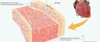
The heart as an organ consists of three membranes: the endocardium, the deepest membrane represented by a connective tissue membrane covered with endothelium, the myocardium - the muscular membrane of the heart and the epicardium - the outer serous membrane of the heart.
The myocardium is built from cardiac striated muscle tissue and has a number of features associated with the function of the heart itself, both as a whole and its parts: - In different parts, the thickness of the heart muscle is not the same, for example, in the left ventricle the wall is thicker than in the right. - The muscles of the atrium are separate from the muscles of the ventricles. — In the ventricles and atria there are common muscle layers. — In the area of the venous mouths of the vestibules there are sphincters. — The presence of two morphofunctional types of muscle fibers in the myocardium.
Under microscopy, cardiac muscle appears similar to skeletal striated muscle. A clearly defined cross-striation and sarcomeric structure are observed.
There are two types of cardiac fibers: 1) typical fibers - the working myocardium, 2) atypical fibers of the conduction system.
Typical fibers:
The working myocardium consists of a chain of muscle cells - sarcomeres connected to each other “end to end” and enclosed in a common sarcoplasmic membrane. United sarcomeres form myofibrils. The contact of sarcomeres is carried out through intercalary disks, due to which the fibers have a characteristic cross-striation.
Structure of sarcomeres:
Sarcomeres consist of alternating dark (myosin) - A, and light (actin) - I stripes. In the center of strip A there is zone H, which has a central T-line. Sarcomeres are connected to each other using intercalary discs - nexuses, which are the true boundaries of cells.
Myosin contained in band A is capable of breaking down ATP to ADP, that is, it is an adenosine triphosphatase, and is also capable of forming a reversible actomyosin complex with myosin (in the presence of Ca++ and the formation of ADP), which is responsible for the contractility of the heart muscle.
Muscle tissue
Muscle tissues are tissues for which the ability to contract is the main property. Muscle tissue constitutes the active part of the musculoskeletal system (the passive part is the bones and bone joints).
The common properties of all muscle tissues are contractility and excitability. This group of tissues includes smooth, striated skeletal and striated cardiac muscle tissue. Muscle tissue cells have a well-developed cytoskeleton and contain many mitochondria.
Smooth (visceral) muscles
This muscle tissue is found in the walls of internal organs (bronchi, intestines, stomach, bladder), in the walls of blood vessels, and gland ducts. Evolutionarily, it is the most ancient type of muscle.
Consists of spindle-shaped myocytes - short mononuclear cells. Between cells there are intercellular contacts - nexuses (Latin nexus - connection). Thanks to nexuses, excitation that arises in one cell spreads in waves to all other cells.
Smooth muscle tissue is distinguished by its ability to undergo prolonged tonic tension, which is very important for the functioning of internal organs (for example, the bladder), it contracts slowly, and practically does not get tired. Skeletal muscle tissue, which we will study a little later, does not have this ability - it contracts and gets tired quickly.
Contraction is carried out with the help of cellular organelles - myofilaments, which are located chaotically in the cell and do not have such an ordered structure as myofibrils in skeletal muscles (everything is learned by comparison, we will soon study them too).
We especially note that in smooth muscle tissue, myofilaments are assembled into myofibrils only during contraction. Such temporary myofibrils cannot have a regular organization, which means that neither such myofibrils nor smooth myocytes can have cross-striations.
Smooth muscle tissue contracts involuntarily (not subject to human will). The work of smooth muscles is ensured by the autonomic nervous system. For example, it is impossible to narrow or expand the bronchi, blood vessels, or pupil at will.
Smooth muscle tissue is called unstriated because it does not have the transverse striations characteristic of striated skeletal and cardiac muscle tissue.
Skeletal (striated) muscle tissue
Skeletal muscle tissue forms the diaphragm (respiratory muscle), the muscles of the torso, limbs, head, and vocal cords.
Unlike smooth muscle, skeletal muscle is formed not by individual mononuclear cells, but by long multinuclear fibers with up to 100 or more nuclei - myosimplasts. Myosimplast (Greek sim - together + plast - formed) is a collection of fused cells, has a length from several millimeters to several centimeters (corresponding to the length of the muscle).
Inside the myosymplast there is sarcoplasm, on the outside the myosymplast is covered with sarcolemma. Contractile elements - myofibrils (lat. fibra - fiber) - long cord-like organelles in the myosymplast (about 1400).
A characteristic feature of this tissue is transverse striation, expressed in a uniform alternation of light and dark stripes on the muscle fiber. This occurs because the boundaries of the sarcomeres in neighboring myofibrils coincide, as a result of which the entire fiber acquires transverse striations. Now is the time to study the microscopic basis of muscle - the sarcomere.
Sarcomere (from the Greek sarco - meat (muscle) + mere - small)
Sarcomere is an elementary contractile unit of striated muscles, a structural unit of myofibril. The composition of the sarcomere (and myofibrils in general) includes two types of myofilaments (Latin filamentum - thread), which ensure the contractility of muscle tissue.
The sarcomere consists of actin (thin) and myosin (thick) filaments, which are formed mainly by the proteins actin and myosin. Contraction occurs due to the mutual movement of myofilaments: they stretch towards each other, the sarcomere shortens (and the muscle as a whole).
The source of energy for contraction is ATP molecules. In addition, it is impossible to imagine muscle contraction without the participation of calcium ions: it is they that bind to troponin, which leads to a change in the conformation of tropomyosin (troponin and tropomyosin are regulatory proteins between actin filaments), due to which the connection of actin and myosin becomes possible. When muscles contract, heat is released (contractile thermogenesis).
I note that rigor mortis (lat. rigor mortis) - post-mortem hardening of muscles - is associated precisely with calcium ions, which rush to an area of low concentration (into the sarcoplasm of the myosymplast), promoting the binding of actin and myosin.
After death, ATP ceases to be synthesized in the muscle, and its level quickly decreases. As a consequence of this, the Ca-ATPase, the pump that pumps Ca ions from the sarcoplasm into the sarcoplasmic reticulum (a membrane organelle of muscle cells (similar to the EPS) in which Ca ions are stored), ceases to function.
In the sarcoplasm, the concentration of Ca ions increases - the bridges between actin and myosin close, but they can no longer open, and therefore persistent muscle contracture is observed (lat. contractura - tightening, narrowing): the limbs are very difficult to straighten or bend.
Let's return to skeletal muscles. There are a number of other important points that you need to know about.
One myosymplast is involved in the process of excitation in isolation; neighboring myosymplasts (fibers) do not excite each other, unlike smooth myocytes, where excitation is transmitted between neighboring cells through nexuses. Skeletal muscles contract quickly and tire quickly (in smooth muscles, the contraction and relaxation phases are extended in time and do not tire much).
Skeletal muscles contract voluntarily: they are controlled by our consciousness. For example, if we wish, we can change the speed of hand movement, the pace of running, or the force of jumping. Muscles are covered with fascia, attached to bones by tendons, and, contracting, set the joints in motion.
Cardiac striated muscle tissue
Cardiac muscle tissue forms the muscular layer of the heart - the myocardium (from ancient Greek μῦς “muscle” + καρδία - “heart”). Myocardium is the middle layer of the heart, making up the bulk of its mass. When working, the cardiac muscle tissue does not get tired.
Cardiac muscle tissue consists of cardiomyocytes - single cells with transverse striations. By connecting with each other, cardiomyocytes form functional fibers.
This type of muscle tissue amazingly combines the properties of the two previous tissues we studied (excitability, contractility) and has one new unique property - automatism.
Automatism is the ability of cardiac muscle tissue to excite and contract spontaneously, without external influences. This can be easily confirmed by observing the contractions of an isolated frog heart in a saline solution: the contractions of the heart in it will continue for several tens of minutes after the heart is separated from the body.
The places of contact of neighboring cardiomyocytes are intercalated discs (they contain nexuses), thanks to which the excitation of one cell is transmitted to neighboring ones, thus being covered in waves by excitation and contracting new areas of the myocardium.
A large number of contacts between cardiomyocytes ensures high efficiency and reliability of excitation through the myocardium. This tissue contracts involuntarily and does not get tired.
In a drawing or microslide, you can recognize this tissue by the central position of the nuclei in the cells, transverse striations, the presence of intercalary discs and anastomoses (Greek anastomosis - hole) - junctions of the lateral surfaces of functional fibers (cardiomyocytes).
Normally, excitation is carried out along the conduction system of the heart from the atria to the ventricles (unidirectionally). The area of the heart muscle in which impulses are generated that determine the heart rate - the heart pacemaker.
Automaticity is possible due to the presence in the myocardium of special pacemaker cells, which are also called pacemakers. They spontaneously generate nerve impulses that cover the entire myocardium, resulting in contraction. It is thanks to the pacemakers that the frog's heart continues to beat, being completely separated from the body.
Muscle response to exercise
Physical activity leads to muscle hypertrophy (from ancient Greek ὑπερ - through, too + τροφή - food, food) - the number of muscle fibers in them increases, the volume of muscle mass increases.
Under conditions of physical inactivity (from the Greek ὑπό - under and δύνᾰμις - strength), that is, reduced activity, muscles decrease until complete atrophy (Greek a - “not” + trophe - nutrition). In the worst case, the muscle fibers degenerate into connective tissue, after which the patient becomes immobilized.
It should be noted that cardiac muscle tissue also responds to excessive load: the heart increases in size and the mass of the myocardium increases. The cause may be genetic diseases or high blood pressure. Cardiac hypertrophy is a condition that requires medical intervention and patient monitoring.
In most cases, cardiac hypertrophy is reversible, and athletes experience so-called physiological hypertrophy (a variant of the norm).
Origin of muscles
Muscles develop from the middle germ layer, the mesoderm.
© Bellevich Yuri Sergeevich 2018-2021
This article was written by Yuri Sergeevich Bellevich and is his intellectual property. Copying, distribution (including by copying to other sites and resources on the Internet) or any other use of information and objects without the prior consent of the copyright holder is punishable by law. To obtain article materials and permission to use them, please contact Yuri Bellevich
.
Atypical fibers.
Thanks to atypical nerve fibers, heart automation is realized.
Automaticity of the heart is the ability of the heart to contract rhythmically under the influence of impulses originating in itself.
Atypical cardiac fibers serve as the morphological substrate of automation. – pacemakers capable of periodic self-generation of membrane potential.
Atypical myocytes are larger than working ones; they contain more sarcoplasm with a high glycogen content, but few myofibrils and mitochondria. In atypical cells, enzymes that promote anaerobic glycolysis predominate.
The atypical cells themselves are located in strictly defined areas and form the sinatrial (Keith-Flerk) and atrioventricular (Aschoff-Tavara) nodes and the bundle of His, dividing into legs that branch like Purkinje fibers.
Diagram of the conduction system of the heart:
Typical myocytes maintain a stable membrane potential during contraction, while the potential of atypical myocytes of the synatrial node slowly decreases due to increased membrane permeability for sodium ions entering the fibers and potassium ions leaving them. When the sodium gate opens, Na+ ions rush into the fibers like an avalanche, causing the propagation of a new potential. (“drift” of potential). After which the process is repeated.
The ability for automaticity in different parts of the heart is not the same and in the atrioventricular node it is already lower, and in the Hiss bundle it is so small that the corresponding frequency of occurrence of the membrane potential is not compatible with life.
Cross-striped fabric
The cells have a thickness from 10 to 100 microns, a length from 10 to 40 cm. The cytoplasm contains a large number of nuclei and myofibrils, occupying a central position (Fig. 2) . Mature cells have hundreds of myofibrils and more than 100 nuclei. Actin and myosin filaments inside myofibrils are linked to each other (Fig. 3) . The ability for rapid contraction of this tissue is higher than that of others.
Rice. 3. Structure of skeletal muscle
Muscle fibers are covered with a membrane called sarcolemma. There are alternating plates of proteins of different densities that have unequal light refractive indices. Under an optical microscope, such muscles appear to be striated across. The contractile elements are united into muscle bundles covered with a connective tissue membrane. Skeletal muscles are well supplied with blood vessels and nerves.
Physiological features of the structure of the heart muscle.
To ensure the normal existence of the body in various conditions, the heart can work in a fairly wide range of frequencies (for example, in a horse while running, the heart rate can increase 4–5 times). This is possible due to some properties, such as:
1 - Automaticity of the heart is the ability of the heart to contract rhythmically under the influence of impulses originating in itself. Described above.
2 – Cardiac excitability is the ability of the heart muscle to be excited by various stimuli of a physical or chemical nature, accompanied by changes in the physical and chemical properties of the tissue.
3 – Cardiac conduction, carried out in the heart electrically due to the formation of an action potential in pacemaker cells. The place where excitation transfers from one cell to another is the nexus.
4 – Cardiac contractility – The force of contraction of the heart muscle is directly proportional to the initial length of the muscle fibers
5 – Myocardial refractoriness is a temporary state of non-excitability of tissues
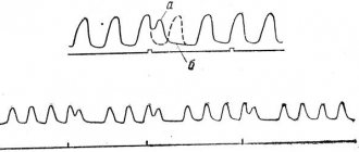
When the heart rhythm fails, fibrillation occurs - rapid asynchronous contractions of the heart, which can lead to death.
Cardiac cycle.
The work of the heart can be divided into several phases (periods):
Tension - systole,
expulsions of blood,
Relaxation - diastole.
The cardiac cycle is the coordinated alternation of systole and diastole of the heart.
The beginning of the cardiac cycle is considered to be atrial systole (and the left one contracts slightly earlier than the right one); when the atria contract, the pressure in them increases, and blood flows into the ventricles of the heart. Blood does not flow into the veins, since at the moment of atrial systole the lumen of the veins is narrowed, and blood flows freely into the ventricles, since the ventricles are relaxed and the atrioventricular valves are free. Cycle time 0.1 s.
The next stage of the cycle is ventricular systole. When they contract, the pressure increases and the blood, trying to flow out, slams the atrioventricular valves and rushes into the lumen of the arteries, opening the semilunar valves. Cycle time 0.4 s.
After the opening of the semilunar valves, the pressure in the ventricles drops, and in the arteries it increases sharply, the semilunar valves slam shut, and ventricular diastole occurs.
Additional definitions
The sound phenomena that accompany the work of the heart are called heart sounds.
The amount of blood ejected by the heart during a unit of time is called the minute volume of blood flow.
The ratio of minute blood volume to the number of heart contractions is called systolic blood volume.
When the heart operates, bioelectric potentials arise that can be detected using special ECG recording equipment.
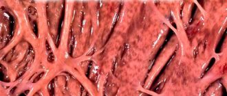
Due to the constant load, the heart is very sensitive to a lack of oxygen and nutrients, so more than 10% of the blood passing through the aorta enters the coronary vessels that supply the heart muscle.
Regulation of the heart takes place both at the humoral and nervous levels. The hormones adrenaline and noradrenaline participate in humoral regulation, and nervous regulation involves the sympathetic and parasympathetic nervous system.
An important role in the movement of blood is played by the so-called peripheral hearts, that is, skeletal muscles. When muscles contract (walking, working), the lumens of the vessels narrow, pressure increases and blood is pushed towards the heart.
Properties
The muscle fiber stretches, but at rest returns to its original size. This property is the result of the interaction of protein filaments of myofibrils in the cytoplasm of cells. Each myofibril consists of protofibrils: thin ones formed by actin, and thicker ones formed by myosin.
Properties of muscle tissue:
- electrical excitability;
- contractility;
- conductivity;
- extensibility;
- elasticity.
Muscle tissue is capable of voluntary or involuntary contractions in response to nerve impulses. There is an interaction between fibrillar proteins - actin and myosin. Inorganic calcium ions necessarily participate in this process. During contraction, thin filaments of actin slide along thick myosin protofibrils.