Multifocal atherosclerosis is a chronic metabolic disease in which fat (lipoproteins, triglycerides) are deposited on the inner wall of the arteries. In the initial stages, the disease is not dangerous, but then these substances form plaques and can cause thrombosis and thromboembolism. Atherosclerosis can affect the aorta, arteries of the heart, brain and limbs, as well as vessels that supply blood to internal organs (kidneys, intestines). It is called multifocal in the presence of several pathological foci, which can be located in any area. It is important to diagnose the disease in time, while its development remains in the early stages - complicated forms either cannot be corrected or require surgical intervention.
Causes and risk factors
Atherosclerosis is one of the most common diseases of the cardiovascular system. The incidence rate ranges from 182 (France) to more than 800 people (Russia) per 100 thousand population. This depends, first of all, on nutrition and the presence of bad habits, on the degree of environmental pollution and other factors. The risk of developing this disease especially increases in the presence of one or more factors:
- bad habits, especially smoking;
- overweight and insufficiently active lifestyle;
- chronic increase in blood pressure;
- consumption of foods high in animal fats;
- diabetes;
- endocrine system disorders and others.
REFERENCE! Cholesterol is found in large quantities in animal fats that undergo heat treatment. It is its components that can be deposited on the inner surface of blood vessels.
Theories of the development of atherosclerosis
Doctors are working to determine the exact cause why multifocal atherosclerosis develops. There are several theories that explain the primary origin of the disease and the mechanism of its development. These include:
- lipoprotein infiltration – accumulation of fats in the walls of blood vessels;
- endothelial dysfunction – deposits are formed due to the weakness of the inner lining of blood vessels and a decrease in its protective properties;
- autoimmune – disruption of the immune system, including increased synthesis of macrophages and leukocytes;
- peroxide – the influence of free radicals and lipid peroxidation reactions, in which cell death occurs;
- hormonal - when the level of certain hormones decreases, the accumulation of cholesterol necessary for their synthesis increases.
And mixed theories of the development of atherosclerosis are also popular. They imply that the disease develops when several factors combine simultaneously, and their results are the destruction of vascular walls.
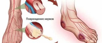
Multifocal motor neuropathy (MMN)
Multifocal motor neuropathy (MMN) (information for patients and doctors)
What is Multifocal Motor Neuropathy?
- “Multifocal” means multiple lesions.
- “Motor” - that the pathological process involves exclusively the motor fibers of the nerves responsible for muscle strength and the execution of movements.
- “Neuropathy” is damage to the peripheral nerves.
How common is this disease in the population? This is a very rare disease. The incidence of MMN in the population is only 1–2 cases per 100,000 population; is diagnosed 3-4 times more often in men, and the onset (onset) of the disease occurs in working age (20-50 years).
What is the mechanism of development of this disease? MMN is a chronic multiple motor neuropathy , the development of which is based on an autoimmune process, when components of the immune system mistakenly begin to work against the structures of their own body. MMN is based on selective dysimmune damage to the nodal zones of motor nerves - areas of thick nerve fibers rich in ion channels and responsible for the transmission of nerve impulses. As a result of such a lesion, a block in the conduction of excitation along the motor nerve develops and movements are impaired.
What worries patients with this disease?
- asymmetrical muscle weakness in the arms or arms > legs
- There is no pain or sensory disturbances!
MMN begins in most patients with the gradual development of muscle weakness, most often in the arms. Sensory impairment and pain are absent. The initial complaint may be weakness in one arm or arms; much less often, the disease begins in the legs. Weakness in the distal arms (hands) is common and is found in approximately 95% of patients with MMN. More than half of patients experience muscle spasms and twitching. There is no damage to the motor cranial nerves responsible for eye movement, facial muscles, chewing, swallowing and speech. Pelvic functions are not impaired. The course of the disease is slowly progressive or wavy in nature. Muscle wasting (hypo- or atrophy) is uncommon and is present in late stages of the disease, often in untreated patients. There are no signs of damage to the central nervous system.
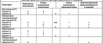
On what basis is the diagnosis made and what examinations can be prescribed for me to confirm it? Currently, clear criteria for the diagnosis of “Multifocal motor neuropathy” have been developed (EFNS/PNS 2011). According to these criteria, an analysis of the history of the development of the disease, a neurological examination and electroneuromyography are sufficient:
- Electroneuromyography makes it possible to detect conduction blocks along the motor fibers of peripheral nerves in places atypical for their compression (outside the tunnels), while the study parameters of sensory fibers are unchanged. In addition, this study excludes damage to other structures of the peripheral neuromotor apparatus (motor neurons, muscles).
- Additionally, neuroimaging may be required - ultrasound of peripheral nerves and MRI of the brachial plexuses with contrast, which are also highly informative and help confirm the diagnosis and exclude other pathologies.
However, in practice, difficulties may arise in making a diagnosis when patients come for consultation years after the onset of the disease with severe motor impairment, muscle atrophy, or an atypical form of the disease. In these situations, a differential diagnosis is required.
When carrying out a differential diagnosis, the list of examinations expands:
- routine laboratory tests of blood and urine, MRI of the spine and spinal cord do not help in making the diagnosis of MMN, but will exclude possible other causes of multiple nerve damage (such as diabetes mellitus, rheumatoid arthritis, vertebrogenic myelopathy, Hiroyama disease, etc.)
- blood test for antibodies to peripheral nerve gangliosides - antibodies to GM1 gangliosides are present in approximately half of cases of MMN
- genetic blood test for mutations of the PMP22 gene - will exclude possible hereditary causes of neuropathy
- etc.
Of course, not all of these examinations are always prescribed. In most cases, only an ENMG study is sufficient. It should be emphasized that the ENMG study must be carried out by a well-trained and experienced specialist in a high-class myorapha. Methodological errors and insufficient size of this study often lead to erroneous diagnoses. Therefore, we recommend performing ENMG in our center.
What treatment is there? MMN is a chronic disease, complete cure is impossible. But effective treatment has now been developed that can control the disease. Intravenous immunoglobulin (IVIG) therapy is the only treatment for MMN that has been shown to be effective in controlled studies.
The effective dose is 2 g of IVIG per 1 kg of patient weight (dose administered over 3-5 days). The vast majority of patients respond positively to this therapy. However, the improvement is short-term and long-term maintenance therapy is required: periodic infusions of IVIG drugs - 1 g of IVIG drug per 1 kg of patient weight every 3-4 weeks (administration 3 days before) or administration of 2 g of the drug per kg of weight every 1-2 months (introduction in 3-5 days).
It should be remembered that not all IVIG drugs are suitable for the treatment of MMN. The main requirements for human immunoglobulin preparations are:
- safety of the drug with respect to transmission of infection;
- IVIG should be well tolerated;
- the IgG content must be at least 95% of the total IgG content;
- IgA content should be minimal;
- the drug should not exhibit thrombogenic (procoagulant) activity;
- possibility of high-dose therapy (no daily dose limitation);
- preference is given to 10% solutions.
Contraindications to IVIG therapy are hypersensitivity (allergy) to IVIG components, as well as IgA deficiency. Therefore, before treatment, a study of IgA levels is recommended. If its deficiency is detected, treatment is carried out with caution, in particular, premedication and special preparation are used. In the absence of therapy, irregular administration of IVIG, or treatment with insufficient doses of the drug, motor deficits can slowly, steadily, and irreversibly progress due to secondary axonal damage.
Glucocorticosteroids and plasmapheresis are ineffective in patients with MMN!
What is the prognosis for this disease? Approximately 80% of patients respond to IVIG treatment. MMN does not lead to death, since the muscles involved in the acts of swallowing and breathing are not involved in the pathological process. Because MMN primarily affects the muscles of the hands, writing and fine motor skills may be limited. In this regard, alternative professional employment options should be considered.
There are 40 patients with MMN under the supervision of cPNS specialists. We are able to diagnose this disease and determine the optimal personalized treatment regimen with IVIG drugs.
Employees of the Center for Peripheral Nervous System Diseases provide consultations to patients on an outpatient basis within the framework of compulsory medical insurance and on a commercial basis. Make an appointment,
Be healthy!!!
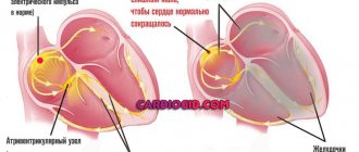
Mechanism of disease development
The process of development of atherosclerosis is called atherogenesis. It consists of several successive stages, as a result of which the vessel loses strength and elasticity. Normally, its wall consists of smooth muscles, and the inside is lined with intima - a single-layer membrane of endothelium. Each of the layers plays its own role, and together they provide the ability of the vascular wall to stretch and contract. With atherosclerosis, tissues become saturated with harmful deposits, and plaques can additionally form on the inner surface of the vessel.
- Accumulation of fat cells. At the first stage of the disease, lipid stains are found on the inner surface of the arteries. Only low and very low density lipoproteins participate in their formation, and their other types do not have atherogenic activity. These compounds form bonds with the components of the intercellular substance and remain in the form of single spots.
- Activation of leukocytes and formation of xanthoma cells. In the area of the lipid stain, the migration of leukocytes, especially lymphocytes and monocytes, increases. Further, monocytes play a major role in the development of atherosclerosis. In the vascular intima they transform into macrophages and participate in the formation of xanthoma (foam) cells.
- Pro- and antiatherogenic factors are compounds that can accelerate and slow down the development of atherosclerosis. Macrophages release cytokines and growth factors. They enhance the division of vascular smooth muscle cells and intercellular substance, which is used as a building material for atherosclerotic plaques. Next, there is a decrease in the processes of synthesis of smooth muscle cells and collagen, which ensures the density and elasticity of the vascular wall.
- The role of smooth muscle cells. They represent the inner lining of the artery wall, and they are synthesized by the intercellular substance. Their proliferation becomes the main point in the development of atherosclerotic plaque. If normally the tissue is capable of contracting and lengthening many times over, in the obvious stages of the disease this ability is lost, the vessels become fragile and brittle.
- Formation of a complicated plaque. This formation begins to gradually sprout with its own vessels. They cause hemorrhage, thrombosis and thromboembolism - a dangerous condition in which the plaque enters the lumen of the blood vessel and circulates with fluid flow.
IMPORTANT! Not every lipid stain goes through all stages of development and transforms into atherosclerotic plaques. However, these processes are interconnected, and the disease can progress.
Atherosclerosis is an acquired disease, the development of which is influenced by lifestyle and bad habits.
Symptoms
Multifocal atherosclerosis develops in stages. It is difficult to diagnose in the early stages, since clinical changes are visible only on test results. The patient has no complaints. In the future, they depend on the type of artery affected.
- With atherosclerosis of the arteries that participate in the nutrition of the myocardium, chest pain, heart rhythm disturbances, and coronary artery disease occur. The patient experiences severe pallor of the skin and mucous membranes, shortness of breath, and trembling of the limbs. These symptoms are especially worse after exercise.
- With atherosclerosis of the arteries of the brain, there is a decrease in performance, deterioration of hearing and vision. In addition, the disease often causes strokes, which are life-threatening and require emergency medical attention.
- Atherosclerosis of the extremities (usually the lower ones) leads to a deterioration in their blood supply. As a result, patients complain of severe pain in the legs, numbness and pins and needles sensation, cramps and trophic ulcers. In the initial stages, the clinical picture is mild and worsens only after prolonged physical activity, and in advanced stages it can even appear during sleep. A typical sign of atherosclerosis of the lower extremities is intermittent claudication, in which the patient experiences pain alternately in the right and then in the left leg.
Advanced forms of multifocal atherosclerosis are accompanied by acute pain. In addition, the formation of aneurysms is observed - pathological dilations of blood vessels, which are formed as a compensatory reaction of the body to insufficient blood circulation. In advanced stages they become noticeable under the skin. In these areas, the vascular walls are thin and fragile, so rupture of an aneurysm is a common and dangerous occurrence that poses a threat to the patient’s life. In areas that lack normal trophism, ulcers appear that do not respond to conventional treatment methods and may not heal for a long time.
Clinical observation
Patient D., 73 years old, pensioner. Diagnosis: coronary heart disease (CHD). Atherosclerosis of the aorta, coronary vessels, carotid arteries and arteries of the lower extremities. Primary polygenic hypercholesterolemia type IIa. Varicose veins of the lower extremities. Thrombophlebitis of the lower extremities without exacerbation. Duodenal ulcer in remission. Sensorineural hearing loss II degree. Condition after angioplasty with stenting of the left common carotid artery (CCA), performed in 2013. Arterial hypertension stage I, degree 1, risk 4 (very high).
The patient first consulted a cardiologist/lipidologist in December 2004 (15.5 years ago). At that time, she complained of a feeling of discomfort in the chest lasting 2–3 minutes (“heart ache”) during emotional and physical stress, radiating to the right hand, occurring 2–3 times a week, as well as attacks of dizziness and hearing loss.
Chest pain first appeared in 2002 (at the age of 55). In the anamnesis, the level of total cholesterol increased to a maximum of 13 mmol/l (according to the patient). Since 2003, she began taking simvastatin 10–20 mg/day, but at the time of the initial consultation (December 2004) she did not receive statins, because she believed that they did not help. In May 2004, after working at the dacha, suddenly, against the background of complete health, numbness in the right hand (lasting 15 minutes) and a headache appeared. I didn’t go to the doctors. Physical activity is low. Doesn't systematically follow the diet. Office work, sedentary. Menopause since 2003. Never smoked.
The family history is burdened: the mother suffered an acute cerebrovascular accident (ACVA) at the age of 60, the onset of IHD in the father occurred before the age of 50, he died of a myocardial infarction at the age of 74, the brother suffered an ACVA at the age of 56.
According to the initial blood test (2 weeks without statins, December 2004): alanine aminotransferase (ALT), aspartate aminotransferase (AST), creatine phosphokinase (CPK), glucose, lactate dehydrogenase (LDH), alkaline phosphatase, γ-glutamyl transpeptidase, urea, creatinine , bilirubin, sodium, potassium - normal, total cholesterol 9.1 mmol/l, triglycerides 1.81 mmol/l, high-density lipoprotein (HDL) cholesterol 1.35 mmol/l, LDL-C 6.93 mmol/l . Fibrinogen level increased to 3.9 g/l. Based on the results of the consultation, a diagnosis of hereditary hyperlipoproteinemia (HLP) type IIa was made and an initial dose of atorvastatin of 20 mg/day was prescribed.
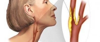
In February 2005, she underwent an outpatient examination at the National Medical Research Center of Cardiology of the Russian Ministry of Health. Duplex scanning revealed multiple stenoses in the carotid arteries with a maximum stenosis of 75% in the left CCA and multiple stenoses in the vessels of the lower extremities with a maximum stenosis of 40% in the left popliteal artery. According to the results of bicycle ergometry (VEM) at the height of the load and at rest, a decrease in the ST segment in leads V4–V6 to 1.5 mm of a horizontal nature was recorded. The test was regarded as positive, exercise tolerance was average. During 24-hour electrocardiography (ECG) monitoring, ventricular extrasystoles (2091) and 22 episodes of ST depression per 1 mm with a total duration of 3 minutes were recorded (according to the exercise diary). Echocardiography (EchoCG), ECG and 24-hour blood pressure (BP) monitoring revealed no significant deviations from the norm, with the exception of thickening of the aortic walls. Acetylsalicylic acid 100 mg/day, isosorbide dinitrate 60 mg/day for the prevention of anginal pain, atorvastatin 20 mg/day were prescribed, it was recommended to continue treatment with betahistine 8 mg/day and come for a second consultation after 6 months. A consultation with an angiosurgeon was also recommended to determine the indications for surgical treatment of left carotid artery stenosis. The patient categorically refused surgical treatment of atherosclerosis of the carotid arteries for a long time, and a decision was made on drug therapy.
Results of an objective examination (2005): heart sounds are rhythmic, no pathological noises. The rhythm is correct with a heart rate (HR) of 64 per minute. Blood pressure 98/60 mm Hg. Art. Breathing is vesicular, no wheezing. Liver along the edge of the costal arch. The symptom of tapping in the lumbar region is negative. The pulsation in the peripheral arteries is satisfactory. Xanthoma, xanthelasma, thickening of the Achilles tendons, and lipoid arch of the cornea were not detected.
In 2010, at the Federal State Budgetary Institution “National Medical Research Center of Siberian Agricultural Sciences named after. A.N. Bakulev" of the Ministry of Health of Russia, the patient underwent multislice computed tomography (MSCT) with contrast. This is an effective non-invasive screening method for studying the coronary bed (especially in asymptomatic patients), which is widely used in domestic [20] and foreign clinical practice and is included in international recommendations for the diagnosis and treatment of lipid metabolism disorders [3, 4]. Conclusion: coronary calcium (equivalent to Agatson Score) 40.6 units. CT picture of atherosclerosis and calcification of the aorta and coronary arteries. Stenosis of the anterior interventricular descending artery (LAD) 40%. Ejection fraction 70%. The thickness of the interventricular septum in diastole is 8 mm, the thickness of the posterior wall of the left ventricle in diastole is 7 mm. Right type of blood supply to the myocardium. No disturbances in left ventricular myocardial contractility were detected.
Repeated MSCT was performed in 2012 at the National Medical Research Center of Cardiology of the Russian Ministry of Health. Conclusion: LAD stenosis 50% at the border of the proximal and middle third, calcified plaque. Comments: no hemodynamically significant stenoses were identified. Compared to MSCT data from 2010, no dynamics were noted.
Over 15 years of follow-up (in 2004, 2006, 2007, 2008, 2010, 2012 and 2020), the patient underwent a series of stress tests (treadmill test) to non-invasively assess the severity of myocardial ischemia and exercise tolerance. As stated above, initially (in 2004) at the height of the load, a decrease in the ST segment in leads V4–V6 to 1.5 mm of a horizontal nature was detected. The test was regarded as positive, exercise tolerance was average. Retests 2006–2008 were positive, and in 2010, for the first time, a stress test showed a negative result (no ischemia). Conclusion based on the results of the treadmill test in 2010: the test for the presence of transient myocardial ischemia is negative. Load tolerance is high. The blood pressure response is adequate. Stop the load when the submaximal heart rate is reached (140 per minute). Estimated MET (metabolic equivalent) value is 10.5. In March 2021, a stress echocardiography was performed, the results of which also revealed no signs of ischemia. Conclusion: the test for identifying hidden coronary insufficiency is negative. Initially and at maximum load zones, no disturbances in local contractility of the left ventricle were noted. Exercise tolerance is average. The response of blood pressure to stress according to the hypertensive type. At maximum load the patient had no complaints.
In January 2013, an operation on the carotid arteries was performed in the neurosurgical department of the Federal State Budgetary Institution National Center for Neurology: transluminal balloon angioplasty with stenting of the left internal carotid artery. The postoperative period proceeded without complications. Data from control duplex scanning of the vessels of the main arteries of the head: the stent is evenly and adequately expanded. After the operation, I was bothered by throbbing pain in the temporal region for about six months. For a year she took clopidogrel 75 mg, which she stopped taking in February 2014 due to the appearance of hematomas of various locations. In November 2014, during an examination at the Federal State Budgetary Institution “National Medical Research Center of Cardiology” of the Ministry of Health of Russia, a VEM test was positive; at the height of the load, a horizontal decrease in the ST segment to 1.5 mm in leads V5–V6 was recorded, load tolerance was average. It was decided to perform coronary angiography. Conclusion: the trunk of the left coronary artery has uneven contours, a pronounced bend is determined in the middle segment. The first and second diagonal arteries with uneven contours. At the mouth of the intermediate branch there is 50% stenosis. In the circumflex artery, the first artery of the blunt edge, the right coronary artery there are uneven contours. There were no indications for surgical treatment of coronary artery disease, and it was decided to continue conservative treatment.
During the entire period of outpatient observation, the patient continuously received treatment with simva-, atorva- or rosuvastatin as monotherapy or, for a longer period of time, a combination of a statin with ezetimibe 10 mg/day (from 2013 to the present time she has been taking the original rosuvastatin 40 mg/day in combinations with ezetimibe).
The patient was and continues to be observed on an outpatient basis at the Federal State Budgetary Institution "National Medical Research Center of Cardiology" of the Ministry of Health of the Russian Federation and the Federal State Budgetary Educational Institution of Further Professional Education RMANPO of the Ministry of Health of the Russian Federation for the last 16 years. In table Tables 1–3 present data from studies of the lipid profile and some biochemical parameters over the course of observation (with the exception of 2009 and 2021), as well as the results of duplex scanning tests of the carotid arteries and arteries of the lower extremities. All ultrasound examinations were performed on expert-class ultrasound systems with linear sensors with a frequency from 9 to 17 MHz (most of the examinations were performed on an iU-22 device, Philips, by one expert).
Diagnostic methods
Diagnosis of multifocal atherosclerosis is an important stage, thanks to which it is possible to determine the localization of the pathological focus and the stage of the disease. For this purpose, instrumental and laboratory techniques are used to monitor the condition of blood vessels. These include:
- initial examination and medical history, identification of typical symptoms;
- palpation (palpation) of all superficial arteries located under the skin, as well as listening to them in areas accessible for diagnosis;
- determining the degree of filling of blood arteries and their integrity;
- blood test with mandatory determination of various types of fats and their percentages, including cholesterol;
- radiography, ultrasound examination – basic techniques;
- Dopplerography is an assessment of the conductivity of blood vessels at any part of their passage, using a contrast agent.
When diagnosing atherosclerosis, it is important to determine the location of the pathological area. And also in the process it is possible to differentiate this disease from many chronic diseases of the heart and blood vessels, which manifest themselves with a similar set of symptoms.
Obliterating atherosclerosis of leg vessels
Diagnostics
First of all, the doctor must collect anamnesis from the patient. Moreover, depending on the location of the process, patients indicate the corresponding symptoms, be it angina or blurred vision, intermittent claudication, headache or urinary disorders. A physical examination reveals frequent changes in such patients: cardiac hypertrophy, the presence of pathological murmurs, pulse surges.
The patient undergoes a general blood and urine test, determines the level of lipoproteins, cholesterol, sugar and many other important parameters. For differential diagnosis, pharmaceutical tests (nitroglycerin and dobutamine) are used.
For the purpose of instrumental diagnostics, the following methods are used:
- electrocardiography and Holter monitoring;
- ECHO-CG and Dopplerography;
- radiography of the OGK;
- Ultrasound of the abdominal organs;
- radioisotope study of the kidneys;
- angiography;
- volumetric sphygmography.
All these measures make it possible to establish an accurate diagnosis, determine the localization of the pathology and its immediate cause, which is important for further treatment.
Treatment
Treatment of multifocal atherosclerosis and its duration depend on the stage of the disease. A mandatory step will be the correction of lifestyle and nutrition. It is not always possible to return the vessels to their previous condition, but they can be maintained in the same condition for many years.
Non-drug methods
The most important stage of atherosclerosis treatment takes place at home. It includes following all the recommendations of doctors, thanks to which you can establish fat metabolism and prevent further deposition of lipids on the inner wall of blood vessels. Treatment at home consists of several stages:
- nutrition correction - exclusion of animal fats, especially during heat treatment of other products;
- giving up bad habits, including smoking and drinking alcohol;
- normalization of body weight;
- physical activity that will correspond to the state of health - it is better to check with your doctor what loads you can bear with atherosclerosis.
Without following simple rules, treatment of multifocal atherosclerosis is not possible. It is important to understand that they will have to be carried out throughout your life. Since poor diet and smoking are considered the main predisposing factors to atherosclerosis, it is important to give up habits to slowly restore all biochemical processes.
Drug treatment
Drugs for the treatment of multifocal atherosclerosis are aimed simultaneously at several targets. They participate in lipid metabolism and gradually normalize it, but they should be taken regularly. The selection of drugs is carried out individually, taking into account the diagnostic results. Four main groups of drugs are used to treat this disease:
- drugs that prevent further absorption of cholesterol;
- substances that inhibit the synthesis of lipoproteins in the liver, thereby reducing their concentration in the blood;
- means for processing and rapid removal of atherogenic compounds;
- additional medications, including agents to strengthen the inner surface of blood vessels.
REFERENCE! Treatment can be carried out at home. However, its timeliness and gradualism are important. Those remedies that will help in the early stages of the disease will be ineffective at further stages.
Surgery
The operation is prescribed for patients diagnosed with vascular aneurysms or other pathologies that are not amenable to conservative treatment. One of the common types of surgery is the removal of a section of a vessel, especially if it has an aneurysm. If previously the process required full access, now operations are easily carried out under the control of an endoscope.
Multifocal atherosclerosis is a dangerous disorder that gradually progresses and can lead to dangerous consequences. The disease occurs with a violation of fat metabolism and has several pathological foci. Treatment must be timely to avoid complications and the need to perform surgery to remove the affected vessel.
Discussion
During the entire observation period, the patient was highly compliant; there were practically no breaks in taking statins.
Increased LDL-C levels are the strongest independent risk factor for the development of atherosclerosis [1–4]. The patient had a history of an increase in the level of total cholesterol to 13 mmol/l, which is typical for familial (hereditary) type IIa HLP. Taking into account a family history and an initial LDL-C level of 6.93 mmol/l, the diagnosis of “familial (hereditary) HLP” is quite likely, since the score according to the Dutch Lipid Clinic Network Score algorithm was 8 points (probable familial HLP) [3]. Since DNA diagnostics for familial heterozygous HLP was not performed and examination did not reveal thickening of the Achilles tendons, xanthomatosis and lipoid arch of the cornea, it was decided to make a diagnosis of primary polygenic hypercholesterolemia type IIa. The polygenic form of familial HLP may not phenotypically differ from monogenic forms [21, 22], and such diagnostic tactics are more justified if there is no evidence of monogenic familial hypercholesterolemia [23].
Since 2004, four revisions of the European guidelines for the diagnosis and treatment of dyslipidaemias have been published, including the latest version in 2021 [3]. In this document, an addition was introduced to the category “very high CV risk”: the presence of plaques in the carotid arteries and arteries of the lower extremities according to ultrasound examination and the diagnosis of “familial hypercholesterolemia”. For this category of patients, target LDL-C levels are set at no more than 1.4 mmol/l and a decrease in LDL-C levels by more than 50% from initial values [3]. The LDL-C content for the entire observation period, taking into account both low (1.39 mmol/l in 2013 after stenting surgery) and high (4.41 mmol/l in 2010) values, averaged 3.14 mmol /l, the average decrease in LDL-C level is 55% from the initial value of 6.93 mmol/l. In recent years, combination therapy with the original rosuvastatin 40 mg/day and ezetimibe has achieved LDL-C levels in the range of 2.4–2.74 mmol/l, which is higher than the existing target values for individuals at very high CV risk [3]. Among the additional risk factors, this patient had a twofold increase in the level of lipoprotein(a), which is an independent factor of atherosclerosis, especially in patients with hereditary dyslipidemia [3, 24]. Along with an increase in triglyceride levels, high lipoprotein(a) levels are one of the main residual risk factors during statin therapy [24]. As lipoprotein(a) levels increase, control of hypercholesterolemia should be even more stringent, according to the principle: the lower the LDL level, the better [3].
Management of the patient for a long time (2004–2013) was complicated by her refusal (for fear of surgical complications and death) from coronary angiography and surgical treatment of severe stenosis. In December 2012, due to a significant increase in the linear velocity of blood flow from 1.8 to 2.5 m/s in the area of 75% stenosis in the bifurcation of the left CCA, based on the totality of clinical data, a council of doctors decided on the need for stenting of the carotid artery in the area stenosis 75%.
At the initial consultation in December 2004, the patient complained of rare chest pain, characteristic of exertional angina, took nitrates, the results of a series of treadmill tests in 2004–2008. were positive. In 2010, a treadmill test showed a submaximal heart rate of 140 per minute (10.5 MET) without pain. Two studies of MSCT of the coronary arteries in 2010 and 2012, as well as coronary angiography, revealed the presence of one stenosis in the intermediary artery and uneven contours in other coronary arteries. The peculiarity of this clinical observation is that it was possible to retrospectively analyze the ultrasound dynamics of stenoses in the carotid and peripheral arteries (the studies were performed by the same researcher on the same equipment; see Tables 2 and 3). Taking into account that the criterion for progression/regression of atherosclerotic lesions is considered to be a change in the diameter of the stenosis ± 15% of the initial values, we can say with a high degree of confidence that over 16 years of observation, long-term and intensive (in the last 8–10 years) lipid-lowering therapy allowed slow down the progression of atherosclerosis in three vascular areas - carotid, coronary and peripheral arteries.
A significant increase in LDL-C levels (6.93 mmol/l) revealed at the initial consultation is the main and, perhaps, the only significant modifiable risk factor for the development of atherosclerosis. For 16 years, blood pressure levels were within normal limits; there were no CV risk factors such as diabetes mellitus, smoking, or obesity (body mass index for 16 years was in the range of 24.4–25.3 kg/m2). Mild reclassifiers of CV risk include menopause [4] and elevated lipoprotein(a) levels [3, 24]. The effect of long-term intensive statin therapy on the state of atherosclerosis in the carotid and coronary arteries has been well studied in various “regression” studies using intima-media thickness monitoring [25, 26], repeated quantitative coronary angiography [27], and intravascular ultrasound [28–32] , magnetic resonance imaging [33], as well as optical coherence tomography [34–38]. In particular, a number of studies have shown that over 24 months. intensive therapy with statins (atorvastatin 80 mg/day, rosuvastatin 40 mg/day or in combination with i-PCSK9) can slow the progression of carotid [26, 33] and coronary [28–32] atherosclerosis. The mechanisms of slowing the progression of atheroma with intensive treatment with statins or the use of combination therapy are well studied and include delipidation of plaques, their subsequent calcification, reduction in inflammatory activity, the number of macrophages, a decrease in the concentration of cytokines, interleukins 1 and 6, and a decrease in the level of high-sensitivity C-reactive protein [35–37 ].
In the ASTEROID study after 24 months. Treatment with rosuvastatin 40 mg/day in the majority (78%) of patients managed to normalize the total volume of plaques and the volume of plaques in the most affected segments; the average reduction in LDL-C was 53% [29]. In another study, SATURN, examining the possibility of slowing the progression of coronary atherosclerosis, rosuvastatin at a dose of 40 mg/day was superior to atorvastatin 80 mg/day in the percentage change in total atheroma volume [30]. According to data from [34], in just 13 months. Treatment with rosuvastatin 40 mg/day in 83 patients after myocardial infarction succeeded in achieving a significant increase in the thickness of the atheroma cap in non-infarction arteries from 64.9 to 87.9 μm (p = 0.008) and reducing the steepness of the macrophage arch (reducing the intensity of inflammation) from 9.6 ° to 6.4° (p<0.0001) [34]. According to recent data, high doses of rosuvastatin increase the efflux of cholesterol from peripheral tissues to the liver through the protein ATP-binding cassette A1 (ABCA1; ATP-binding cassette transporter A1) regardless of the decrease in lipid levels [39]. As already indicated, in patients with a history of ischemic stroke/transient ischemic attack and in a subgroup of patients with carotid atherosclerosis, intensive therapy with atorvastatin 80 mg/day was well tolerated and improved the prognosis (SPARCL study [6]).
During the entire observation period, there was not a single episode of poor tolerability or clinically significant deviations from normal laboratory parameters (AST, ALT, CPK, bilirubin, glucose; see Table 1). Two international consensus statements on the safety of statins indicate their good tolerability and safety across the entire range of registered doses in the nervous system, liver, kidneys, muscles, eyes, etc. [16, 17]. However, rosuvastatin has an advantage in lowering LDL-C levels (analysis of the VOYAGER study database) and a better safety profile compared to other statins (especially compared to atorvastatin) at initial and high doses [40–43]. Given the good current tolerability of combination lipid-lowering therapy, the patient underwent lipid tests and safety indicators once a year, as specified in the EAS/EAS 2021 Guidelines [3].
The patient's concomitant diseases included: a long-term duodenal ulcer in remission, sensorineural hearing loss, osteochondrosis of the cervicothoracic spine, and varicose veins of the lower extremities. For a long time, the patient took courses of troxerutin, omeprazole, and 2-3 times a year droppers with sodium citicoline and ethylmethylhydroxypyridine succinate - with good effect and tolerability.
At the time of consultation in March 2021, the patient was doing well. There are still complaints of dizziness, decreased hearing and numbness of the hands, more often at night. Blood pressure according to the self-monitoring diary is 105/55 mm Hg. Art., but recently there has been an increase in blood pressure to 135/65 mm Hg. Art. and 145/75 mm Hg. Art. The degree of physical activity is high - 8–12 thousand steps 4–5 times a week. Continues therapy with the original rosuvastatin in combination with ezetimibe 10 mg/day, acetylsalicylic acid 100 mg; it is recommended to add metoprolol succinate 50 mg to the treatment under the control of blood pressure and pulse. According to the latest ultrasound Dopplerography of the carotid and peripheral arteries, compared with tests in 2021, no progression of atherosclerosis was noted. The result of stress echocardiography for myocardial ischemia is negative. It is recommended to continue current therapy and monitor blood pressure more often at home.
The main result of long-term observation and treatment of the patient can be considered the absence of serious complications (heart attacks, unscheduled hospitalizations, transient ischemic attacks and strokes, with the exception of planned stenting surgery of the left CCA in 2013, indications for which had been available since 2004). Over the entire observation period, it was possible to slow down the progression of atherosclerosis in three vascular territories and avoid the progression of angina pectoris. The quality of life has significantly improved, the physical activity regime has expanded (Nordic walking and swimming pool). Current problems in 2020 include clarification of the diagnosis, examination and selection of therapy for frequent episodes of increased blood pressure.