What is total peripheral resistance?
Total peripheral resistance (TPR) is the resistance to blood flow present in the body's vascular system.
It can be understood as the amount of force opposing the heart as it pumps blood into the vascular system. Although total peripheral resistance plays a critical role in determining blood pressure, it is solely an indicator of cardiovascular health and should not be confused with the pressure exerted on arterial walls, which is an indicator of blood pressure.
Components of the vascular system
The vascular system, which is responsible for the flow of blood from and to the heart, can be divided into two components: the systemic circulation (systemic circulation) and the pulmonary vascular system (pulmonary circulation).
The pulmonary vascular system delivers blood to and from the lungs, where it is oxygenated, and the systemic circulation is responsible for transporting this blood to the body's cells through the arteries, and returning the blood back to the heart after being supplied.
Diagnosis of vascular diseases of the lower extremities
To identify obliterating diseases of the arteries of the lower extremities (LOAD) and aneurysms, the patient is referred to a vascular surgeon. After a physical examination, the patient may be prescribed the following tests:
- determination of the ankle-brachial index;
- Doppler and duplex ultrasound scanning;
- digital angiography;
- CT angiography and MRI angiography;
- blood tests for the level of lipoproteins VP, NP and SNP, SC myoglobin and C-reactive protein, the level of total cholesterol and triglycerides, sugar, atherogenicity coefficient.
To find out the root cause of the disease and prevent complications, the patient may be recommended to consult other highly specialized specialists: cardiologist, neurologist, endocrinologist, hematologist, ophthalmologist. If necessary, the examination of the patient is supplemented by ECG, Echo-CG, Dopplerography of the vessels of the head and neck, renal arteries, etc.
Varicose veins are usually detected during an objective examination and special tests. If necessary, the examination is supplemented with duplex ultrasound scanning of the leg veins.
What is opps in cardiology
Total peripheral resistance influences the functioning of this system and can ultimately significantly affect the blood supply to organs.
The total peripheral resistance is described by the partial equation:
OPS = change in pressure/cardiac output
The change in pressure is the difference between mean arterial pressure and venous pressure.
Mean arterial pressure equals diastolic pressure plus one-third of the difference between systolic and diastolic pressure. Venous blood pressure can be measured using an invasive procedure using special instruments that physically detects the pressure inside the vein.
Cardiac output is the amount of blood pumped by the heart in one minute.
Factors influencing the components of the OPS equation
There are a number of factors that can significantly influence the components of the OPS equation, thereby changing the values of the total peripheral resistance itself.
These factors include vessel diameter and the dynamics of blood properties. The diameter of blood vessels is inversely proportional to blood pressure, so smaller blood vessels increase resistance, thus increasing OPS. Conversely, larger blood vessels correspond to a less concentrated volume of blood particles exerting pressure on the vessel walls, meaning lower pressure.
Hydrodynamics of blood
Blood hydrodynamics can also significantly contribute to an increase or decrease in total peripheral resistance.
Behind this is a change in the levels of coagulation factors and blood components that can change its viscosity. As one might expect, more viscous blood causes greater resistance to blood flow.
Less viscous blood moves more easily through the vascular system, resulting in lower resistance.
An analogy is the difference in force required to move water and molasses.
Phlebeurysm
Varicose veins are a pathological expansion of the lumen of superficial venous vessels, accompanied by incompetence of the venous valves and leading to impaired blood flow. It is usually not possible to determine the underlying cause of vascular damage. Such a disorder is usually caused by primary valvular insufficiency and reflux, primary dilatation of the venous walls due to weakness of vascular tissues, chronic venous hypertension or insufficiency.
Varicose veins are more often detected in women. It often occurs during pregnancy or difficult childbirth. There are usually no risk factors for its development, but sometimes its occurrence is explained by hereditary predisposition. As a rule, it is the veins of the lower extremities that are affected.
Symptoms of varicose veins are not always visually noticeable. At the beginning, the veins may be tense and palpable. As the disease progresses, they enlarge, protrude above the surface of the skin and become visible. The patient complains of leg pain, discomfort, fatigue, tension and pressure. Particularly pathological dilated vessels are noticeable in a standing position.
When a vein thromboses, the patient experiences severe pain, and the superficial veins can form venous bullae, which rupture and bleed at the slightest physical impact. Sometimes such bleeding goes unnoticed (for example, during sleep), and leads to death.
Ulcers and other dermatological disorders with varicose veins rarely occur. They can manifest as eczema or pigmentation that appears in the ankle area. Ulcerative lesions usually occur after injury and are small in size and superficial.
Hemodynamic parameters
At the same time, depending on the greater or lesser severity of changes in regional vascular resistance, they will accordingly receive a smaller or larger volume of blood ejected by the heart.
This mechanism is the basis for the effect of “centralization” of blood circulation in warm-blooded animals, which ensures redistribution of blood, primarily to the brain and myocardium, in difficult or life-threatening conditions (shock, blood loss, etc.).
Resistance, pressure difference and flow are related by the basic equation of hydrodynamics: Q=AP/R.
Since the flow (Q) must be identical in each of the successive sections of the vascular system, the drop in pressure that occurs throughout each of these sections is a direct reflection of the resistance that exists in that section.
Thus, a significant drop in blood pressure as blood passes through the arterioles indicates that the arterioles have significant resistance to blood flow. The average pressure decreases slightly in the arteries, as they have little resistance.
Likewise, the moderate pressure drop that occurs in the capillaries is a reflection of the fact that capillaries have moderate resistance compared to arterioles.
The flow of blood flowing through individual organs can change tenfold or more.
Since mean arterial pressure is a relatively stable indicator of the activity of the cardiovascular system, significant changes in the blood flow of an organ are a consequence of changes in its general vascular resistance to blood flow. Consistently located vascular sections are combined into certain groups within the organ, and the total vascular resistance of the organ must be equal to the sum of the resistances of its sequentially connected vascular sections.
Since arterioles have significantly greater vascular resistance compared to other parts of the vascular bed, the total vascular resistance of any organ is determined to a large extent by the resistance of the arterioles.
Arteriolar resistance is, of course, largely determined by arteriolar radius. Therefore, blood flow through the organ is primarily regulated by changes in the internal diameter of the arterioles through contraction or relaxation of the muscular wall of the arterioles.
When the arterioles of an organ change their diameter, not only does the blood flow through the organ change, but the drop in blood pressure that occurs in that organ also undergoes changes.
Arteriolar constriction causes a greater drop in arteriolar pressure, resulting in an increase in blood pressure and a concomitant decrease in changes in arteriolar resistance to vascular pressure.
(The function of arterioles is somewhat similar to that of a dam: closing the dam gates reduces the flow and raises the dam level in the reservoir behind the dam and lowers the level downstream.)
On the contrary, an increase in organ blood flow caused by the dilation of arterioles is accompanied by a decrease in blood pressure and an increase in capillary pressure.
Due to changes in hydrostatic pressure in the capillaries, arteriolar constriction leads to transcapillary fluid reabsorption, while arteriolar dilation promotes transcapillary fluid filtration.
Peripheral vascular resistance refers to the resistance to blood flow created by blood vessels. The heart, as a pumping organ, must overcome this resistance in order to pump blood into the capillaries and return it back to the heart.
Peripheral resistance determines the so-called subsequent cardiac load. It is calculated by the difference in blood pressure and CVP and by MOS. The difference between mean arterial pressure and CVP is designated by the letter P and corresponds to a decrease in pressure within the systemic circulation.
To convert the total peripheral resistance to the DSS system (length•s•cm-5), it is necessary to multiply the obtained values by 80. The final formula for calculating peripheral resistance (Pk) looks like this:
To determine P, it is necessary to recalculate the CVP values in centimeters of water column into millimeters of mercury.
For such a recalculation there is the following relationship:
1 cm water. Art. = 0.74 mm Hg. Art.
In accordance with this ratio, it is necessary to multiply the values in centimeters of water column by 0.74. So, the central venous pressure is 8 cm of water. Art. corresponds to a pressure of 5.9 mmHg. Art. To convert millimeters of mercury to centimeters of water, use the following ratio:
1 mmHg Art. = 1.36 cm water. Art.
CVP 6 cm Hg.
Art. corresponds to a pressure of 8.1 cm water. Art. The value of peripheral resistance, calculated using the above formulas, reflects the total resistance of all vascular sections and part of the resistance of the systemic circle.
Peripheral vascular resistance is therefore often referred to in the same way as total peripheral resistance.
DYNAMICS OF TOTAL PERIPHERAL VASCULAR RESISTANCE DURING THE YEAR IN WOMEN (4-9)
UDC 57.573
DYNAMICS OF TOTAL PERIPHERAL VASCULAR RESISTANCE DURING THE YEAR IN WOMEN
Kozhukhova Vera Konstantinovna
Candidate of Biological Sciences, Associate Professor of the Department of Physical Education
State Technical University, Yaroslavl
Annotation. The issue related to changes in total peripheral vascular resistance (TPVR) during the year in women is considered. OPSS is a calculated indicator depending on pressure, heart rate and minute blood volume (MBV). Heart rate and blood pressure were obtained using the device, and the IOC was determined by calculation.
Annotation. The question connected with change of the General peripheral resistance of vessels (OPSS) within a year at women is considered. HRV is a calculated indicator that depends on blood pressure, heart rate and minute blood volume (IOC). Heart rate and blood PRESSURE were obtained using the device, and the IOC was established by calculation.
Key words: women, year, total peripheral vascular resistance.
Key words: women, year, total peripheral resistance of patients.
METHODS
All studies were conducted with the participation of clinically healthy middle-aged women (40 - 49 years old), 160 - 169 cm tall and weighing about 60 kg, leading an active lifestyle.
The work of collecting material was carried out in laboratory conditions at an air temperature of + 22 º – + 24 ºС.
The organization of the study included recording parameters of the circulatory system for one year.
To record SBP, DBP and HR, a semi-automatic device for measuring blood pressure MT-30 (10016, New York, USA) was used, which consisted of the following parts: a display showing the values of SBP, DBP and HR; a special cuff connecting to the display, which helps to record central hemodynamic parameters; air blower built into the cuff.
To obtain objective indicators, the following conditions were met:
1.The sleeve of the garment should not compress the shoulder.
2.For thirty minutes before measuring blood pressure and pulse rate, the subjects did not perform physical activity.
3. For five to eight minutes before measuring pressure, the body position did not change.
4. During the registration of parameters, the brachial artery of the arm, on which the measurement was carried out, was at the level of the heart.
5. A 14 cm wide cuff was placed on the bare shoulder without squeezing it [19].
6. For repeated measurements, the arm rested for at least ten minutes between registrations of parameters (with the cuff removed from the arm).
7. Measurement of blood pressure in test subjects should be carried out on the same arm.
8. The cuff was placed on the left humerus, located at an angle of 45˚ to the horizontal surface.
As is known, the functional capabilities of the body are largely determined by the state of the cardiovascular system [15]. The cardiovascular system (CVS) largely ensures a fairly rapid adaptation of the body’s activity to changing environmental conditions and contributes to the preservation of certain parameters of homeostasis (blood flow velocity, blood gas composition, etc.) [26]. Thanks to blood circulation, energy and plastic substances are delivered to the tissues and metabolic products are removed. In this case, the circulatory system, with a certain convention, can be imagined as consisting of two pumps combined in one organ - the heart, and a complex of vessels interconnected in a certain way [23]. As a result, the main physiological function of the heart is to pump blood into the vascular system [3]. In this regard, the most important characteristics of the heart are heart rate and blood pressure [5]. When talking about blood pressure, we always mean pressure measured relative to atmospheric pressure. It is usually assumed that the pressure in the body tissues, immediately at the outer wall of the artery, is equal to atmospheric pressure, so the blood pressure is considered as transmural, which is equal to the pressure difference on both sides of the wall of a large artery [14].
From a physics point of view, pressure is considered as the ratio of force to surface area and is expressed in N/m² (SI system) [29]. However, in practice, blood pressure recorded by the auscultation method (or listening) by N.S. Korotkov is obtained in mm Hg. This unit was introduced in 1828 by Poiseuille [14], which is still used today (1 mm Hg = 133.32 N/m² = 1 Pa; [29]. The value of blood pressure in different parts of the vascular bed is not the same. Blood pressure decreases in the direction from the left half of the heart to the right. Thus, the average pressure in the aorta is approximately 100 mm Hg, and in arterioles - 35 - 70, capillaries - 20 - 25, venules - 10 - 20, large veins - 10 - 15 mm Hg, and in the right atrium it is close to zero [20]. Due to the presence of a pressure gradient, blood moves through the vessels of the body [10]. The greater the difference in pressure drops at the ends of the vessels, the higher the speed of blood flow [5].
Under normal conditions, in clinically healthy adults the blood pressure parameter is in the range of 120 – 140/80 – 90 mmHg. [6].
The difference between SBP and DBP, that is, the amplitude of pressure fluctuations, is called pulse pressure and is calculated by the formula [2,23]:
ADP = SBP – DBP, where: (1)
PAP – pulse blood pressure (mm Hg), SBP – systolic blood pressure (mm Hg), DBP – diastolic blood pressure. ADP, other things being equal, is proportional to the amount of blood ejected by the heart at each systole. Normally, the ADP value is 40–70 mmHg. Art. and increases with an increase in SBP or a decrease in DBP [19]. Pulse pressure decreases in small arteries and, consequently, the difference between systolic and diastolic pressure decreases. There are no pulse waves of blood pressure in the capillaries [27,28] and conditions are created for the diffuse exchange of gases in these vessels.
Thus, fluctuations in blood pressure are caused by the pulsating nature of the blood flow, high distensibility and elasticity of the vascular wall [11,17].
Mean pressure (BPav) was determined using the following equation [2]:
ADsr = DBP + 1/3 ADp, where: (2)
MAP – mean arterial pressure; PAP – pulse arterial pressure; DBP – diastolic blood pressure (mm Hg). MAP represents the average value at which, in the absence of pulse waves, the same hemodynamic effect is observed, that is, mean arterial pressure is the resultant of all changes in pressure in the vessels. According to [28], blood pressure is closer to diastolic pressure indicators, since the duration of the decrease in pressure during diastole is longer than the increase during ventricular systole.
HR – heart rate (beats/min). It is assumed that there is a linear relationship between DP and the amount of oxygen absorption by the myocardium [25]. DP can be used to judge the aerobic capacity of the heart. Under the same conditions, both at rest and after physical activity, the lower the DP, the higher the level of functional state [21].
Systolic blood volume (SBV, ml) was calculated using the Starr formula [16]:
SOC = 90.97 + 0.51 ADP + 0.57 DBP – 0.61 V, where: (3)
B – age in years (we took the age of the subjects for the formula as 48 years). PAP – pulse arterial pressure (mm Hg). DBP – diastolic blood pressure (mm Hg).
Minute blood volume (MBV) was obtained as follows [16]:
IOC = SV x HR, where: (4)
MOC – minute blood volume (l/min); SVR – systolic blood volume (ml); HR – heart rate (beats/min); IOC - the amount of blood ejected by the left ventricle into the aorta in 1 minute depends on: the amount of blood flowing to the right atrium; pumping function of the heart, determined mainly by the contractility of the myocardium, peripheral vascular resistance.
Total peripheral vascular resistance (TPVR) can be obtained by calculation [4]:
ADSR x 60 x 1333
OPSS = ————————- din x s x cm ‾5 (5)
IOC
MAP – mean arterial pressure (mm Hg); MOC – minute blood volume (in l/min).
Total peripheral vascular resistance (TPVR) is the resistance of blood vessels to blood flow, resulting from blood viscosity, friction against the walls of blood vessels and vortex movements. Basically, peripheral vascular resistance is determined by the degree of narrowing of resistive vessels, which include arterioles and venules located in the pre- and post-capillary areas of the vascular bed. An increase in peripheral vascular resistance leads to a significant increase in systolic and especially diastolic pressure and a decrease in pulse pressure [20].
All obtained material was processed statistically to determine the standard deviation, average values and error of the average value (Q, M, m). Testing the statistical hypothesis showed that in most cases the sample size allowed the distribution of values to be normal, so the significance of the differences was determined using the t-Student criterion. Statistical analysis of the results was carried out on an electronic calculator Sadar (SL - v88 (number: 9230499130), made in China, as well as on a computer.
RESULTS AND ITS DISCUSSION
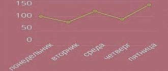
In each of the twelve months of the year, two maximum values (acrophases) of OPSS were identified, namely: at 8 and 22 o'clock (in September - at 20 o'clock). Significant decreases in OPSS were recorded at 22 o'clock relative to 8 o'clock in April (P < 0 .05; - 5, 10%) and October (P<0.05; - 8, 75%), respectively (Table 1). Regarding 8 hours and partly 10 hours, decreases in OPSS were found in each of the 12 months of the year (Table 2).
The minimum significant decrease in OPSS was found in July at 16 o'clock relative to 10 o'clock (BD; - 4.99%; P <0.05), and the maximum reliable decrease in OPSS was detected in June at 10 o'clock relative to 8 o'clock (AB; - 10, 71%; P <0.001).
Table 1
Dynamics of biological rhythms of total peripheral vascular resistance (TPVR) during the year in women sitting (M ± m), n = 28
| Time | M E S I C Y YEARS. | |||||
| days. | January | February | March. | April | May | June |
| 8 hours A | 999891,0 | 1046236,3 | 1019501 | 1023213,6 | 1063265 | 1001922 |
| ± 23222,5 | ± 20593,7 | ± 20165 | ±16949, 1 | ±14352,4 | ±21037,4 | |
| 10 hours B | 923444,4 | 904083,9 | 969778,6 | 949307,6 | 910582,1 | 894683,2 |
| ± 20714,4 | ± 21065,0 | ± 27473 | ± 19410,5 | ±22054,1 | ±20627,7 | |
| 12 o'clock B | 903233,5 | 914510,8 | 947619,6 | 894643,0 | 849343,8 | 823754,8 |
| ± 21711,0 | ± 14048,8 | ± 22753 | ± 21314,8 | ±16805,0 | ±15266,4 | |
| 14 hours G | 896551,6 | 954562, 3 | 923951,7 | 927954,4 | 875441,4 | 802844,1 |
| ± 19916,7 | ± 17185,3 | ± 23281 | ±17974, 7 | ±15091,3 | ±17579,8 | |
| 16 hours D | 895579, 0 | 979136,1 | 959824,3 | 911608, 8 | 895447,5 | 868866,8 |
| ± 19942,8 | ± 18106,1 | ± 20493 | ± 20052,0 | ±19445,7 | ±17118,9 | |
| 18 hours E | 921579,7 | 973428,4 | 951111,9 | 925200, 3 | 898099,3 | 864648,8 |
| ± 19084,7 | ± 12568,6 | ± 12815 | ± 20524,4 | ±19305,6 | ±19681,1 | |
| 20 h F | 916030, 1 | 951615,4 | 1002972 | 955037,3 | 906519,1 | 849804,1 |
| ± 29550,4 | ± 15653,8 | ± 18468 | ± 16889,4 | ±17341,6 | ±17294,1 | |
| 22 h. W | 960420,7 | 1004257,0 | 1028325 | 971127,0 | 945946,6 | 879794,2 |
| ± 24043,6 | ± 19537,6 | ± 15560 | ± 15645,8 | ±19472,1 | ±16219,9 | |
| 1 | AB* | AG * * * | AB * | AB * * * | BV* | AB * ** |
| 2 | AB * * * | BP * | AG * * * | AJ * * * | VD * | BV * * * |
| 3 | AG * * * | AE * * * | BP * | AZ * | BE* | BG * * * |
| 4 | BP * * * | AJ * * * | AE * * * | BV* | VJ * | VD * |
| 5 | AE * * | BG * * | BZ * | VJ * | GZ * * * | VZ * * |
| 6 | AJ * | DB * * | VJ * | VZ * * * | DZ * | GD * * |
| 7 | VZ * | BE * * * | VZ * * * | GZ * | EZ * | GE* |
| 8 | GZ * | BZ * | GJ * * | DZ * | GZh * | |
| 9 | DZ * | BZ * * * | DZ * * | EZ * | GZ * * * | |
| 10 | VG * | EJ * | ||||
| 11 | VD * * * | |||||
| 12 | BE * * * | |||||
| 13 | VJ * | |||||
| 14 | GZ * | |||||
| 15 | ZhZ * | |||||
Continuation of Table 1
| Time | M E S I C Y YEARS. | |||||
| days. | July | August | September | October | November | December |
| 8 hours A | 986308,0 | 983034,3 | 956361,4 | 993369,0 | 957324,6 | 922226,6 |
| ± 12053,8 | ± 11342,8 | ± 12804,9 | ±24041,9 | ± 18400,8 | ± 17930,0 | |
| 10 hours B | 882106,0 | 870030,5 | 882574,9 | 895259,2 | 857419,4 | 844827,2 |
| ± 17301,1 | ± 14167,3 | ± 17181,3 | ± 19956,2 | ± 19717,8 | ± 17246,5 | |
| 12 o'clock B | 825022,0 | 832275,1 | 812661,9 | 846989,1 | 859717,8 | 859097,8 |
| ± 21163,8 | ± 12179,4 | ± 16320,8 | ± 13512,4 | ± 17150,2 | ± 16071,1 | |
| 14 o'clock G. | 845260,6 | 803676,4 | 856465,1 | 856787,5 | 874889,3 | 871938,4 |
| ± 16022,2 | ± 10398,9 | ± 23729,2 | ±17377,1 | ± 15056,0 | ± 13663,4 | |
| 16 hours D. | 838149,0 | 855635,4 | 857536,4 | 849386,8 | 881624,8 | 864236,9 |
| ± 16072,3 | ± 12552,9 | ± 11920,4 | ± 17448,8 | ± 19343,4 | ± 17060,7 | |
| 18 hours E. | 859356,4 | 850843,6 | 886949,6 | 872388,8 | 866200,7 | 881744,3 |
| ± 14870,1 | ± 11199,5 | ± 13741,8 | ±17613,5 | ± 11988,4 | ± 16672,1 | |
| 20 hours F | 851830,4 | 850986,8 | 890024,3 | 891481,6 | 891973,3 | 885551,3 |
| ± 14102,5 | ± 7719,84 | ± 10231,7 | ± 13104,4 | ± 20644,7 | ± 15023,1 | |
| 22 h. W | 875350,5 | 874575,0 | 886774,0 | 906450,0 | 938368,3 | 925549,5 |
| ± 12277,2 | ± 10712,6 | ± 13021,9 | ± 27630,2 | ± 23688,3 | ± 26597,3 | |
| 1 | BV* | BV* | AB * ** | AB * * * | AB * * * | AB * * * |
| 2 | DB * | VG * | BV * * * | AZ * | AG * * * | AB * * |
| 3 | VZ * | VZ * * | VD * | BV* | BP * * * | AG* |
| 4 | DZ * | GD * * * | BE * * * | DB * | AJ * | BP * |
| 5 | GE * * * | VZ * * * | VJ * | BZ * * | BZ * | |
| 6 | GZ * * * | J* | VZ * | VZ * * | BZ * * | |
| 7 | ZhZ * | J* | GZ * | VZ * | ||
| 8 | DZ * | DZ * | GZ * | |||
| 9 | EZ * | DZ * | ||||
Note: * - Р ˂ 0.05 or Р ˂ 0.02; * * — Р ˂ 0.01; * * * — Р ˂ 0.001Acrophases (or maximum values) are highlighted in bold with underlining. In order to show a more complete picture of the reliability of changes in results, time periods were given letter designations.
table 2
Significant changes in OPSS as a percentage between time periods during the year in women sitting (M ± m), n = 28
| Time | MONTHS OF THE YEAR | |||||
| segments. | January | February | March | April | May | June |
| 8-10 AB | — 7, 65 | — 7, 23 | — 10, 71 | |||
| 8-12 AB | — 9, 67 | — 7, 06 | ||||
| 8-14 AG | — 10, 34 | — 8, 77 | — 9, 38 | |||
| 8-16 BP | — 10, 44 | — 6, 42 | — 5, 86 | |||
| 8-18 AU | — 7, 84 | — 6, 96 | — 6, 71 | |||
| 8-20 AJ | — 8, 39 | — 9, 05 | — 6, 67 | |||
| 8-22 AZ | — 5, 10 | |||||
| 10-12 BV | — 5, 76 | — 6, 73 | — 7, 93 | |||
| 10-14 BG | + 5, 58 | — 10, 27 | ||||
| 10-16 DB | + 8, 90 | |||||
| 10-18 BU | + 7, 67 | |||||
| 10-20BJ | + 5, 25 | |||||
| 10-22 BZ | + 11, 08 | + 6, 03 | ||||
| 12-14 VG | + 4, 37 | |||||
| 12-16 VD | + 7, 06 | + 5, 42 | + 5, 47 | |||
| 12-18 EE | + 6, 44 | + 5, 74 | ||||
| 12-20VZH | + 4, 05 | + 5, 84 | + 6, 75 | + 6, 73 | ||
| 12-22VZ | + 6, 33 | + 8, 51 | + 8, 54 | + 6, 80 | ||
| 14-16GD | + 8, 22 | |||||
| 14-18GE | + 7, 69 | |||||
| 14-20GZH | + 8, 55 | + 5, 84 | ||||
| 14-22 GZ | + 7, 12 | + 5, 20 | + 4, 65 | + 8, 05 | + 9, 58 | |
| 16-18DE | ||||||
| 16-20J | ||||||
| 16-22DZ | + 7, 24 | + 7, 13 | + 6, 52 | + 5, 63 | ||
| 18-20EZh | + 5, 45 | |||||
| 18-22EZ | — 4, 96 | + 5, 32 | ||||
| 20-22ZhZ | + 5, 53 | |||||
Continuation of Table 2
| Time | MONTHS OF THE YEAR | |||||
| segments. | July | August | September | October | November | December |
| 8-10 AB | — 7, 72 | — 9, 88 | — 10, 44 | — 8, 40 | ||
| 8-12 AB | — 6, 85 | |||||
| 8-14 AG | — 8, 62 | — 5, 46 | ||||
| 8-16 BP | — 7, 94 | — 6, 29 | ||||
| 8-18 AU | ||||||
| 8-20 AJ | — 6, 83 | |||||
| 8-22 AZ | — 8, 75 | |||||
| 10-12 BV | — 6, 48 | — 4, 34 | — 7, 93 | — 5, 40 | ||
| 10-14 BG | ||||||
| 10-16 DB | — 4, 99 | — 5, 13 | ||||
| 10-18 BU | ||||||
| 10-20BJ | + 4, 82 | |||||
| 10-22 BZ | + 9, 44 | + 9, 55 | ||||
| 12-14 VG | — 3, 44 | |||||
| 12-16 VD | + 5, 52 | |||||
| 12-18 EE | + 9, 14 | |||||
| 12-20VZH | + 5, 25 | |||||
| 12-22VZ | + 6, 10 | + 5, 08 | + 9, 11 | + 7, 02 | + 9, 14 | + 7, 73 |
| 14-16GD | + 6, 46 | |||||
| 14-18GE | + 5, 86 | |||||
| 14-20GZH | + 5, 88 | |||||
| 14-22GZ | + 7, 25 | + 6, 14 | ||||
| 16-18DE | ||||||
| 1620J | + 3, 78 | + 4, 95 | ||||
| 16-22DZ | + 4, 43 | + 6, 71 | + 6, 43 | + 7, 09 | ||
| 18-20EZh | ||||||
| 18-22EZ | + 8, 33 | |||||
| 20-22ZhZ | + 2, 77 | |||||
The OPSS parameter has rhythm. As noted in the literature [1,7,18,30,31], OPSS in the daytime was found to be lower compared to morning values, and the capillaries are maximally dilated at 18 hours. In our work, lower values of TPSS were recorded throughout the year at 14-16 hours compared to the morning (Table 1), which coincides with the above mentioned literature data. It is assumed that the maximum release of adrenaline and norepinephrine from the adrenal medulla occurs in the morning, namely at 9-12 am [1.7, 12, 18, 31], and the minimum is at 18 pm [31]. According to [1], the maximum level of physiological and mental activity of various human functional systems is observed during a period of high levels of adrenaline in the blood, namely in the first half of the day. It can be assumed that classes in an educational institution are most useful from 10 to 12 hours.
CONCLUSIONS:
1. The maximum morning and evening values of OPSS were recorded during the year in women.
2. Seasonal changes in OPSS were identified.
Bibliography:
1 Agadzhanyan N.A. "Biological rhythms". - M. - Medicine. — 1967.- 120 p.
2.Aulik, I.V. “Determination of physical performance in the clinic and sports.” // M.: Medicine. - 1990. - 192 P.
3. Babsky R.M., Berseneva A.P. “Adaptive capabilities of the body and the concept of physiological norm.” // Collection of materials of the 18th Congress of the Physiological Society. - Kazan. - 2001. - P. 304.
4. Belousov Yu.B., Akhadov Sh.V., Volkov M.G. “Comparative effect of calcium antagonists on hemodynamics in patients with a stable form of essential hypertension.” // Ter. archive. - 1994. - No. 3. - P. 72 - 77.
5. Bisyarina V.P. Yakovlev V.M., Kuksa P.Ya. Chapter 7. “Age-related characteristics of blood pressure.” // In the book: Arterial vessels and age. - M. - Medicine. - 1986. - P. 174 - 199.
6. Glazyrina I.P. “This different medicine” - M.: Znanie. - 1992. - 96 pp.
7.Johnson P. “Peripheral circulation” - M.: Medicine, 1982. - 440 pp.
8. Dligach D.L. and Kulaev B.S. “Vascular reflexes as they are” // In the book. Life and vessels.- M.- Knowledge -1989.- P.153 –
9.Doskin V.A., Lavrentieva N.A. “Daily rhythms and their role in human physiology and pathology” // Soviet Medicine. - 1972. - No. 4. - P. 67 - 70.
10, Caro K. Pedley T., Schroter R. et al. “Mechanics of Blood Circulation.” - M -Mir - 1981. - 624 P.
11. Karpman V.L., Lyubina B.G. “Dynamics of blood circulation in athletes.” -
M - F and S - 1982.- 135 S.
12.Kozinets, V.I. Physiological systems of the human body, main indicators.” M: Triad - X, 2000.- 336s
13. Krylova V.I. and Soboleva T.M. “Human microvasculature.” - M. - Published at the Peoples' Friendship University. - 1986. - 63 p.
14. Kupriyanovich L.I. "Location of biological clocks in living organisms and their regulation." / In the book: Biological rhythms and sleep. - M. - Nauka. - 1976. - P. 38 - 45.
15.Trainer's medical reference book. / Comp. Geselevich V.A. M.- FiS.- 1981.- P.271.
16. Minkin R.B. “Fundamentals of anatomy and physiology of the cardiovascular system.” - St. Petersburg Academy. - 1994. - pp. 8 - 46.
17.Mityanin Yu.P. Shuvalov M.A., Rubtsov A.T. "Robinson's double product as a method for determining the functional state of the cardiovascular system of students." // Materials of the 9th international conference. - M.-MSU. - 2006. - Part 1. - P. 200-202.
18. Seleznev S.A., Vashtina S.M., Mazurkevich G.S. “Comprehensive assessment of blood circulation in experimental pathology” - L - d. - Medicine. - 1976. - 207 p.
19. Smirnov A.D. ,Churina S.K., “Double work” in the diagnosis of the state of the cardiovascular system.//Human Physiology.- 1991.- T.17.- No. 3.- P. 64 - 66.
20. Tikhvinsky S.B. “Current problems of pediatric sports medicine.” // In the book: Children's sports medicine (edited by S.B. Tikhvinsky and S.V. Khrushchev). - M. - Medicine. - 1991. - P. 20 -25.
21. Human physiology. In 2 volumes. T.1.// Under the general editorship. V.M.Pokrovsky and G.F. Briefly. - M. - Medicine. - 1997. - 448 p.
22./Fomin N.A. Chapter 7. Blood circulation. // in the book: Human Physiology.- M.- Enlightenment.- 1992.- P. 164 - 193.
23.Chertov A.G. "Physical quantities". - M. - Higher School. - 1990. - 335 P.
24. Shurygin D.Ya., Alekseeva N.M., Karlov V.A. “Changes in indicators of hormonal activity of the adrenal glands, sympathetic-adrenal system and hemodynamics in healthy people at different times of the day” // Problems of endocrinology. - 1977. - T .23.- No. 3.- P. 50 - 57.
25.Emme A. M. “Watch of nature”. M.-Soviet Russia.- 1962.-150 p.